. 2022 Jul 5:13:914899.
doi: 10.3389/fimmu.2022.914899. eCollection 2022.

RGD-Labeled Hemocytes With High Migration Activity Display a Potential Immunomodulatory Role in the Pacific Oyster Crassostrea gigas

Affiliations

RGD-Labeled Hemocytes With High Migration Activity Display a Potential Immunomodulatory Role in the Pacific Oyster Crassostrea gigas

Zhao Lv et al. Front Immunol. .

Abstract

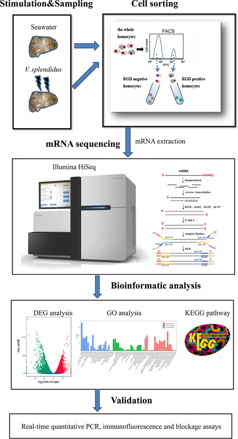
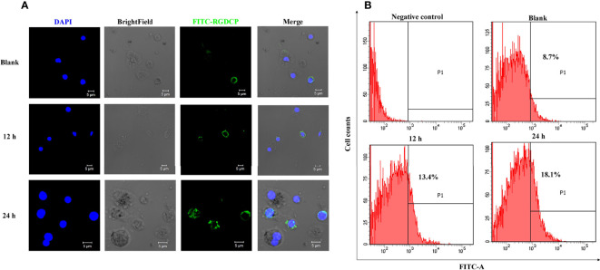
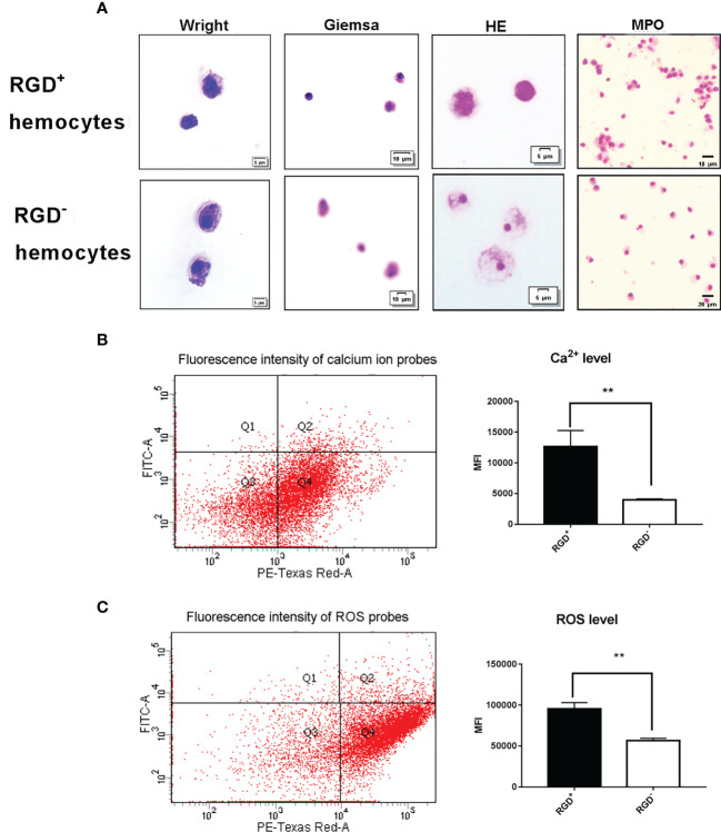
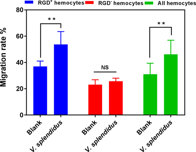
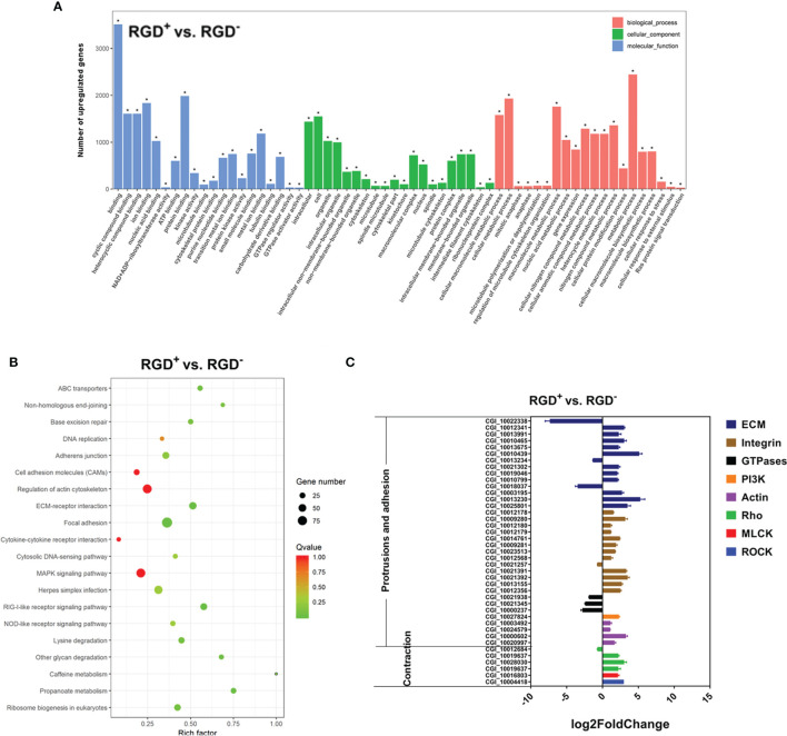
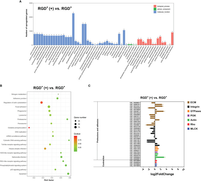
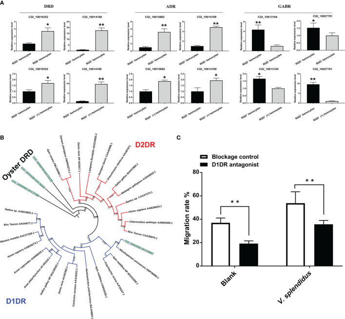
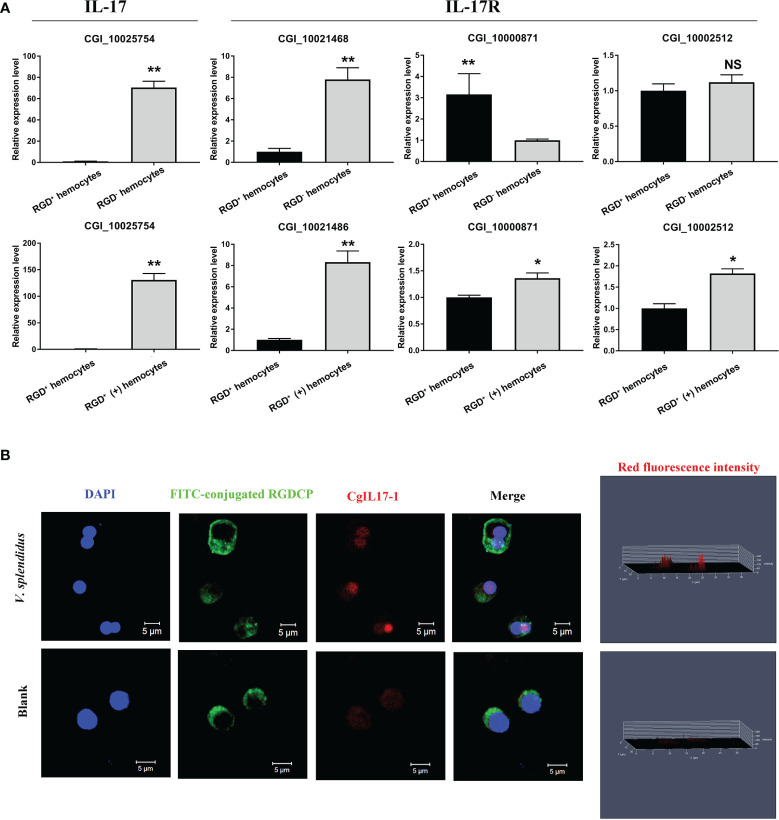
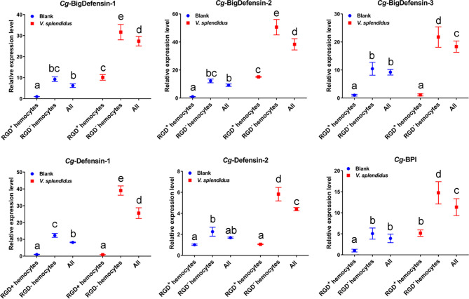
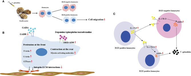
Immunocyte migration to infection sites is important for host cellular defense, but the main types of migrating hemocytes and their mechanisms against pathogen invasions are unclear in invertebrates. In the present study, a population of hemocytes in the Pacific oyster Crassostrea gigas labeled with a fluorescein isothiocyanate (FITC)-conjugated Arg-Gly-Asp (RGD)-containing peptide was sorted. RGD+ hemocytes were characterized by a smaller cell size and cytoplasmic-nucleo ratio, fewer cytoplasmic granules, and higher levels of myeloperoxidase, reactive oxygen species, and intracellular free calcium concentration. RGD+ hemocytes exhibited a high level of migration activity, which was further induced after V. splendidus infection. Transcriptome analysis revealed that RGD+ hemocytes highly expressed a series of migration-related genes, which together with migration-promoting genes were significantly upregulated after V. splendidus infection. The neuroendocrine system was also proven to regulate the migration activity of RGD+ hemocytes, especially with the excitatory neuroendocrine factor dopamine, which promoted migration activity as confirmed by receptor blocking assays. Meanwhile, RGD+ hemocytes could highly express immunomodulatory factor interleukin (IL)-17s and their receptor genes, which was positively related to the production of antimicrobial peptides in whole hemocytes after V. splendidus infection. Collectively, this study identified a specific hemocyte population, i.e., RGD+ hemocytes, that shows high migration activity in response to pathogen infection and exerts a potential immunomodulatory role by highly expressing IL-17s that might enhance the hemocytes' antimicrobial peptide production in oysters.

Keywords: C. gigas; RGD labeled hemocytes; antimicrobial immunity; immunomodulatory; migration activity.


Conflict of interest statement

The authors declare that the research was conducted in the absence of any commercial or financial relationships that could be construed as a potential conflict of interest.

Figures

Figure 1
Figure 1
Pipeline overview of analyses of the molecular mechanisms for the mediation of high migration activity and regulation of antimicrobial immunity of C. gigas RGD+ hemocytes. The hemocytes were collected, incubated with FITC-conjugated RGDCP, and sorted using FACS to prepare both RGD+ hemocytes and RGD- hemocytes. The differential expression in RGD+ hemocytes before or after V. spelendidus infection was revealed by transcriptome bioinformatic analyses, including DEG, GO term, and KEGG pathway analyses. The molecular mechanisms for the mediation of high migration activity and regulation of antimicrobial immunity were revealed based on DEGs and further validated using qPCR, receptor blocking assay, and confocal microscopy.
Figure 2
Figure 2
Fluorescent labeling of C. gigas RGD+ hemocytes and their response to V. splendidus challenge. (A) Immunofluorescence analysis of numerary changes in C. gigas RGD+ hemocytes after stimulation with V. splendidus. FITC-conjugated RGDCP-labeled oyster integrins on cell membranes are indicated in green, and the cell nucleus stained with DAPI is indicated in blue. Bar = 5 μm. (B) Flow cytometry analysis of numerary changes in C. gigas RGD+ hemocytes after stimulation with V. splendidus. FITC-positive cells and RGD+ hemocytes were gated as P1.
Figure 3
Figure 3
Morphological and cytochemical features of C. gigas RGD+ hemocytes. (A) The cytochemical staining of resting RGD+/RGD- hemocytes. Resting RGD+/RGD- hemocytes were stained for Wright, Giemsa, HE, and MPO. The upper panel and lower panel show cytochemical staining of sorted resting RGD+ hemocytes and RGD- hemocytes, respectively. The differences in (B) intracellular Ca2+ and (C) ROS levels between resting RGD+ and RGD- hemocytes. The double asterisk (**) stands for statistical significance at p < 0.01.
Figure 4
Figure 4
The migration activity of RGD+ hemocytes before and after V. splendidus stimulation. The double asterisk (**) stands for statistical significance at p < 0.01, and “NS” represents no statistical significance.
Figure 5
Figure 5
Transcriptome analysis of resting RGD+ hemocytes. (A) Top 60 GO terms enriched in the significantly upregulated genes in the resting RGD+ hemocytes compared to the resting RGD- hemocytes. (B) Top 20 KEGG pathway items enriched in the significantly upregulated genes in the resting RGD+ hemocytes compared to the resting RGD- hemocytes. (C) qPCR validation of the 40 selected DEGs involved in the formation of protrusions and firm adhesions at the front and contractions at the rear in resting RGD+ hemocytes compared to resting RGD- hemocytes. RGD+ and RGD- represent the resting RGD+ and RGD- hemocytes, respectively. The asterisk (*) stands for statistical significance at p < 0.01.
Figure 6
Figure 6
Transcriptome analysis revealing the molecular basis of the enhanced C. gigas RGD+ hemocyte migration in response to V. splendidus infection. (A) Top 60 GO terms enriched in the significantly upregulated genes in the activated RGD+ hemocytes compared to the resting RGD+ hemocytes. (B) Top 20 KEGG pathway items enriched in the significantly upregulated genes in the activated RGD+ hemocytes compared to the resting RGD+ hemocytes. (C) qPCR validation of the selected 38 DEGs involved in the formation of protrusions and firm adhesions at the front and contractions at the rear in the activated RGD+ hemocytes compared to the resting RGD+ hemocytes. RGD+ and RGD+ (+) represent the resting RGD+ and the activated RGD+ hemocytes, respectively. The asterisk (*) stands for statistical significance at p < 0.01.
Figure 7
Figure 7
The involvement of the neuroendocrine system in regulating migration activity in response to V. splendidus. (A) qPCR validation of differentially expressed neuroendocrine factor receptor genes in resting RGD+ hemocytes compared to resting RGD- hemocytes and in activated RGD+ (+) hemocytes compared to resting RGD+ hemocytes. (B) The dendrogram tree of the four differentially expressed DRDs. (C) Changes in migration activity after RGD+ hemocytes were blocked by a D1DR receptor inhibitor. The double asterisks (**) indicate statistical significance at p < 0.01, and the asterisk (*) indicates statistical significance at p < 0.05.
Figure 8
Figure 8
The expression profiles of IL-17s and their receptor genes. (A) qPCR validation of differentially expressed IL-17s and their receptor genes in resting RGD+ hemocytes compared to resting RGD- hemocytes and in activated RGD+ hemocytes compared to resting RGD+ hemocytes. (B) Immunofluorescence validation of CgIL17-1 (CGI_10025754) in activated RGD+ hemocytes compared to resting RGD+ hemocytes. FITC-conjugated RGDCP-labeled oyster integrins on cell membranes are indicated in green, the cell nucleus stained by DAPI is indicated in blue, and the CgIL17-1 antibody conjugated with Alexa Fluor 594 is indicated in red. Bar = 5 μm. The asterisk (*) and the double asterisk (**) stand for statistical significance at p < 0.05 and p < 0.01 respectively and “NS” represents no statistical significance.
Figure 9
Figure 9
Changes in the mRNA expression of representative AMPs in different hemocyte populations before and after V. splendidus challenge. The relative expression levels of the reported AMPs, including Cg-BigDefensin-1, Cg-BigDefensin-2, Cg-BigDefensin-3, Cg-Defensin-1, Cg-Defensin-2, and Cg-BPI, in all hemocytes, RGD+ hemocytes, and RGD- hemocytes were detected by qPCR. The letters “a, b, c, etc.” indicate statistical significance at p < 0.05.
Figure 10
Figure 10
A supposed path to enhance antimicrobial immunity mediated by RGD+ hemocytes in C. gigas. (A) The migration activity of C. gigas RGD+ hemocytes was enhanced by stimulation with V. splendidus. (B) The molecular mechanisms for the mediation of high migration activity in C. gigas RGD+ hemocytes. The downregulation of integrin–ECM interactions and the upregulation of migration-promoting genes and migration-related genes enhanced the migration activity of RGD+ hemocytes in response to V. splendidus infection. In addition, the migration activity of RGD+ hemocytes was regulated by neuroendocrine factors, among which the excitatory neuroendocrine factor dopamine especially promoted the migration of RGD+ hemocytes. (C) The molecular mechanisms for the regulation of antimicrobial immunity of C. gigas RGD+ hemocytes. RGD+ hemocytes highly expressed the immunomodulatory factor IL-17s and their receptor genes, which might promote the production of AMPs in whole hemocytes to enhance antimicrobial immunity.

Similar articles

Cited by

References

    1. Bhusal RP, Foster SR, Stone MJ. Structural Basis of Chemokine and Receptor Interactions: Key Regulators of Leukocyte Recruitment in Inflammatory Responses. Protein Sci (2020) 29(2):420–32. doi: 10.1002/pro.3744 - DOI - PMC - PubMed
    1. Nourshargh S, Alon R. Leukocyte Migration Into Inflamed Tissues. Immunity (2014) 41(5):694–707. doi: 10.1016/j.immuni.2014.10.008 - DOI - PubMed
    1. Teng TS, Ji AL. Neutrophils and Immunity: From Bactericidal Action to Being Conquered. J Immunol Res (2017) 2017:9671604. doi: 10.1155/2017/9671604 - DOI - PMC - PubMed
    1. van Rees DJ, Szilagyi K, Kuijpers TW, Matlung HL, van den Berg TK. Immunoreceptors on Neutrophils. Semin Immunol (2016) 28(2):94–108. doi: 10.1016/j.smim.2016.02.004 - DOI - PMC - PubMed
    1. Oishi Y, Manabe I. Macrophages in Inflammation, Repair and Regeneration. Int Immunol (2018) 30(11):511–28. doi: 10.1093/intimm/dxy054 - DOI - PubMed

Publication types