Review
. 2022 Jul 5:10:928113.
doi: 10.3389/fcell.2022.928113. eCollection 2022.

Chromatin Ubiquitination Guides DNA Double Strand Break Signaling and Repair

Affiliations
Review

Chromatin Ubiquitination Guides DNA Double Strand Break Signaling and Repair

Ksenia G Kolobynina et al. Front Cell Dev Biol. .

Abstract

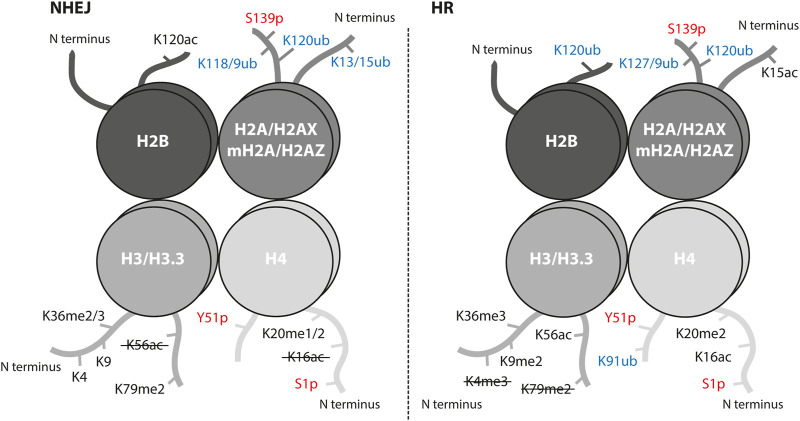
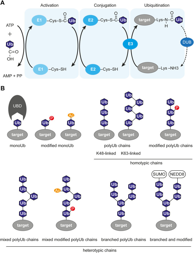
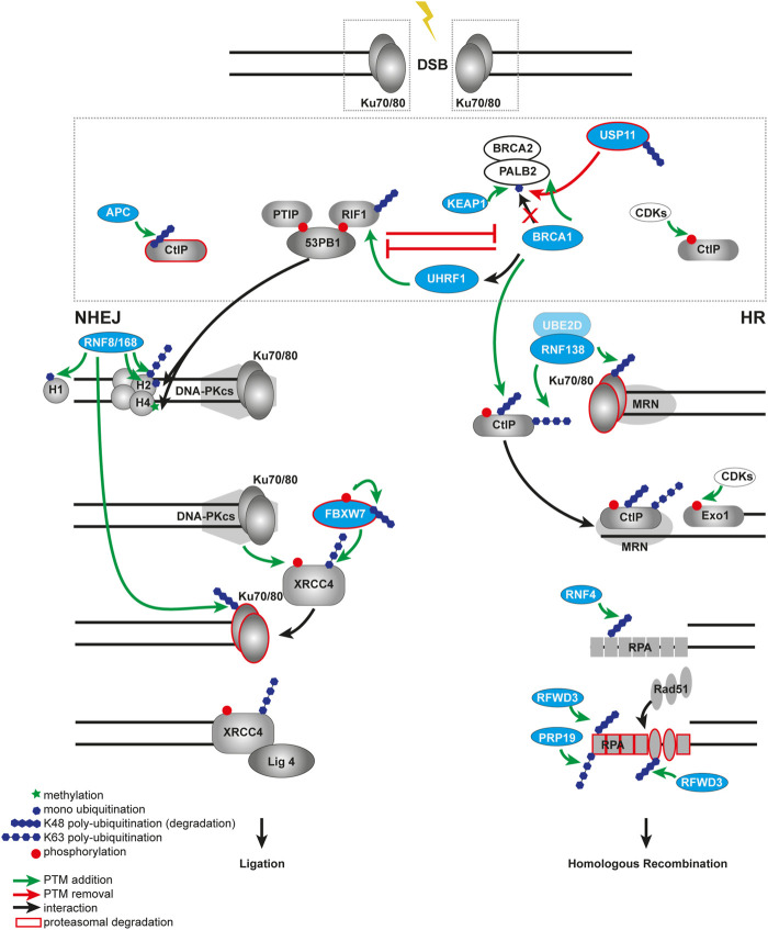
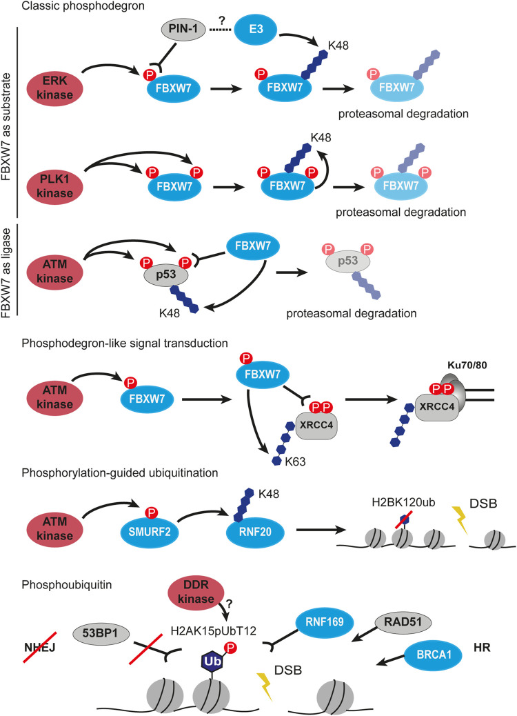
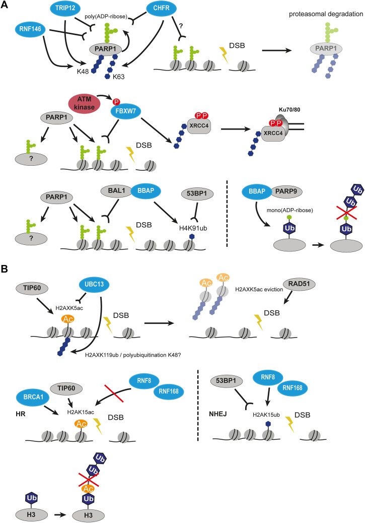
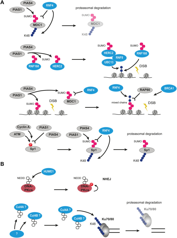
Chromatin is the context for all DNA-based molecular processes taking place in the cell nucleus. The initial chromatin structure at the site of the DNA damage determines both, lesion generation and subsequent activation of the DNA damage response (DDR) pathway. In turn, proceeding DDR changes the chromatin at the damaged site and across large fractions of the genome. Ubiquitination, besides phosphorylation and methylation, was characterized as an important chromatin post-translational modification (PTM) occurring at the DNA damage site and persisting during the duration of the DDR. Ubiquitination appears to function as a highly versatile "signal-response" network involving several types of players performing various functions. Here we discuss how ubiquitin modifiers fine-tune the DNA damage recognition and response and how the interaction with other chromatin modifications ensures cell survival.

Keywords: DNA damage response; DNA repair; chromatin; double-strand breaks; ubiquitination.


Conflict of interest statement

The authors declare that the research was conducted in the absence of any commercial or financial relationships that could be construed as a potential conflict of interest.

Figures

FIGURE 1
FIGURE 1
Histone post-translational modifications associated with NHEJ and HR repair pathways. Interaction of proteins and successful crosstalk between damage signaling and repair mechanism at the sites of DSBs is achieved by a highly controlled network of PTMs. Histone PTMs play a role in DSB repair pathway choice and serve as binding sites for repair proteins. Ubiquitination marks in blue, phosphorylation marks in red; ub, ubiquitination; p, phosphorylation; ac, acetylation; me, methylation. The crossed-out mark means it is removed in the repair pathway.
FIGURE 2
FIGURE 2
Ubiquitin-dependent signaling system. (A) Enzymatic cascade of ubiquitination involving three enzymes. The first step is activation of ubiquitin and covalent attachment to the E1 enzyme. The second step is conjugation, where ubiquitin is transferred to the E2 enzyme. The last step is the transfer of (poly)-ubiquitination to it’s target molecule mediated by a E3 ligase. The ubiquitination process is opposed by deubiquitination, which is the removal of the ubiquitin mark from the target protein by deubiquitinase (DUB). (B) Variety of ubiquitin chains, with or without modifications. P, phosphorylation; Ac, acetylation; SUMO, SUMOylation; NEDD8, NEDDylation; Ub, ubiquitin; UBD, ubiquitin-binding domain; target, target protein harboring ubiquitin modification.
FIGURE 3
FIGURE 3
Ubiquitination in DSB repair pathway choice. Open DNA ends are rapidly recognized by the Ku70/80 heterodimer. The choice between NHEJ and HR at the DSB is controlled by the competition between 53BP1 and BRCA1. In a NHEJ-permissive environment, 53BP1 builds a complex via ATM in a phosphorylation-dependent manner with its binding partners RIF1 and PTIP. The 53BP1-PTIP-RIF1 complex prevents recruitment of BRCA1 to the break and, therefore, antagonizes HR. The E3 ubiquitin ligase KEAP1 ubiquitinates PALB2 at the BRCA1 binding site and prevents assembling of the BRCA1-BRCA2-PALB2 complex, thus, promoting NHEJ. This process is balanced by the DUB USP11 and controlled by the cell cycle. BRCA1 antagonizes NHEJ by impairing retention of RIF1 at DSB sites in a CtIP- or UHRF1-dependent manner. Ubiquitin-dependent degradation of CtIP by the APC/C in NHEJ prevents resection process, in contrast to HR, where CtIP is not degraded but phosphorylated by the CDKs. The RNF8-RNF168-mediated ubiquitination on chromatin provides a binding platform for both 53BP1 and BRCA1. Later RNF8 also ubiquitinates Ku70/80 to facilitate its proteasomal degradation and removal from the broken DNA ends. In a HR-permissive environment the E3 ubiquitin ligase RNF138 ubiquitinates Ku to remove it from DSB much earlier compared to NHEJ, allowing end-resection factors to gain access to the deprotected ends. RNF138 also ubiquitinates CtIP, enhancing its retention on damaged chromatin. At the later steps of NHEJ the ligation of broken ends promoted by FBXW7 that recognizes phosphorylated XRRC4 and subsequently ubiquitinates it. In contrast during the later steps of HR, RPA SUMOylation-guided ubiquitination by RNF4 promotes RPA turnover and RAD51 recruitment. Multi-site ubiquitination of RPA by the ubiquitin ligases RFWD3 and PRP19 also facilitates HR.
FIGURE 4
FIGURE 4
Crosstalk between phosphorylation and ubiquitination in DSB signaling and repair. First three panels describe the classic phosphodegron situations highlighting FBXW7 as substrate to phosphorylation and as phosphorylation reading ubiquitin ligase. Next, phosphodegron-like signal transduction by FBXW7 is shown, which results in ubiquitination and stabilization of XRCC4 at Ku70/80 covered DSB. The next panel depicts phosphorylation-guided ubiquitination involving sequential signal transfer by two ubiquitin ligases SMURF2 and RNF20. The last example of the crosstalk between phosphorylation and ubiquitination is the phosphorylation of the ubiquitin moiety. The H2AK15pUbT12 is recognized by the HR factors such as RNF169, RAD51 and BRCA1, but prevents binding of 53BP1 at DSB. P, phosphorylation, Ub, ubiquitin.
FIGURE 5
FIGURE 5
Crosstalk between PARylation and ubiquitination, as well as acetylation and ubiquitination in DSB signaling and repair. (A) Crosstalk between PARylation and ubiquitination. PARP1 auto-PARylates itself followed by recognition of PAR chains by several ubiquitin ligases such as CHFR, RNF146, TRIP12 and subsequent polyubiquitination. This results in proteasomal degradation of PARP1. PARP1-mediated modification of histones at DSBs can be recognized by FBXW7, which polyubiquitinates XRRC4 and leads to its interaction with Ku70/80. On the other hand, PARP1-mediated modification of histones can also be recognized by BAL1-BBAP which leads to ubiquitination of H4K91 creating a binding site for 53BP1. Interaction of BBAP with PARP9 leads to mono(ADP-ribosyl)ation of ubiquitin moiety preventing elongation of ubiquitin chain. (B) Crosstalk between acetylation and ubiquitination. TIP60 acetylates H2AXK5 creating a recognition site for UBC13, which then polyubiquitinates H2AXK119 resulting in eviction of H2AX. H2AK15 can be acetylated or ubiquitinated. When acetylated by TIP60, it is recognized by BRCA1 but prevents the action of RNF8/RNF168, thus promoting HR. When ubiquitinated by RNF8/RNF168, it is recognized by 53BP1 promoting NHEJ. Acetylation of the ubiquitin moiety similarly to mono(ADP-ribosyl)ation prevents elongation of ubiquitin chain. P, phosphorylation, Ac, acetylation, Ub, ubiquitin.
FIGURE 6
FIGURE 6
Crosstalk between SUMOylation and ubiquitination, as well as NEDDylation and ubiquitination in DSB signaling and repair. (A) Crosstalk between SUMOylation and ubiquitination. MDC1 can be SUMOylated by PIAS1 and PIAS4 and then recognized by RNF4 leading to its polyubiquitination and degradation. PIAS1 and PIAS4 can also SUMOylate RNF168 and HERC2 leading to their activation. Activated HERC2 interacts with RNF8 and UBC13 and ubiquitinates histones together with activated RNF168. PIAS1 and PIAS4-mediated SUMOylation of MDC1 and histones recruits RNF4, which builds mixed SUMO-ubiquitin chains recognized by RAP80. Phosphorylated Sp1 is recognized PIAS1 and PIAS4 leading to its SUMOylation. SUMOylated Sp1 is recognized by RNF4 and gets polyubiquitinated and degraded. (B) Crosstalk between NEDDylation and ubiquitination. HUWE1 ubiquitin ligase can NEDDylate DNA-PKcs leading to its autophosphorylation and promoting NHEJ. NEDDylation of potentially cullins leads to polyubiquitination of Ku70/80 and their degradation. The enzyme performing NEDDylation is unknown. P, phosphorylation; SUMO, SUMOylation; NEDD8, NEDDylation; U, ubiquitin.

Similar articles

Cited by

References

    1. Acs K., Luijsterburg M. S., Ackermann L., Salomons F. A., Hoppe T., Dantuma N. P. (2011). The AAA-ATPase VCP/p97 Promotes 53BP1 Recruitment by Removing L3MBTL1 from DNA Double-Strand Breaks. Nat. Struct. Mol. Biol. 18, 1345–1350. 10.1038/nsmb.2188 - DOI - PubMed
    1. Ahmed S. F., Buetow L., Gabrielsen M., Lilla S., Chatrin C., Sibbet G. J., et al. (2020). DELTEX2 C-Terminal Domain Recognizes and Recruits ADP-Ribosylated Proteins for Ubiquitination. Sci. Adv. 6. 10.1126/sciadv.abc0629 - DOI - PMC - PubMed
    1. Alagoz M., Katsuki Y., Ogiwara H., Ogi T., Shibata A., Kakarougkas A., et al. (2015). SETDB1, HP1 and SUV39 Promote Repositioning of 53BP1 to Extend Resection during Homologous Recombination in G2 Cells. Nucleic Acids Res. 43, 7931–7944. 10.1093/nar/gkv722 - DOI - PMC - PubMed
    1. Alcón P., Shakeel S., Chen Z. A., Rappsilber J., Patel K. J., Passmore L. A. (2020). FANCD2-FANCI Is a Clamp Stabilized on DNA by Monoubiquitination of FANCD2 during DNA Repair. Nat. Struct. Mol. Biol. 27, 240–248. 10.1038/s41594-020-0380-1 - DOI - PMC - PubMed
    1. Ali M. A. M., Strickfaden H., Lee B. L., Spyracopoulos L., Hendzel M. J. (2018). RYBP Is a K63-Ubiquitin-Chain-Binding Protein that Inhibits Homologous Recombination Repair. Cell Rep. 22, 383–395. 10.1016/j.celrep.2017.12.047 - DOI - PubMed

LinkOut - more resources