Review
. 2022 Jul 5:10:913809.
doi: 10.3389/fcell.2022.913809. eCollection 2022.

Microtubules in Microorganisms: How Tubulin Isotypes Contribute to Diverse Cytoskeletal Functions

Affiliations
Review

Microtubules in Microorganisms: How Tubulin Isotypes Contribute to Diverse Cytoskeletal Functions

Abesh Bera et al. Front Cell Dev Biol. .

Abstract

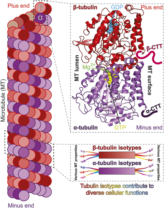
The cellular functions of the microtubule (MT) cytoskeleton range from relatively simple to amazingly complex. Assembled from tubulin, a heterodimeric protein with α- and β-tubulin subunits, microtubules are long, hollow cylindrical filaments with inherent polarity. They are intrinsically dynamic polymers that utilize GTP binding by tubulin, and subsequent hydrolysis, to drive spontaneous assembly and disassembly. Early studies indicated that cellular MTs are composed of multiple variants, or isotypes, of α- and β-tubulins, and that these multi-isotype polymers are further diversified by a range of posttranslational modifications (PTMs) to tubulin. These findings support the multi-tubulin hypothesis whereby individual, or combinations of tubulin isotypes possess unique properties needed to support diverse MT structures and/or cellular processes. Beginning 40 years ago researchers have sought to address this hypothesis, and the role of tubulin isotypes, by exploiting experimentally accessible, genetically tractable and functionally conserved model systems. Among these systems, important insights have been gained from eukaryotic microbial models. In this review, we illustrate how using microorganisms yielded among the earliest evidence that tubulin isotypes harbor distinct properties, as well as recent insights as to how they facilitate specific cellular processes. Ongoing and future research in microorganisms will likely continue to reveal basic mechanisms for how tubulin isotypes facilitate MT functions, along with valuable perspectives on how they mediate the range of conserved and diverse processes observed across eukaryotic microbes.

Keywords: cytoskeleton; microorganism; microtubule; tubulin; tubulin isotype.


Conflict of interest statement

The authors declare that the research was conducted in the absence of any commercial or financial relationships that could be construed as a potential conflict of interest.

Figures

FIGURE 1
FIGURE 1
Isotypes of α- and β-tubulin construct functionally diverse microtubules. Microtubules (MTs) polymerize from “head-to-tail” binding of tubulin, a heterodimer of α- and β-tubulin subunits (left). Both α- and β-tubulin subunits bind GTP but only the β-tubulin bound GTP is hydrolysable and exchangeable [top-right; cryo-EM structure of S. cerevisiae tubulin, polymerized with GTP in vitro; PDB ID: 5W3F (Howes et al., 2017)]. The carboxy terminal tails (CTTs) are normally unstructured and are only drawn representatively and not to scale. The α- and β-tubulin subunits can be encoded by multiple genes to produce variants, or isotypes (bottom-right; color gradient represents different isotypes). Tubulin isotypes can each possess common and/or unique molecular properties which expands the ability of MTs to optimally perform diverse and specialized functions (represented by arms/hands differentially extending from isotypes).
FIGURE 2
FIGURE 2
Microorganisms express one or more α- and β-tubulin isotypes to construct various MT-organelles and to perform diverse functions with a range of complexity. Example microorganisms are represented in either different cell cycle (blue text) or life cycle (orange text) stages, along with MTs (dark and light green lines), mitotic spindle (thick green line), nuclei (blue) and corresponding MTOCs (red). Top row (from left to right, clockwise): schematics of mitotic S. cerevisiae in anaphase; mitotic S. pombe in anaphase; tetranucleate, interphase Aspergillus nidulans germling (germinated from conidia, an asexual, uninucleate spore produced during vegetative life cycle. Septa are not formed until third karyokinesis) and infectious F. graminearum multicellular macroconidia, the translucent, canoe-shaped asexual spores possessing 4–5 septa and derived from phialides, the conidium producing cells. Middle row (left): T. thermophilus undergoing cell division and demonstrating distinct localization of β-tubulin isotypes. Btu2 is enriched in somatic cilia (green lines on cell surface) and basal bodies (black dots beneath each cilium on cell surface; not all cilia are shown). Blt1 and Blt4 construct the spindle (red lines) in the mitotic division of the micronucleus (smaller blue circle) and also assemble MTs (red lines) during amitotic division of the macronucleus (large blue circle). Middle row (right): C. reinhardtii in interphase, showcasing diverse MT organelles including the two apical flagella (thick green lines projecting outward) and 4 MT rootlets (thinner green and light green lines emerging from basal bodies within the cell), which contains stable, acetylated α-tubulin. The mother (red) and daughter (dark red) basal bodies, several nucleus-basal body connectors (black lines), eyespot (brown) and chloroplast (grey object at cell posterior) are also shown. Bottom row (left): P. polycephalum depicted in respective amoeba and flagellate stages. The uninucleate amoeba (left) have unorganized MTs (green lines), randomly positioned nucleus (blue circle) with filopods (blue lines extending from the cytoplasm) that contributes to multidirectional movements. It reversibly transitions to the uninucleate, comma-shaped, flagellated, sexual spore myxamoeba (right). This phase demonstrates an anterior and posterior flagellum (thick green lines; anterior longer than posterior) that emanates from the basal body (red) as well as a flagellar cone of MTs (thinner green lines in conical arrangement, emanating from the apical basal body) that can extend to the dorsal side of the organism. The beak-shaped nucleus (blue) is positioned underneath the cone. Bottom row (right): T. gondii tachyzoite demonstrates a diverse array of complex MT-organelles. The spindle MTs (green lines) and the corset of 22 subpellicular MTs respectively nucleate from the centrioles (orange cylinders above the blue nucleus) and the Apical Polar Ring (APR) MTOC (orange circle at apical part of the cell). Also shown are the tubulin-based hollow cylindrical conoid (green cylinder above the APR), two preconoidal rings (grey circles) above it and two intraconoid MTs (green lines) within its circumference.

References

    1. Adachi Y., Toda T., Niwa O., Yanagida M. (1986). Differential Expressions of Essential and Nonessential Alpha-Tubulin Genes in Schizosaccharomyces pombe . Mol. Cell. Biol. 6, 2168–2178. 10.1128/mcb.6.6.2168-2178.1986 - DOI - PMC - PubMed
    1. Aiken J., Moore J. K., Bates E. A. (2018). TUBA1A Mutations Identified in Lissencephaly Patients Dominantly Disrupt Neuronal Migration and Impair Dynein Activity. Hum. Mol. Genet. 28, 1227–1243. 10.1093/hmg/ddy416 - DOI - PMC - PubMed
    1. Aiken J., Sept D., Costanzo M., Boone C., Cooper J. A., Moore J. K. (2014). Genome-wide Analysis Reveals Novel and Discrete Functions for Tubulin Carboxy-Terminal Tails. Curr. Biol. 24, 1295–1303. 10.1016/j.cub.2014.03.078 - DOI - PMC - PubMed
    1. Asakawa K., Kume K., Kanai M., Goshima T., Miyahara K., Dhut S., et al. (2006). The V260I Mutation in Fission Yeast α-Tubulin Atb2 Affects Microtubule Dynamics and EB1-Mal3 Localization and Activates the Bub1 Branch of the Spindle Checkpoint. MBoC 17, 1421–1435. 10.1091/mbc.e05-08-0802 - DOI - PMC - PubMed
    1. Ayaz P., Ye X., Huddleston P., Brautigam C. A., Rice L. M. (2012). A TOG:αβ-tubulin Complex Structure Reveals Conformation-Based Mechanisms for a Microtubule Polymerase. Science 337, 857–860. 10.1126/science.1221698 - DOI - PMC - PubMed