

A visualized pulmonary arterial thrombus by using a modified echocardiographic view in an intermediate-risk acute pulmonary embolism patient: A case report
- PMID: 35865766
- PMCID: PMC9295681
- DOI: 10.1002/ccr3.6105
A visualized pulmonary arterial thrombus by using a modified echocardiographic view in an intermediate-risk acute pulmonary embolism patient: A case report
Abstract
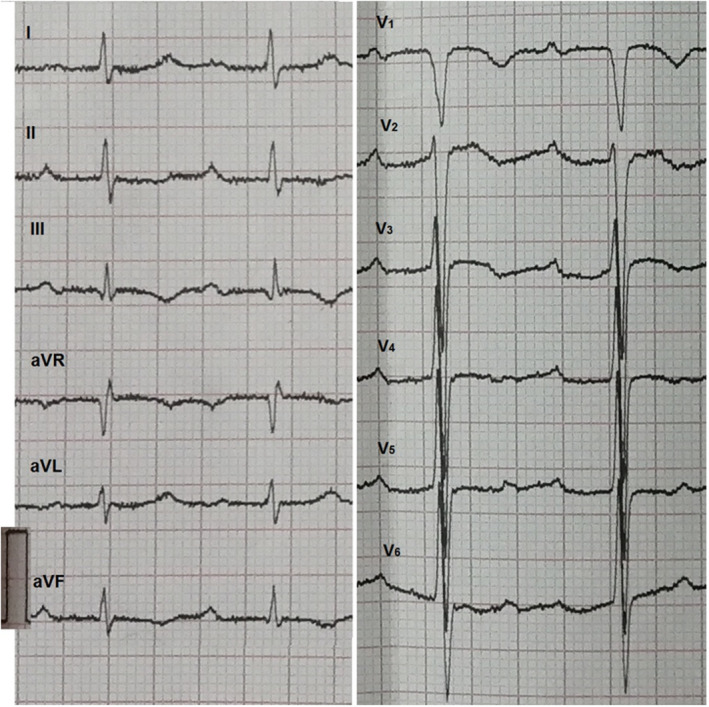
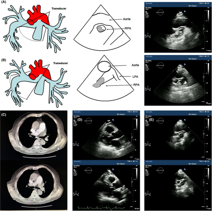
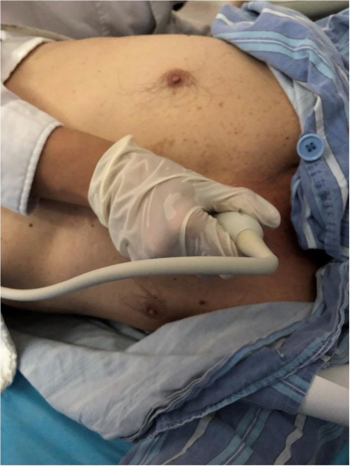
Acute pulmonary embolism (APE) is a life-threatening disease with nonspecific clinical signs and symptoms. Rapid and accurate diagnosis is crucial for the clinical management of patients with acute pulmonary embolism. A recommended echocardiography view may be of further help in the diagnosis and evaluation of the change in thrombosis and treatment. We reported a case of a 74-year-old man with a 12-day history of decreased exercise capacity and dyspnea. The patient was diagnosed with intermediate-risk APE as several pulmonary emboli in pulmonary artery were seen in multidetector computed tomographic pulmonary angiography with normal blood pressure and echocardiographic right ventricular overload. And we found a pulmonary artery clot in the right pulmonary artery through transthoracic echocardiography. After 11-days anticoagulation, the patient underwent a reassessment, showed a decrease in RV diameter and pulmonary artery thrombus. This case highlights the significant role that echocardiography played in a patient who presented pulmonary embolism with a stable hemodynamic situation and normal blood pressure. The modified echocardiographic view could provide correct diagnosis by identifying the clot size and location visually. Knowledge of the echocardiography results of APE would aid the diagnosis.
Keywords: acute pulmonary embolism; modified echocardiographic view; pulmonary artery clot.
© 2022 The Authors. Clinical Case Reports published by John Wiley & Sons Ltd.
Conflict of interest statement
The authors declare that they have no competing interests.
Figures
Similar articles
-
Clinical Application of Echocardiography in Evaluating Left Ventricular Diastolic Function in Patients with Acute Pulmonary Embolism.Comput Math Methods Med. 2022 May 11;2022:3483390. doi: 10.1155/2022/3483390. eCollection 2022. Comput Math Methods Med. 2022. Retraction in: Comput Math Methods Med. 2023 Sep 27;2023:9791415. doi: 10.1155/2023/9791415. PMID: 35602343 Free PMC article. Retracted.
-
Role of echocardiography in a patient with suspected acute pulmonary embolism: a case report.J Med Case Rep. 2019 Feb 19;13(1):37. doi: 10.1186/s13256-019-1994-y. J Med Case Rep. 2019. PMID: 30777120 Free PMC article.
-
High-risk pulmonary embolism assessed by transthoracic echocardiography: A case report.Medicine (Baltimore). 2018 May;97(18):e0545. doi: 10.1097/MD.0000000000010545. Medicine (Baltimore). 2018. PMID: 29718846 Free PMC article.
-
What echocardiographic findings differentiate acute pulmonary embolism and chronic pulmonary hypertension?Am J Emerg Med. 2023 Oct;72:72-84. doi: 10.1016/j.ajem.2023.07.011. Epub 2023 Jul 10. Am J Emerg Med. 2023. PMID: 37499553 Review.
-
ST-segment elevation in V1-V4 in acute pulmonary embolism: a case presentation and review of literature.Eur Heart J Acute Cardiovasc Care. 2016 Dec;5(8):579-586. doi: 10.1177/2048872615604273. Epub 2015 Sep 15. Eur Heart J Acute Cardiovasc Care. 2016. PMID: 26373811 Review.
Cited by
-
Saddle Pulmonary Embolism Detected by Transthoracic Echocardiography in a Patient With Suspected Myocardial Infarction.CASE (Phila). 2023 Dec 15;8(2):54-57. doi: 10.1016/j.case.2023.11.006. eCollection 2024 Feb. CASE (Phila). 2023. PMID: 38425574 Free PMC article.
References
-
- Konstantinides SV, Meyer G, Becattini C, et al. 2019 ESC guidelines for the diagnosis and management of acute pulmonary embolism developed in collaboration with the European Respiratory Society (ERS). Eur Heart J. 2019;40:3453‐3455. - PubMed
-
- Raskob GE, Angchaisuksiri P, Blanco AN, et al. Thrombosis: a major contributor to global disease burden. Arterioscler Thromb Vasc Biol. 2014;34(11):2363‐2371. - PubMed
-
- van der Hulle T, Dronkers CE, Klok FA, Huisman MV. Recent developments in the diagnosis and treatment of pulmonary embolism. J Intern Med. 2016;279(1):16‐29. - PubMed
Publication types
LinkOut - more resources
Full Text Sources
Research Materials
Miscellaneous
