. 2022 Jul 5:13:914153.
doi: 10.3389/fphar.2022.914153. eCollection 2022.

Clemastine Promotes Differentiation of Oligodendrocyte Progenitor Cells Through the Activation of ERK1/2 via Muscarinic Receptors After Spinal Cord Injury

Affiliations

Clemastine Promotes Differentiation of Oligodendrocyte Progenitor Cells Through the Activation of ERK1/2 via Muscarinic Receptors After Spinal Cord Injury

Lu-Yao Tong et al. Front Pharmacol. .

Abstract

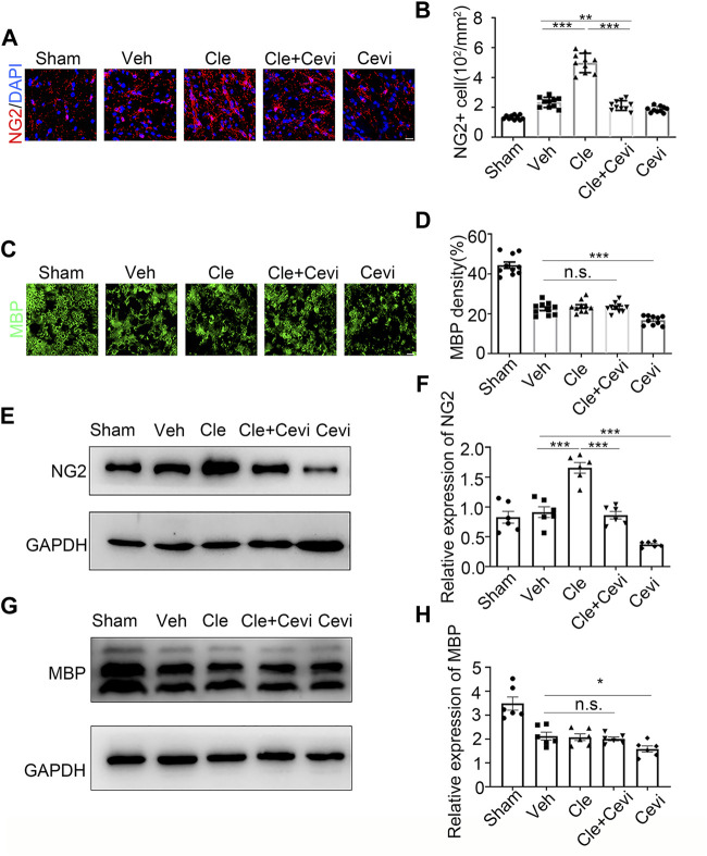
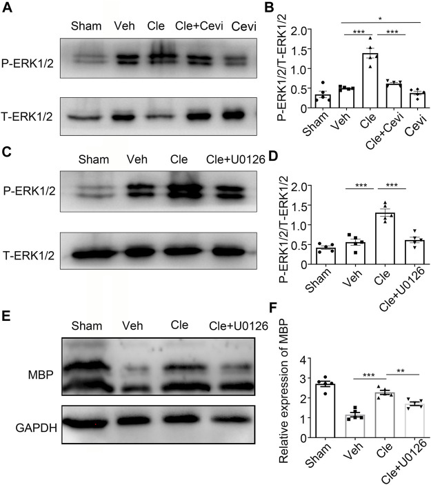
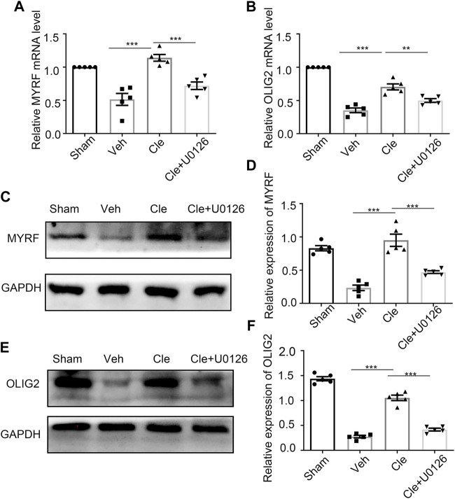
The recovery of spinal cord injury (SCI) is closely associated with the obstruction of oligodendrocyte progenitor cell (OPC) differentiation, which ultimately induces the inability to generate newly formed myelin. To address the concern, drug-based methods may be the most practical and feasible way, possibly applying to clinical therapies for patients with SCI. In our previous study, we found that clemastine treatment preserves myelin integrity, decreases the loss of axons, and improves functional recovery in the SCI model. Clemastine acts as an antagonist of the muscarinic acetylcholine receptor (muscarinic receptor, MR) identified from a string of anti-muscarinic drugs that can enhance oligodendrocyte differentiation and myelin wrapping. However, the effects of clemastine on OPC differentiation through MRs in SCI and the underlying mechanism remain unclear. To explore the possibility, a rat model of SCI was established. To investigate if clemastine could promote the differentiation of OPCs in SCI via MR, the expressions of OPC and mature OL were detected at 7 days post injury (dpi) or at 14 dpi. The significant effect of clemastine on encouraging OPC differentiation was revealed at 14 dpi rather than 7 dpi. Under pre-treatment with the MR agonist cevimeline, the positive role of clemastine on OPC differentiation was partially disrupted. Further studies indicated that clemastine increased the phosphorylation level of extracellular signal-regulated kinase 1/2 (p-ERK1/2) and the expressions of transcription factors, Myrf and Olig2. To determine the relationship among clemastine, ERK1/2 signaling, specified transcription factors, and OPC differentiation, the ERK1/2 signaling was disturbed by U0126. The inhibition of ERK1/2 in SCI rats treated with clemastine decreased the expressions of p-ERK 1/2, Myrf, Olig2, and mature OLs, suggesting that ERK1/2 is required for clemastine on promoting OPC differentiation and that specified transcription factors may be affected by the activity of ERK1/2. Moreover, the impact of clemastine on modulating the level of p-ERK 1/2 was restricted following cevimeline pre-injecting, which provides further evidence that the role of clemastine was mediated by MRs. Altogether, our data demonstrated that clemastine, mediated by MRs, promotes OPC differentiation under the enhancement of Myrf and Olig2 by activating ERK1/2 signaling and suggests a novel therapeutic prospect for SCI recovery.

Keywords: ERK1/2; clemastine; muscarinic receptor; myelin regulatory factor; oligodendrocyte progenitor cells; oligodendrocyte transcription factor 2.


Conflict of interest statement

The authors declare that the research was conducted in the absence of any commercial or financial relationships that could be construed as a potential conflict of interest.

Figures

FIGURE 1
FIGURE 1
Effect of clemastine on oligodendrocyte progenitor cells at 7 dpi in rats. (A) Immunofluorescence staining for NG2 (red) displays oligodendrocyte precursor cells in the spinal cord white matter tracts in the Sham group, Veh group, Cle group, Cle + Cevi group, and Cevi group. DAPI (blue) was used as a nuclear counterstain. Scale bar, 50 µm (B) Number of NG2-positive cells in the spinal cord white matter tracts was quantified. (C) Representative images showing MBP (green) in the spinal cords from the Sham group, Veh group, Cle group, Cle + Cevi group, and Cevi group at 7 dpi. Scale bar, 50 µm (D) Quantification of MBP density in the spinal cord white matter tracts. (E) Expression of NG2 at 7 dpi was evaluated by Western blotting. GAPDH expression was served as an internal control. (F) Quantitative analysis of NG2 protein levels. (G) MBP protein expression was assessed by Western blot analysis. (H) Quantitative analysis of MBP protein levels. The protein levels of MBP were normalized to GAPDH. Data are shown as the mean ± SEM. *p < 0.05, **p < 0.01, ***p < 0.001 (n = 6 for each group at 7 dpi) was identified by the Student unpaired t-test or one-way ANOVA followed by Tukey’s post-hoc test.
FIGURE 2
FIGURE 2
Clemastine promotes the differentiation of oligodendrocyte progenitor cells at 14 dpi in SCI. (A,C) Representative images showing NG2-positive OPCs (A) and area of MBP-positive mature OLs (C) in the spinal cords of Sham, Veh, Cle, and Cle + Cevi rats at 14 dpi. Scale bar, 50 µm. (B,D) Quantification of NG2+ cell numbers and MBP density from each group (n = 4). (E,G) Representative immunoblots of NG2 (E) and MBP (G) in the indicated situation. (F,H) Quantitative analysis of the expression of NG2 (F) and MBP (H) in the indicated situation. GAPDH expression from the same sample acts as an internal control. Error bars represent mean ± SEM, significance based on Student’s t-test with the respective controls (*p < 0.05, **p < 0.01, and ***p < 0.001, n = 5 for each group at 14 dpi).
FIGURE 3
FIGURE 3
Clemastine promotes OPC differentiation through the upregulation of p-ERK1/2 via MR. (A,C) Protein levels of phosphorylated ERK1/2 and total ERK1/2 were accessed by Western blotting. (B,D) Intensities of p-ERK1/2 were quantified. Total ERK1/2 expression from the same sample acts as an internal control. (E) Representative Western blots of MBP were performed. (F) Quantitative analysis of protein levels of MBP. The MBP protein levels were normalized to GAPDH. Error bars represent the mean ± SEM (n = 5 for each group). *p < 0.05, **p < 0.01, and ***p < 0.001 by Student’s t-test with the respective controls.
FIGURE 4
FIGURE 4
Influences of ERK1/2 activity on Myrf and Olig2 expression (A,B) qRT-PCR analysis of Myrf (A) and Olig2 (B) expression in the spinal cords from the Sham, Veh, Cle, and Cle + U1026 group. (C,E) Western blot analysis of Myrf (C) and Olig2 (E) was evaluated. The expression of Myrf and Olig2 is normalized to GAPDH expression. (D,F) Quantitative analysis of the protein levels of Myrf and Olig2. *p < 0.05, **p < 0.01, and ***p < 0.001 (n = 5 for each group).

References

    1. Ahnstedt H., Mostajeran M., Blixt F. W., Warfvinge K., Ansar S., Krause D. N., et al. (2015). U0126 Attenuates Cerebral Vasoconstriction and Improves Long-Term Neurologic Outcome after Stroke in Female Rats. J. Cereb. Blood Flow. Metab. 35 (3), 454–460. 10.1038/jcbfm.2014.217 - DOI - PMC - PubMed
    1. Ahuja C. S., Nori S., Tetreault L., Wilson J., Kwon B., Harrop J., et al. (2017a). Traumatic Spinal Cord Injury-Repair and Regeneration. Neurosurgery 80 (3S), S9–S22. 10.1093/neuros/nyw080 - DOI - PubMed
    1. Ahuja C. S., Wilson J. R., Nori S., Kotter M. R. N., Druschel C., Curt A., et al. (2017b). Traumatic Spinal Cord Injury. Nat. Rev. Dis. Prim. 3, 17018. 10.1038/nrdp.2017.18 - DOI - PubMed
    1. Alizadeh A., Karimi-Abdolrezaee S. (2016). Microenvironmental Regulation of Oligodendrocyte Replacement and Remyelination in Spinal Cord Injury. J. Physiol. 594 (13), 3539–3552. 10.1113/JP270895 - DOI - PMC - PubMed
    1. Almad A., Sahinkaya F. R., McTigue D. M. (2011). Oligodendrocyte Fate after Spinal Cord Injury. Neurotherapeutics 8 (2), 262–273. 10.1007/s13311-011-0033-5 - DOI - PMC - PubMed