. 2022 Jul 9;8(7):e09878.
doi: 10.1016/j.heliyon.2022.e09878. eCollection 2022 Jul.

Integrated analysis of the clinical consequence and associated gene expression of ALK in ALK-positive human cancers

Affiliations

Integrated analysis of the clinical consequence and associated gene expression of ALK in ALK-positive human cancers

Saifullah et al. Heliyon. .

Abstract

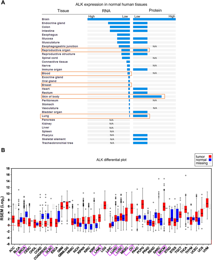
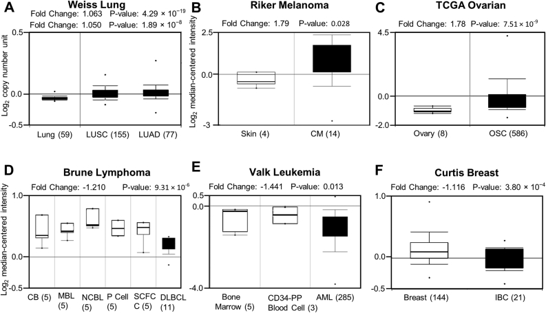
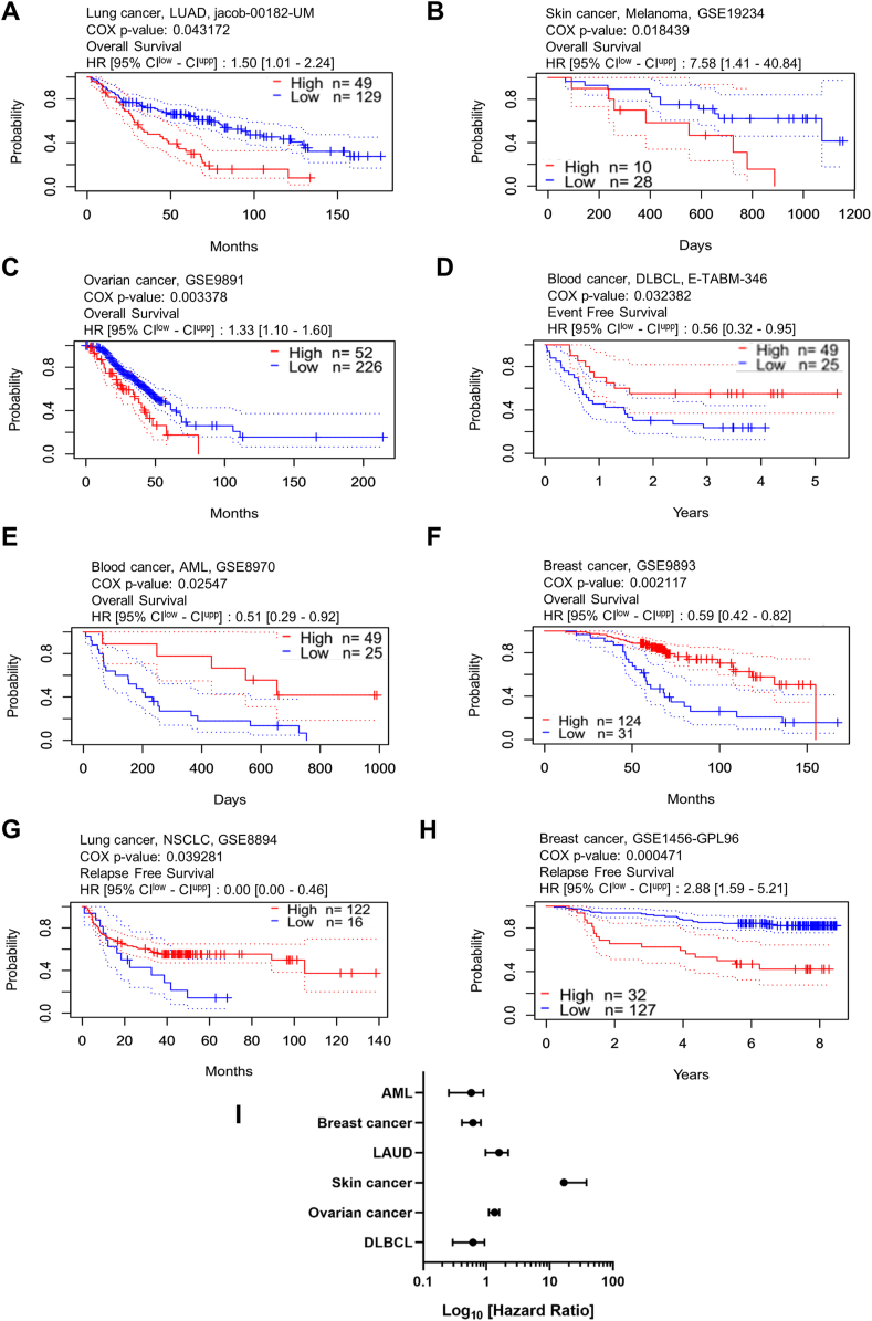
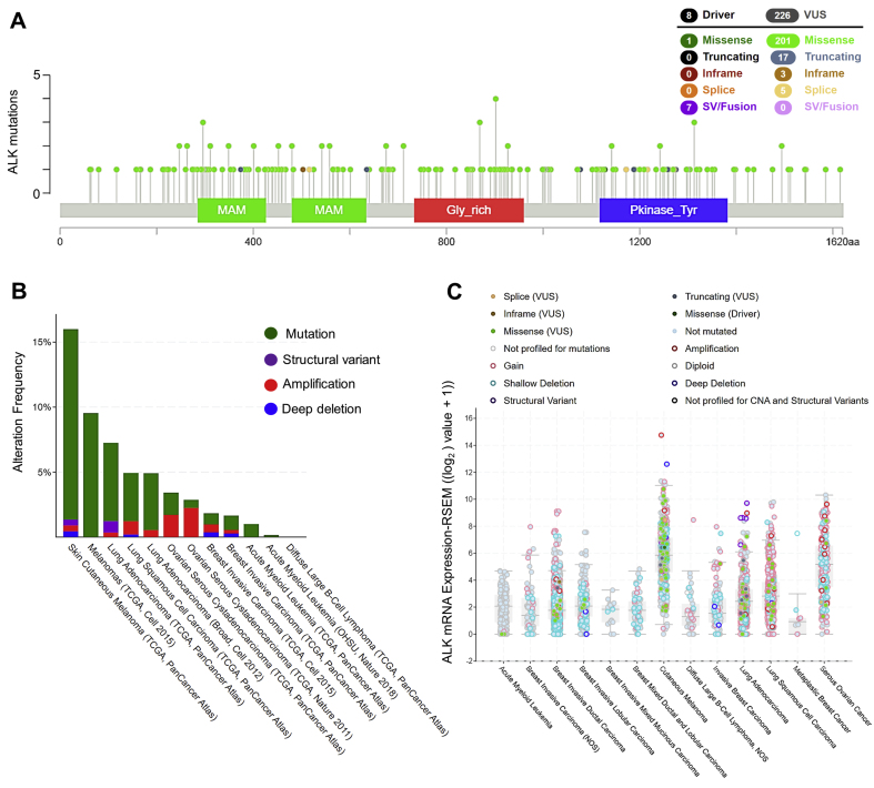
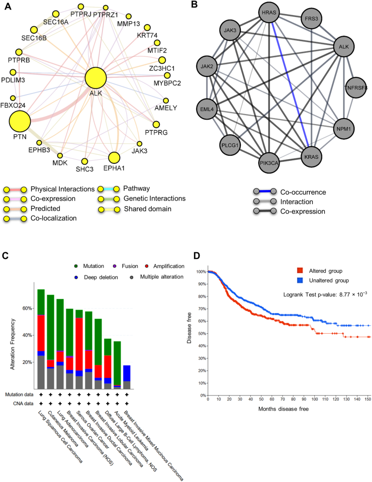
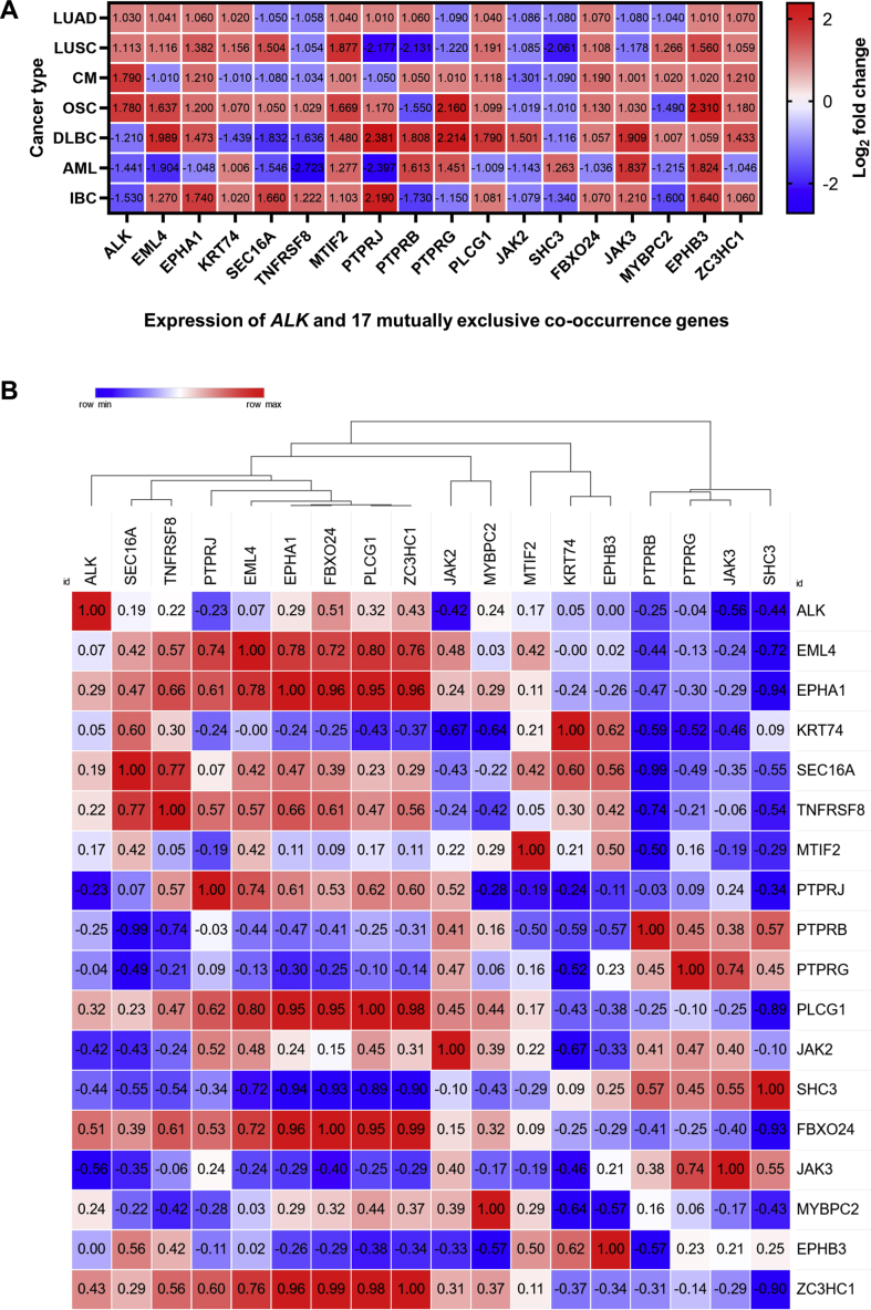
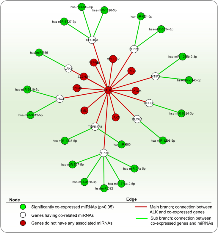
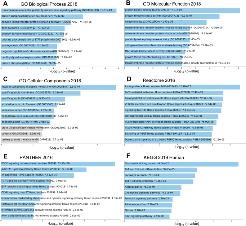
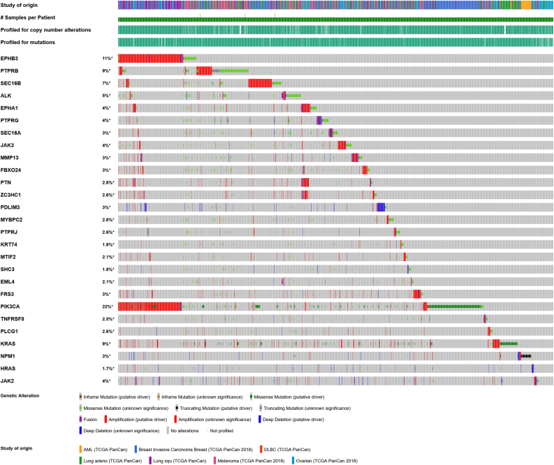
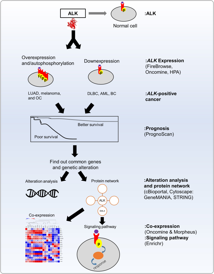
Anaplastic lymphoma kinase (ALK) is a tyrosine kinase receptor that is genetically altered in several cancers, including NSCLC, melanoma, lymphoma, and other tumors. Although ALK is associated with various cancers, the relationship between ALK expression and patient prognosis in different cancers is poorly understood. Here, using multidimensional approaches, we revealed the correlation between ALK expression and the clinical outcomes of patients with LUAD, melanoma, OV, DLBC, AML, and BC. We analyzed ALK transcriptional expression, patient survival rate, genetic alteration, protein network, and gene and microRNA (miRNA) co-expression. Compared to that in normal tissues, higher ALK expression was found in LUAD, melanoma, and OV, which are associated with poor patient survival rates. In contrast, lower transcriptional expression was found to decrease the survival rate of patients with DLBC, AML, and BC. A total of 202 missense mutations, 17 truncating mutations, 7 fusions, and 3 in-frame mutations were identified. Further, 17 genes and 19 miRNAs were found to be exclusively co-expressed and echinoderm microtubule-associated protein-like 4 (EML4) was identified as the most positively correlated gene (log odds ratio >3). The gene ontology and signaling pathways of the genes co-expressed with ALK in these six cancers were also identified. Our findings offer a basis for ALK as a prognostic biomarker and therapeutic target in cancers, which will potentially contribute to precision oncology and assist clinicians in identifying suitable treatment options.

Keywords: ALK expression; Cancers; Co-expression; LUAD; Patient prognosis.


Conflict of interest statement

The authors declare no competing interests.

Figures

Figure 1
Figure 1
Expression profile of ALK across human normal and cancer tissues. A. Outline of ALK mRNA and protein expression in normal human tissues based on Human Protein Atlas normal tissue immunohistochemistry, Expression Atlas data, and RNA-seq expression data derived from Open Targets Genetics platform. B. RSEM (RNA-Seq by Expectation-Maximization) RNA-Seq expression profiles for each cancer and corresponding normal tissue were obtained from The Cancer Genome Atlas (TCGA) using the FireBrowse datasets. The boxes denote the median, and the 25th and 75th % dots symbolize outliers. The red boxes represent tumor tissues and the blue boxes represent corresponding normal tissues. The gray boxes signify that no normal samples exist for that disease cohort. Full form of each cancer type is presented in Table 1.
Figure 2
Figure 2
ALK expression pattern in various cancer types. A–F.ALK expression in six cancer types derived from the Oncomine cancer microarray database. The left box plot represents ALK expression in normal tissue, while the box on the right represents cancer tissue. Statistically significant differences between normal and cancerous tissues are indicated by p < 0.05. LUSC, lung squamous cell carcinoma; LUAD, lung adenocarcinoma; CM, cutaneous melanoma; OSC, ovarian serous carcinoma CB, Centroblast; MBL, Memory B-lymphocyte; NCBL, Naive Pre-germinal Center B-lymphocyte; P cell, Plasma Cell; SCFC, Small Cleaved Follicle Center Cell; DLBCL, diffuse large B-cell lymphoma; CD-34 PP, CD34-Positive Peripheral Blood cell; AML, acute myeloid leukemia; and IBC, invasive breast carcinoma. ALK was found to be substantially upregulated in lung, skin, and ovarian cancers, and considerably downregulated in blood and breast cancers. The sample numbers are indicated in parentheses.
Figure 3
Figure 3
ALK expression and clinical prognosis in six different cancers retrieved from PrognoScan microarray cancer database. Kaplan–Meier patient survival estimate of A. lung adenocarcinoma, B. melanoma, C. ovarian cancer, D–E. blood cancer: DLBCL and AML, F. breast cancer for ALK expression, G. NSCLC, and H. breast cancer for ALK expression. The survival curve was determined as the threshold of the Cox p-value < 0.05 and p-value < 0.01. The red line denotes high expression and the blue line denotes low expression. The dotted line represents the maximum and minimum values of the average survival. HR, hazard ratio; CI, confidence interval; and n, number of patients. I. The statistically significant hazard ratio for the six different cancers was determined from Figure A–F and expressed as a forest plot.
Figure 4
Figure 4
Data on genetic alteration of ALK and consequent cancers in patients derived from TCGA PanCancer Atlas database through cBioPortal. A. Schematic of the ALK protein and its associated functional domains with the physical location of mutations. A total of 234 alterations were found from 1–1620 amino acid sequences, where 8 are driver and 226 VUS mutations respectively. The highest alteration type was a missense mutation (202) and the lowest was an in-frame mutation (3). B. The alteration frequencies of ALK and their associated cancers. The Y-axis shows the alteration frequency percentage, and the X-axis represents the type of cancers resulting from the alterations, including mutation and copy number alterations (CAN). The highest alteration frequency was found in melanoma (gene-altered 15.9% in 444 cases). C. RNA seq v2 results of genetically altered ALK mRNA expression in 12 cancer studies obtained from the cBioPortal web. The mutations included missense mutations in all cancer types (green dots), seven oncofusion in LUAD and melanoma (violet dots), nine truncating (gray dots), three slicing (light brown dots), and one in-frame mutation (deep brown dot). The frequency of copy number gain was the highest and distributed in all cancer types, followed by copy number diploid, shallow deletion, amplification, and deep deletion as indicated in different colors.
Figure 5
Figure 5
Protein network of ALK and their clinical significance in six cancers. A–B. Predicted functional protein partners of ALK generated by considering physical interaction, co-expression, co-localization, genetic interactions, and shared protein domains from GeneMANIA and STRING web servers using Cytoscape_v3.8.2 software. C. The genetic alteration frequency of 30 gene signatures in panels A–B was generated for lung, melanoma, ovarian, DLBC, AML, and breast cancers using the cBioPortal platform. The highest alteration percentage was reported in lung cancer, followed by melanoma and breast cancer, while the lowest was in AML. D. Kaplan–Meier patient survival estimation of patients with genetically altered and unaltered ALK and 17 partner genes (Table 3) in six types of cancers generated from cBioPortal. Patients with altered forms of ALK and 17 correlated genes showed significantly less survival than those with unaltered forms.
Figure 6
Figure 6
Co-expression and hierarchical clustering analyses. A. Co-expression analysis of ALK and its 17 mutually associated functional protein partner genes in six cancer types. The heat map was generated from the log2 fold-change expression value of ALK and mutually exclusive protein partner genes retrieved from the cancer microarray database (Oncomine). B. Hierarchical clustering and similarity matrix analysis of ALK and the expression of correlated genes in six cancer types. The heat map was generated from the log2 fold-change expression value of ALK and common genes retrieved from Oncomine. The outcome of the correlation was visualized using the similarity matrix and hierarchical clustering tools in the Morpheus server.
Figure 7
Figure 7
miRNA co-expressed with ALK and its mutually exclusive genes in humans. The miRNAs were identified from the Enrichr web portal, and interactions with common genes were analyzed using Cytoscape_v3.8.2 software. The green nodes indicate significantly enriched terms (p < 0.05). The white and red nodes denote genes with and without miRNA co-expression, respectively.
Figure 8
Figure 8
Gene ontology and pathway analyses of ALK and its co-related functional protein partners. A. Biological process, B. Molecular function, C. Cellular components, D. Reactome, E. PANTHER, and F. KEGG pathways of ALK and 18 partner proteins were obtained from the Enrichr web tool. The bar chart shows the top 10 enriched terms in the chosen library, along with their corresponding p-values. Colored bars correspond to terms with significant p-values (<0.05). ∗ indicates statistically significant values with p < 0.05.
Figure 9
Figure 9
Oncoprint of ALK and common genes. The genetic alterations of ALK and 17 common genes in six cancers were evaluated using cBioPortal. The highest alteration was observed in PIK3CA (22%), followed by EPHB3 (11%), KRAS, and PTPRB (9%), while the lowest was observed in HRAS (1.7%).
Figure 10
Figure 10
Schematic diagram of this study.

Similar articles

Cited by

References

    1. Golding B., Luu A., Jones R., Viloria-Petit A.M. The function and therapeutic targeting of anaplastic lymphoma kinase (ALK) in non-small cell lung cancer (NSCLC) Mol. Cancer. 2018;17:52. - PMC - PubMed
    1. Soda M., Choi Y.L., Enomoto M., Takada S., Yamashita Y., Ishikawa S., Fujiwara S.-i., Watanabe H., Kurashina K., Hatanaka H., et al. Identification of the transforming EML4–ALK fusion gene in non-small-cell lung cancer. Nature. 2007;448:561–566. - PubMed
    1. Saifullah, Tsukahara T. Integrated analysis of ALK higher expression in human cancer and downregulation in LUAD using RNA molecular scissors. Clin. Transl. Oncol. 2022 - PubMed
    1. Morris S., Kirstein M., Valentine M., Dittmer K., Shapiro D., Saltman D., Look A. Fusion of a kinase gene, ALK, to a nucleolar protein gene, NPM, in non-Hodgkin's lymphoma. Science. 1994;263:1281–1284. - PubMed
    1. Cesi G., Philippidou D., Kozar I., Kim Y.J., Bernardin F., Van Niel G., Wienecke-Baldacchino A., Felten P., Letellier E., Dengler S., et al. A new ALK isoform transported by extracellular vesicles confers drug resistance to melanoma cells. Mol. Cancer. 2018;17:145. - PMC - PubMed

LinkOut - more resources