

Urinary Podocyte Excretion Predicts Urinary Protein Selectivity and Renal Prognosis
- PMID: 35866051
- PMCID: PMC9296344
- DOI: 10.1155/2022/2702651
Urinary Podocyte Excretion Predicts Urinary Protein Selectivity and Renal Prognosis
Abstract
Background: Urinary podocyte excretion is related to a reduction in glomerular podocyte numbers, glomerulosclerosis, and urinary protein selectivity. To elucidate the role of urinary podocytes in proteinuria and renal prognosis and to identify the factors that cause podocyte detachment, we examined urinary podocytes in 120 renal biopsy patients.
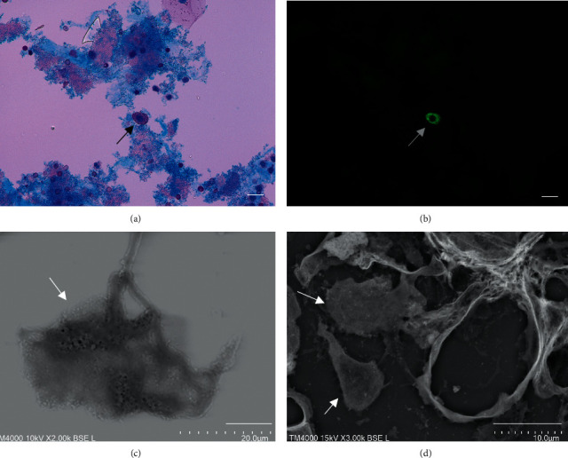
Methods: Podocytes were identified in urinary sediments stained with fluorescent-labeled anti-podocalyxin antibodies in ten high power fields. The amounts of protein bands, separated by SDS-polyacrylamide gel electrophoresis, were calculated using an image software program and the correlation with urinary podocytes was analyzed. Podocyte surface pores were observed using a low-vacuum scanning electron microscope. The renal prognosis, including induction of hemodialysis or 30% reduction in eGFR, was investigated.
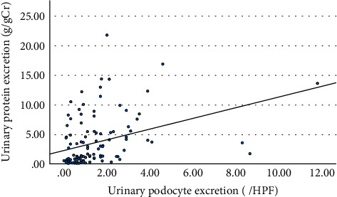
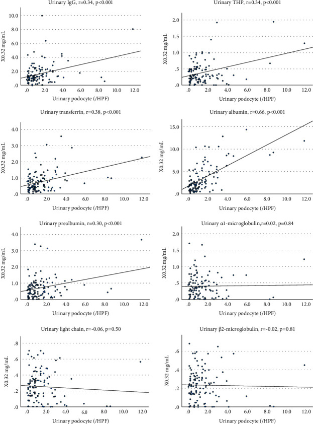
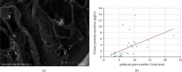
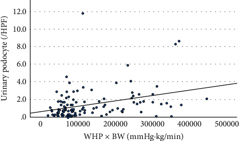
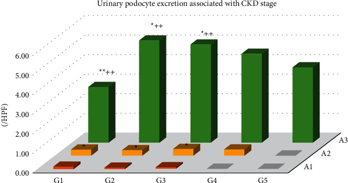
Results: Urinary podocyte excretion showed a higher positive correlation with albumin excretion compared to IgG, prealbumin, and transferrin. There were no significant correlations between urinary podocyte count and low molecular weight proteins, including β2-microglobulin and α1-microglobulin. The number of podocyte surface pores was positively correlated with proteinuria, suggesting enhanced albumin transcytosis. The hemodynamic pressure on the glomerular capillary wall, including products of pulse pressure and pulse rate (water hammer pressure), was positively correlated with urinary podocyte excretion. Urinary podocyte excretion and Tamm-Horsfall protein (THP) were independent risk factors for renal prognosis but were not related to response to treatment.
Conclusion: Urinary podocyte excretion was correlated with urinary albumin excretion, indicating specific albumin transport by podocytes. Podocytes were detached from the glomerular capillaries by water hammer pressure and THP was involved in the renal prognosis.
Copyright © 2022 Makoto Abe et al.
Conflict of interest statement
The authors declare that there are no conflicts of interest regarding the publication of this paper.
Figures
Similar articles
-
Urinary and glomerular podocytes in patients with chronic kidney diseases.Clin Exp Nephrol. 2014 Feb;18(1):95-103. doi: 10.1007/s10157-013-0814-8. Epub 2013 May 15. Clin Exp Nephrol. 2014. PMID: 23670304
-
Urine synaptopodin excretion is an important marker of glomerular disease progression.Korean J Intern Med. 2016 Sep;31(5):938-43. doi: 10.3904/kjim.2015.226. Epub 2016 Sep 1. Korean J Intern Med. 2016. PMID: 27604800 Free PMC article.
-
Urinary podocyte can be an indicator for the pathogenetic condition of patients with IgA nephropathy.Clin Lab. 2014;60(10):1709-15. doi: 10.7754/clin.lab.2014.131225. Clin Lab. 2014. PMID: 25651718
-
[Composition of proteinuria in primary glomerulonephritides: association with tubolo-interstitial damage, outcome and response to therapy].G Ital Nefrol. 2003 Jul-Aug;20(4):346-55. G Ital Nefrol. 2003. PMID: 14523895 Review. Italian.
-
How many ways can a podocyte die?Semin Nephrol. 2012 Jul;32(4):394-404. doi: 10.1016/j.semnephrol.2012.06.011. Semin Nephrol. 2012. PMID: 22958494 Review.
Cited by
-
Direct observation of epoxy resin blocks for renal biopsy by low-vacuum scanning electron microscopy.Med Mol Morphol. 2023 Sep;56(3):206-216. doi: 10.1007/s00795-023-00356-x. Epub 2023 May 10. Med Mol Morphol. 2023. PMID: 37165248 Free PMC article.
References
LinkOut - more resources
Full Text Sources
Research Materials
Miscellaneous
