. 2022 Jun 9;3(7):972-982.
doi: 10.1039/d2cb00091a. eCollection 2022 Jul 6.

Monitoring GAPDH activity and inhibition with cysteine-reactive chemical probes

Affiliations

Monitoring GAPDH activity and inhibition with cysteine-reactive chemical probes

Sarah E Canarelli et al. RSC Chem Biol. .

Abstract

Glyceraldehyde 3-phosphate dehydrogenase (GAPDH) is a central enzyme in glycolysis that regulates the Warburg effect in cancer cells. In addition to its role in metabolism, GAPDH is also implicated in diverse cellular processes, including transcription and apoptosis. Dysregulated GAPDH activity is associated with a variety of pathologies, and GAPDH inhibitors have demonstrated therapeutic potential as anticancer and immunomodulatory agents. Given the critical role of GAPDH in pathophysiology, it is important to have access to tools that enable rapid monitoring of GAPDH activity and inhibition within a complex biological system. Here, we report an electrophilic peptide-based probe, SEC1, which covalently modifies the active-site cysteine, C152, of GAPDH to directly report on GAPDH activity within a proteome. We demonstrate the utility of SEC1 to assess changes in GAPDH activity in response to oncogenic transformation, reactive oxygen species (ROS) and small-molecule GAPDH inhibitors, including Koningic acid (KA). We then further evaluated KA, to determine the detailed mechanism of inhibition. Our mechanistic studies confirm that KA is a highly effective irreversible inhibitor of GAPDH, which acts through a NAD+-uncompetitive and G3P-competitive mechanism. Proteome-wide evaluation of the cysteine targets of KA demonstrated high selectivity for the active-site cysteine of GAPDH over other reactive cysteines within the proteome. Lastly, the therapeutic potential of KA was investigated in an autoimmune model, where treatment with KA resulted in decreased cytokine production by Th1 effector cells. Together, these studies describe methods to evaluate GAPDH activity and inhibition within a proteome, and report on the high potency and selectivity of KA as an irreversible inhibitor of GAPDH.


Conflict of interest statement

The authors declare no competing financial interests.

Figures

Fig. 1
Fig. 1. SEC1 is a phosphopeptide probe that covalently modifies GAPDH (A) Scheme for the GAPDH catalyzed conversion of glyceraldehyde 3-phosphate (G3P) to 1,3 bisphosphoglycerate (1,3 BPG). (B) Structure of chloroacetamide-bearing peptide probes, SEC1 and NJP14. (C) In-gel fluorescence of HeLa lysates labeled with SEC1 (100 μM, 1 h) shows labeling of one major protein at ∼37 kDa. (D) Protein targets of SEC1 identified in Jurkat cells with a MW between 30 and 45 kDa (light:heavy log2R > 1). Unfiltered MS data is provided in Table S1 (ESI†). (E) HEK293T cells were transiently transfected with a pcDNA3.1 plasmid encoding GAPDH to identify C152 as the site of modification. In-gel fluorescence (top panel) of mock transfected, GAPDH WT, and GAPDH C152S mutant-expressing cells labeled with SEC1. Anti-myc (middle panel) and anti-GAPDH (bottom panel) western blots to confirm overexpression. The lower band * represents endogenous protein and the upper band represents the overexpressed myc/His-tagged GAPDH. Western blotting with an anti-myc antibody verified equal protein expression for mutant and WT proteins.
Fig. 2
Fig. 2. The phosphoserine of SEC1 is essential for GAPDH binding (A) Library of chloroacetamide bearing peptide probe analogs used to investigate critical residues essential for GAPDH binding. (B) In-gel fluorescence of HeLa lysates labeled with library members (100 μM, 1 h). The Coomassie-stained gel confirms equal protein loading in all lanes. (C) Chemical structures of SEC1 and glyceraldehyde 3-phosphate (G3P), the substrate of GAPDH.
Fig. 3
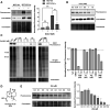
Fig. 3. SEC1 reports on changes in GAPDH activity associated with oncogenic transformation, reactive oxygen species and covalent inhibitors. (A) MCF10A and MCF10CA1a cells were treated with vehicle or KA (10 μM, 1 h) and cell lysates were labeled with SEC1 (100 μM, 1 h). Protein labeling was evaluated by in-gel fluorescence following CuACC with Rh-N3, and protein loading was evaluated by Coomassie stain. Western blot analysis with an anti-GAPDH antibody was used to evaluate GAPDH expression. Band intensities were analyzed and compared using ImageJ software and GraphPad Prism version 9. All values are means  ±  S.E. (error bars) from three replicates (n = 3). *Significantly different p  <  0.05, **p  <  0.01, ***p  <  0.001. ns., not significant. (B) Jurkat cell lysates were treated with increasing concentrations of H2O2 prior to labeling with SEC1, and protein labeling was visualized by in-gel fluorescence. Protein loading was evaluated by Coomassie stain and western blot analysis with an anti-GAPDH antibody was used to evaluate GAPDH expression. (C) Jurkat cell lysates were incubated with two different concentrations (5 and 10 μM) of GAPDH inhibitors: 3-bromopyruvate (3-BP), iodoacetate (IA), KA, and dimethyl fumarate (DMF). Treated lysates were labeled with SEC1 (100 μM, 1 h) and IA-light (100 μM, 1 h) and labeled proteins were visualized by in-gel fluorescence. Band intensities were analyzed and compared using ImageJ software and GraphPad Prism version 9 for IA-light. (D) The chemical structure of KA, a previously reported GAPDH inhibitor. (E) Jurkat cells were incubated with increasing concentrations of KA and lysates were labeled with SEC1 (100 μM, 1 h). Protein labeling was evaluated by in-gel fluorescence and protein loading was evaluated by Coomassie stain. Band intensities were analyzed and compared using ImageJ software and GraphPad Prism version 9. All values are means  ±  S.E. (error bars) from two replicates (n = 2).
Fig. 4
Fig. 4. KA is a potent, irreversible, NAD+ uncompetitive, and G3P competitive inhibitor of GAPDH. (A) Global (left) and local (right) fits of varying concentrations of NAD+ (8-point, 2-fold dilution) and KA (12-point, 3-fold dilution). αKi = 0.70 ± 0.02 μM (global) and αKi = 0.68 ± 0.02 μM (local). (B) Global (left) and local (right) fits of varying concentrations of G3P (8-point, 2-fold dilution) and KA (12-point, 3-fold dilution). Ki = 0.06 ± 0.003 μM (global) and Ki = 0.08 ± 0.02 μM (local). Each data point is an average of 3 replicates. (C) Recovery of GAPDH biochemical activity after rapid dilution of the enzyme-inhibitor complex; (blue) DMSO and (red) KA. GAPDH and KA were pre-incubated for 30 minutes followed by dilution to a negligible concentration of free compound. Enzymatic activity was quantified by generation of NADH in a bioluminescent readout. Error bars for each point represent the standard deviation from two replicates. (D) KA is an extremely effective inactivator of GAPDH. An excess of enzyme was preincubated with varying concentrations of KA, diluted 100×, and the remaining catalytic activity measured. The partition ratio was calculated to be approximately 0 (−0.27 ± 0.01) resulting in completely efficient inactivation of GAPDH (KA is sequestered by the enzyme upon each binding event forming the covalent complex). Standard deviation was calculated based on two independent measurements of the partition ratio.
Fig. 5
Fig. 5. KA is a highly selective GAPDH inhibitor. (A) isoTOP-ABPP workflow used to study KA-induced inhibition of GAPDH. The proteomes of vehicle and KA treated (1, 5, 10 μM) cells were labeled with cysteine-reactive IA-light and IA-heavy probes, respectively, and compared to identify cellular cysteine targets of KA. (B) Heavy:light log2R for all identified cysteines in vehicle (heavy) and 10 μM KA (light) treated Jurkat cells (n = 3, s.d. <25%). (C) Representative traces for the C152-containing peptide and C247 containing peptide of GAPDH from the Jurkat reactivity data. (D) Crystal structure of GAPDH with NAD+ bound (adapted from PDB:4wnc). (E) Thermal shift assay data for WT GAPDH showing stabilization by the addition of its physiological substrate NAD+, and destabilization upon KA binding. Protein stability in the presence of ligand was assessed using SYPRO Orange with increasing temperature (20 to 95 °C). (F) Thermal shift assay data for the C152S mutant GAPDH showing no change in stability upon addition of NAD+ and KA.
Fig. 6
Fig. 6. GAPDH activity and inhibition in activated immune cells. (A) Jurkat cells were activated with PMA (20 ng mL−1) for the listed time. In gel fluorescence (top panel) evaluation of activated and un-activated Jurkat lysates labeled with SEC1 (100 μM, 1 h). Anti-pMAPK (middle panel) was used to verify activation of Jurkat cells and anti-GAPDH was used to evaluate protein expression. Band intensities were analyzed and compared using ImageJ software and GraphPad Prism version 9. All values are means  ±  S.E. (error bars) from three replicates (n = 3) for fluorescence data and two replicates (n = 2) for western blots. *Significantly different p  <  0.05, **p  <  0.01, ***p  <  0.001. ns., not significant. (B) Percentage reduction in cytokine production of Th1 effector cells following addition of KA with no loss in cellular viability. Representative IC50 curves measuring cytokine reduction with varying concentrations of KA following stimulation by CD3/CD28/CD2 after 24 h. Mean IC50 values were calculated by a three-parameter fit constraining the top to 100% inhibition; IFNγ (red) IC50 = 1.4 ± 0.3 μM, TNFα (blue) IC50 = 2.1 ± 0.4 μM, IL-2 (green) IC50 = 3.7 ± 0.7 μM. All values are means  ±  S.E. (error bars) from three replicates (n = 3).

Similar articles

Cited by

References

    1. Warburg O. Wind F. Negelein E. The metabolism of tumors in the body. J. Gen. Physiol. 1927;8:519–530. doi: 10.1085/jgp.8.6.519. - DOI - PMC - PubMed
    1. Cori C. A. Cori G. T. The carbohydrate metabolism of tumours. J. Biol. Chem. 1925;65:397–405. doi: 10.1016/S0021-9258(18)84849-9. - DOI
    1. Al-Ziaydi A. G. Al-Shammari A. M. Hamzah M. I. Kadhim H. S. Jabir M. S. Hexokinase inhibition using d-mannoheptulose enhances oncolytic newcastle disease virus-mediated killing of breast cancer cells. Cancer Cell Int. 2020;20:420. doi: 10.1186/s12935-020-01514-2. - DOI - PMC - PubMed
    1. Abboud G. Choi S. Kanda N. Zeumer-Spataro L. Roopenian D. C. Morel L. Inhibition of glycolysis reduces disease severity in an autoimmune model of rheumatoid arthritis. Front. Immunol. 2018;9:1973. doi: 10.3389/fimmu.2018.01973. - DOI - PMC - PubMed
    1. Liberti M. V. Dai Z. Wardell S. E. Baccile J. A. Liu X. Gao X. Baldi R. Mehrmohamadi M. Johnson M. O. Madhukar N. S. Shestov A. A. Chio I. Elemento O. Rathmell J. C. Schroeder F. C. McDonnell D. P. Locasale J. W. A Predictive model for selective targeting of the Warburg effect through GAPDH inhibition with a natural product. Cell Metab. 2017;26(4):648–659. doi: 10.1016/j.cmet.2017.08.017. - DOI - PMC - PubMed