

Transperineal Ultrasound Before and After Prostatectomy: Technical Approach and Description
- PMID: 35866181
- PMCID: PMC9796877
- DOI: 10.1002/jum.16064
Transperineal Ultrasound Before and After Prostatectomy: Technical Approach and Description
Abstract
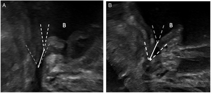
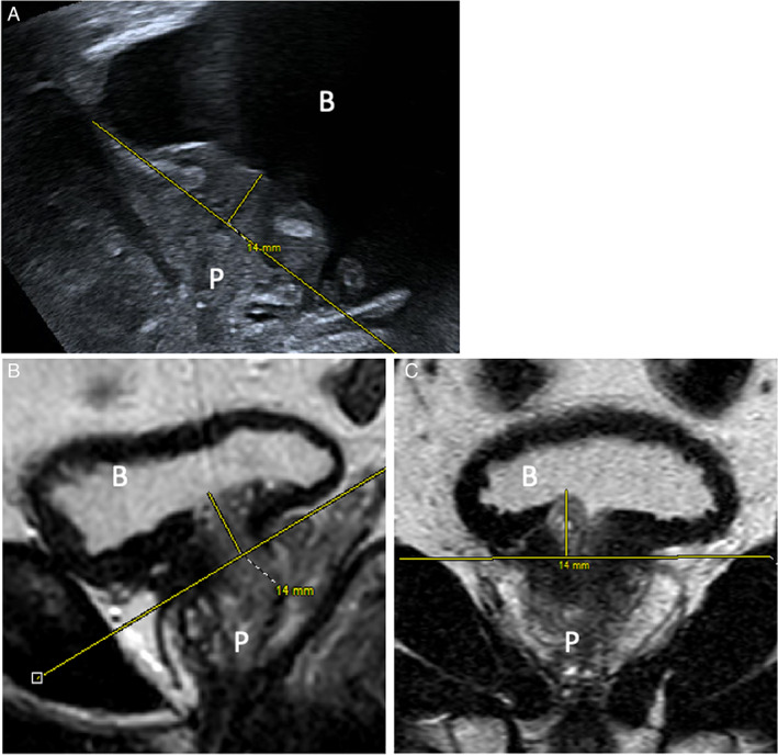
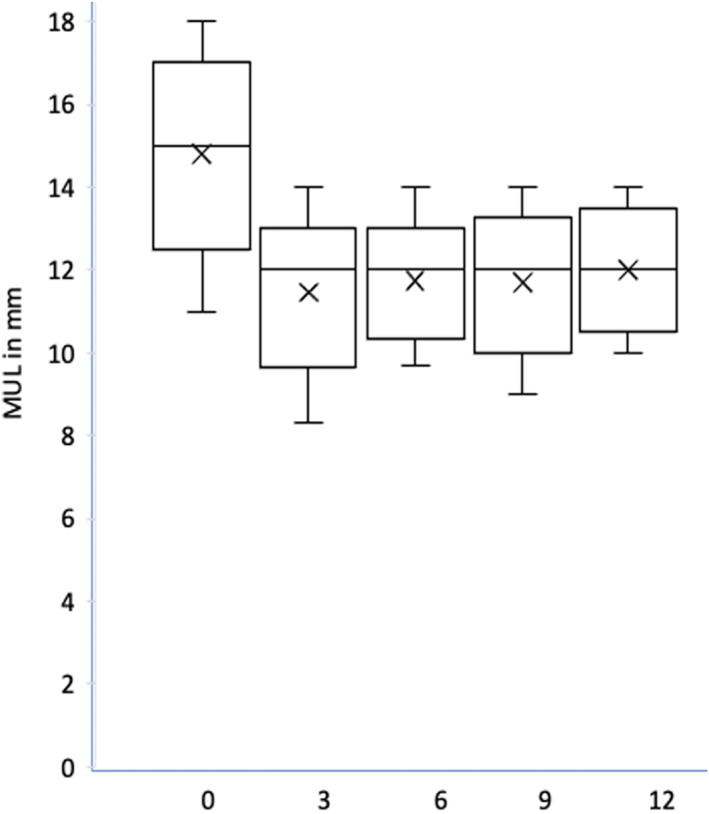
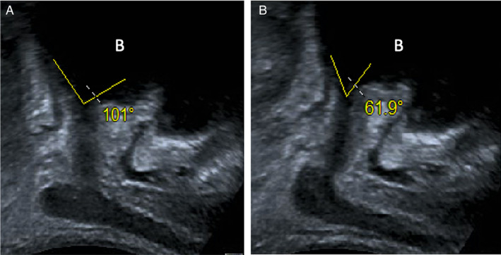
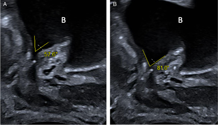
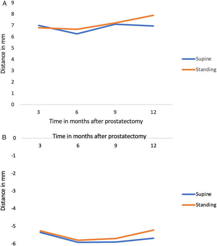
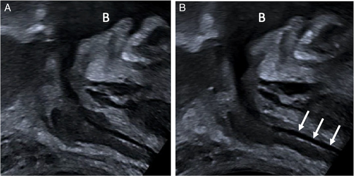
This study assessed the feasibility of dynamic transperineal ultrasound (TPUS) pre/post-radical prostatectomy (RP). Ninety-eight patients were scanned pre-operatively and at four time-points post-operatively. TPUS was performed in 98 patients using an abdominal transducer at rest, during pelvic floor contraction (PFC) and Valsalva (VS) maneuver in supine and standing positions. Urodynamic evaluations included bladder neck angle at rest/PFC/VS, and degree of bladder neck movement. Pre-operative and post-operative measurements were technically feasible in >85% (supine) and >90% (standing) of patients. TPUS offers a reliable non-invasive dynamic assessment of the pelvic floor post-prostatectomy and may prove a useful adjunct for guiding exercises to preserve continence.
Keywords: Valsalva maneuver; membranous urethral length; pelvic floor contraction; post-prostatectomy urinary incontinence; transperineal ultrasound.
© 2022 The Authors. Journal of Ultrasound in Medicine published by Wiley Periodicals LLC on behalf of American Institute of Ultrasound in Medicine.
Figures

Similar articles
-
Influence of body position on dynamics of the pelvic floor measured with transperineal ultrasound imaging in men.Neurourol Urodyn. 2020 Mar;39(3):954-961. doi: 10.1002/nau.24301. Epub 2020 Feb 6. Neurourol Urodyn. 2020. PMID: 32027772
-
May perioperative ultrasound-guided pelvic floor muscle training promote early recovery of urinary continence after robot-assisted radical prostatectomy?Neurourol Urodyn. 2019 Jan;38(1):158-164. doi: 10.1002/nau.23811. Epub 2018 Oct 30. Neurourol Urodyn. 2019. PMID: 30375062
-
Real-time assessment of the behaviour of the bladder neck and proximal urethra during urine leaking in the cough stress test (CST) in supine and standing positions using transperineal ultrasound.Int Urogynecol J. 2020 Dec;31(12):2515-2519. doi: 10.1007/s00192-020-04273-w. Epub 2020 Apr 14. Int Urogynecol J. 2020. PMID: 32291473 Free PMC article.
-
Utility of 2D-ultrasound in pelvic floor muscle contraction and bladder neck mobility assessment in women with urinary incontinence.J Gynecol Obstet Hum Reprod. 2020 Jan;49(1):101629. doi: 10.1016/j.jogoh.2019.101629. Epub 2019 Sep 6. J Gynecol Obstet Hum Reprod. 2020. PMID: 31499282 Review.
-
Evaluation of image-based prognostic parameters of post-prostatectomy urinary incontinence: A literature review.Int J Urol. 2021 Sep;28(9):890-897. doi: 10.1111/iju.14609. Epub 2021 Jun 8. Int J Urol. 2021. PMID: 34101272 Review.
References
Publication types
MeSH terms
LinkOut - more resources
Full Text Sources
Miscellaneous
