. 2022 Jul 22;7(14):e158107.
doi: 10.1172/jci.insight.158107.

A conserved annexin A6-mediated membrane repair mechanism in muscle, heart, and nerve

Affiliations

A conserved annexin A6-mediated membrane repair mechanism in muscle, heart, and nerve

Alexis R Demonbreun et al. JCI Insight. .

Abstract

Membrane instability and disruption underlie myriad acute and chronic disorders. Anxa6 encodes the membrane-associated protein annexin A6 and was identified as a genetic modifier of muscle repair and muscular dystrophy. To evaluate annexin A6's role in membrane repair in vivo, we inserted sequences encoding green fluorescent protein (GFP) into the last coding exon of Anxa6. Heterozygous Anxa6gfp mice expressed a normal pattern of annexin A6 with reduced annexin A6GFP mRNA and protein. High-resolution imaging of wounded muscle fibers showed annexin A6GFP rapidly formed a repair cap at the site of injury. Injured cardiomyocytes and neurons also displayed repair caps after wounding, highlighting annexin A6-mediated repair caps as a feature in multiple cell types. Using surface plasmon resonance, we showed recombinant annexin A6 bound phosphatidylserine-containing lipids in a Ca2+- and dose-dependent fashion with appreciable binding at approximately 50 μM Ca2+. Exogenously added recombinant annexin A6 localized to repair caps and improved muscle membrane repair capacity in a dose-dependent fashion without disrupting endogenous annexin A6 localization, indicating annexin A6 promotes repair from both intracellular and extracellular compartments. Thus, annexin A6 orchestrates repair in multiple cell types, and recombinant annexin A6 may be useful in additional chronic disorders beyond skeletal muscle myopathies.

Keywords: Cardiology; Muscle; Muscle Biology; Neurological disorders.


Conflict of interest statement

Conflict of interest: Northwestern University filed a provisional patent 62/783,619 on behalf of the authors (ARD and EMM) and provisional patent 63/309,925 on behalf of the authors (ARD, EMM, KRS, and RV) related to the content of this work.

Figures

Figure 1
Figure 1. Generation of genomically encoded annexin A6GFP using CRISPR/Cas9 genome editing.
(A) Targeting strategy for generating genomically encoded annexin A6GFP at the endogenous annexin A6 locus. Red lettering indicates protospacer adjacent motif sequence. Lowercase lettering indicates synonymous mutations in targeted allele. (B) Anxa6gfp mouse generation strategy. Genotyping schematic and PCR screening of Anxa6gfp of 6 heterozygous N1 offspring lines. (C) Representative sequence chromatograms of in-frame GFP insertion into the annexin A6 locus in Anxa6gfp CRISPR/Cas9-edited mouse line 46.
Figure 2
Figure 2. Genomic A6GFP protein localizes to the site of muscle membrane injury.
(A) Quantitative PCR demonstrates reduced Anxa6 levels in quadriceps from heterozygous and homozygous Anxa6gfp mice compared with wild-type (WT) controls. (BD) Anti–annexin A6 immunoblots demonstrate reduced ANXA6 protein levels in quadriceps muscles from heterozygous and homozygous Anxa6gfp mice. The loading control is a 42 kDa band detected by MemCode reversible protein stain. (E) Anti–annexin A6 (shown in green) immunofluorescence imaging of extensor digitorum longus myofibers from WT and homozygous Anxa6gfp mice. ANXA6 and annexin A6GFP protein localize in a similar punctate, sarcomeric pattern and at the sarcolemma. Scale bar: 10 μm. (F) Upon laser-induced membrane injury, annexin A6GFP localized to the repair cap (white arrow) with a visible clearance zone (orange arrow) beneath the membrane lesion in heterozygous Anxa6gfp myofibers. (G) Genomically encoded annexin A6GFP membranous blebs (white arrow) erupt from the site of membrane injury. Z-stack images from an injured myofiber. Scale bar: 5 μm. n = 6 mice per genotype. n > 10 myofibers. *P < 0.05 by 1-way ANOVA.
Figure 3
Figure 3. Genomically encoded annexin A6GFP localizes at the site of cardiomyocyte membrane injury.
(A) Quantitative PCR demonstrates reduced Anxa6 levels in heart lysates from heterozygous and homozygous Anxa6gfp mice compared with WT controls. (BD) Anti–annexin A6 immunoblots demonstrate reduced ANXA6 protein levels in cardiac ventricle lysates from heterozygous and homozygous Anxa6gfp mice. The loading control is a 42 kDa band detected by MemCode reversible protein stain. (E) Adult ventricular cardiomyocytes were isolated from homozygous Anxa6gfp mice and subsequently laser-damaged. annexin A6GFP (shown in green) quickly localizes to the cardiomyocyte repair cap (white arrow). (F) Z-projection of a homozygous Anxa6gfp cardiomyocyte illustrating annexin A6GFP repair cap (white arrow) above the annexin-free zone at the site of injury 250 seconds after cardiomyocyte wounding. Scale bar: 5 μm. n = 6 mice per genotype. n > 12 cells from 5 isolations. *P < 0.05 by 1-way ANOVA.
Figure 4
Figure 4. Annexin A6GFP localizes at the site of neuron membrane injury.
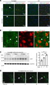
(A) Anti-GFP (shown in green, indicated by arrows) antibody detects genomically encoded annexin A6GFP protein in Anxa6gfp adult cortex and midbrain but not in WT mice. DAPI (blue) marks nuclei. Anti-NeuN (red) marks mature neurons. (B) Anti-GFP (green, arrow) antibody, which detects genomically encoded annexin A6GFP protein, localized to the peripheral membrane of NeuN+ cortical neurons as visualized with high-magnification confocal imaging. (C) Embryonic neurons were isolated from Anxa6gfp mice. Genomically encoded annexin A6GFP expression increases with maturation. After 10 days, neurons expressed 2-fold more annexin A6GFP than at 4 days. (D) Isolated neurons were injured with a confocal laser. Genomically encoded annexin A6GFP (green) quickly localized into a repair cap (white arrow) visible 4 seconds postinjury. Multiple cells from n = 3 mice. *P < 0.05 by 1-way ANOVA.
Figure 5
Figure 5. Recombinant annexin A6 binds PS.
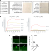
(A) Recombinant annexin A6 preferentially bound PS and phosphatidylinositol 5-phosphate on membrane lipid arrays. (B) SPR sensorgrams showing that recombinant annexin A6 (rA6) binds PS-containing liposomes at approximately 100 nM over a range of Ca2+ concentrations ranging from 0 to 2500 μM (0, 25, 52, 104, 208, 417, 833, and 2500 μM). (C) Wortmannin treatment depleted PIP2 in myofibers as visualized by reduced PLC-PH-EGFP signal (top panel). Additionally, wortmannin treatment reduced genomically encoded annexin A6GFP cap area after laser-induced injury. (n = 10 from 5 isolations; *P < 0.002 by t test).
Figure 6
Figure 6. Recombinant annexin A6 associates with injured myoblasts and myofibers.
(A) Rat L6 myoblasts were injured with LLO and then incubated with recombinant annexin A6 conjugated to 488 (rA6-488). The percentage of rA6-488–positive cells increased with increasing concentrations of annexin protein. (B) Total fluorescence intensity of rA6-488–positive cells increased with increasing concentrations of rA6-488, normalized to 1.0 μg/mL. (C) Increasing rA6-488 incubation time increased fluorescence signal of injured cells but not noninjured control cells. (D) Incubation of myofibers in recombinant annexin A6 (rANXA6) for either 5 or 60 minutes both reduced FM 4-64 dye uptake after injury compared with BSA-treated control myofibers. Scale bar: 5 μm. n ≥ 4 cell platings. n ≥ 6 myofibers from n = 4 isolations. *P < 0.05 by 1-way ANOVA.
Figure 7
Figure 7. Recombinant annexin A6 cap size increases in a dose-dependent fashion, correlating with improved repair capacity.
(A) Myofibers were isolated from Anxa6gfp mice and laser-damaged in the presence of rA6-tdTomato. rA6-tdTomato (shown in red) colocalized with genomically encoded annexin A6GFP (green) at the site of muscle membrane injury (white arrow). (B) rA6-tdTomato cap size increased with increasing concentrations of rA6-tdTomato, 1.3–130 μg/mL. Genomically encoded annexin A6GFP cap size did not change with increasing concentrations of rA6-tdTomato. (C) rA6-tdTomato formed membranous blebs at the site of membrane injury. (D) Dose-dependent reduction of FM 4-64 dye (red) uptake, a marker of membrane injury, with increasing concentrations of recombinant annexin A6. (E) Anxa6gfp mice were crossed with mdx mice to generate mdx mice expressing genomically encoded annexin A6GFP. (F) Dystrophic histopathology is present in Anxa6gfp mdx muscle. Scale bar: 100 μm. (G) Serum creatine kinase (CK) was elevated in Anxa6gfp mdx mice compared to Anxa6gfp controls (n = 7). (H) Increased FM 4-64 dye (red) in injured Anxa6gfp mdx myofibers compared with Anxa6gfp controls. (I and J) In Anxa6gfp mdx myofibers, genomically encoded annexin A6GFP formed a repair cap at the site of membrane injury. rA6-tdTomato cap size increased with increasing concentrations of rA6-tdTomato, 1.3–130 μg/mL. Genomically encoded annexin A6GFP cap size did not change significantly with varying concentrations of rA6-tdTomato in Anxa6gfp mdx myofibers. (K) Increasing concentrations of recombinant annexin A6 resulted in a dose-dependent reduction of FM 4-64 dye (red) uptake in dystrophic myofibers. Scale bar: 5 μm. A total of 4–9 myofibers from n ≥ 4 isolations. *P < 0.05 by 1-way ANOVA.
Figure 8
Figure 8. Recombinant annexin A6 binds neuronal membrane lesions.
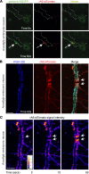
(A) Embryonic neurons were isolated from Anxa6gfp mice, matured, and laser damaged in the presence of rA6-tdTomato. rA6-tdTomato (shown in red) colocalizes with genomically encoded annexin A6GFP (green) at the site of muscle membrane injury (white arrow). Neuron outlined in white dotted line. (B) After transection of Anxa6gfp neuronal processes, rA6-tdTomato (red) localizes at the stumps of the severed process (white arrows). WGA-350 (blue) outlines the neuron. (C) rA6-tdTomato fluorescence signal increases at the process stumps with time (white arrows). Multiple neurons from n ≥ 3 mice. Scale bar: 5 μm.
Figure 9
Figure 9. Model of annexin A6–mediated membrane repair in skeletal muscle, cardiomyocytes, and neurons.
Upon plasma membrane breach, extracellular Ca2+ enters the damaged cell. Annexin A6 (A6) binds Ca2+, translocates to the site of membrane injury targeting exposed phospholipids such as PS, and forms a repair cap at the lesion. Extracellular recombinant annexin A6 (rA6) localizes to the repair cap at the site of injury, enhancing repair capacity.

Similar articles

Cited by

References

    1. Dias C, Nylandsted J. Plasma membrane integrity in health and disease: significance and therapeutic potential. Cell Discov. 2021;7(1):4. doi: 10.1038/s41421-020-00233-2. - DOI - PMC - PubMed
    1. Ammendolia DA, et al. Plasma membrane integrity: implications for health and disease. BMC Biol. 2021;19(1):71. doi: 10.1186/s12915-021-00972-y. - DOI - PMC - PubMed
    1. Andrews NW, Corrotte M. Plasma membrane repair. Curr Biol. 2018;28(8):R392–R397. doi: 10.1016/j.cub.2017.12.034. - DOI - PubMed
    1. Cooper ST, McNeil PL. Membrane repair: mechanisms and pathophysiology. Physiol Rev. 2015;95(4):1205–1240. doi: 10.1152/physrev.00037.2014. - DOI - PMC - PubMed
    1. Koerdt SN, et al. Annexins and plasma membrane repair. Curr Top Membr. 2019;84:43–65. doi: 10.1016/bs.ctm.2019.07.006. - DOI - PubMed

Publication types