. 2022 Oct;6(10):2812-2826.
doi: 10.1002/hep4.2037. Epub 2022 Jul 22.

Activin B promotes the initiation and progression of liver fibrosis

Affiliations

Activin B promotes the initiation and progression of liver fibrosis

Yan Wang et al. Hepatol Commun. 2022 Oct.

Abstract

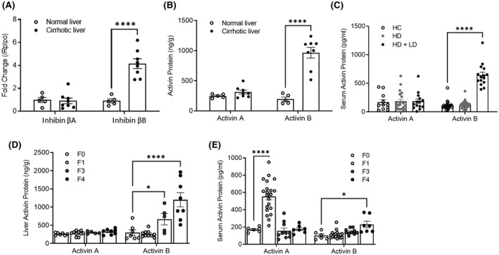
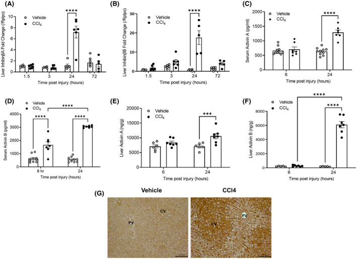
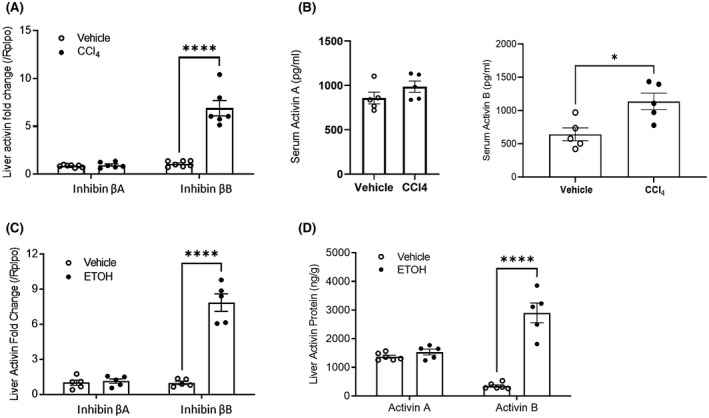
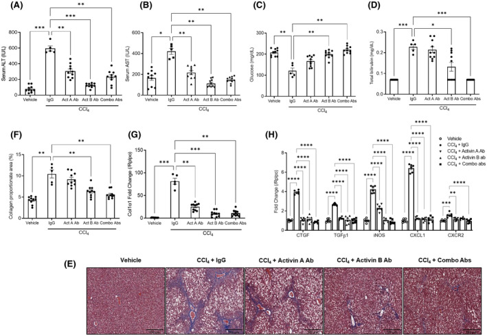
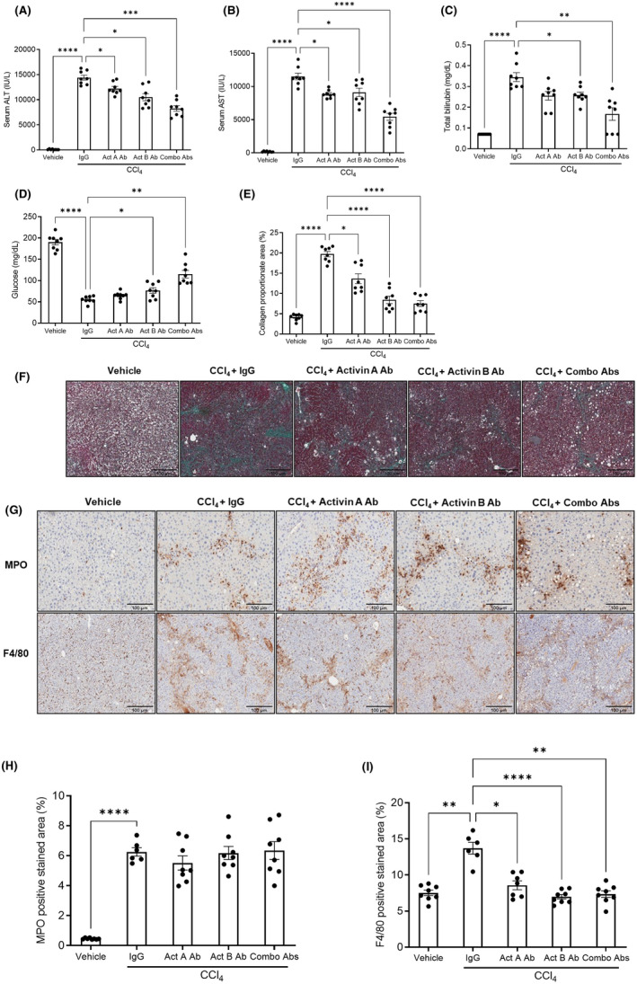
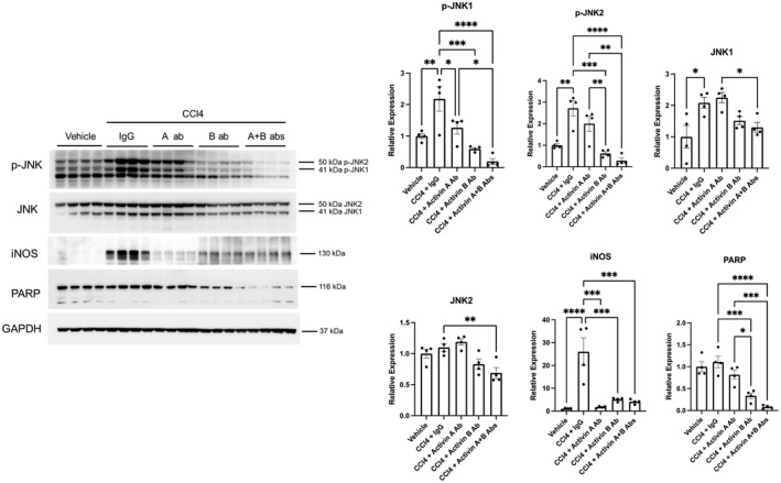
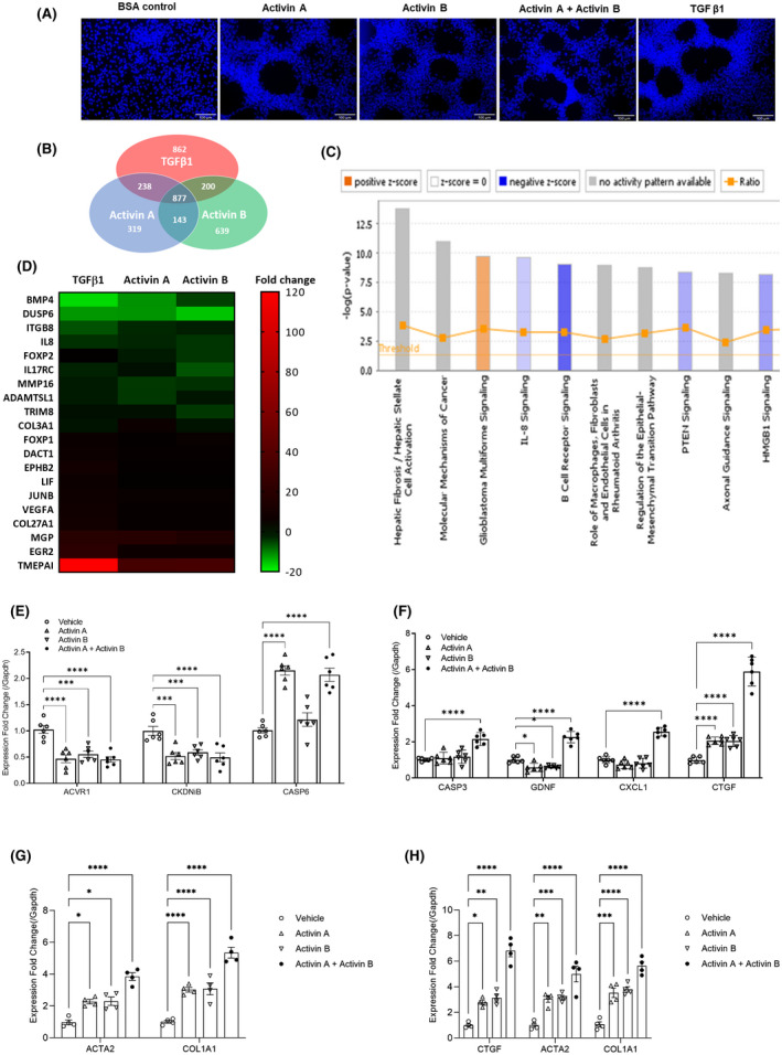
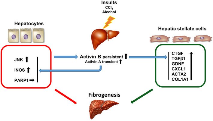
The role of activin B, a transforming growth factor β (TGFβ) superfamily cytokine, in liver health and disease is largely unknown. We aimed to investigate whether activin B modulates liver fibrogenesis. Liver and serum activin B, along with its analog activin A, were analyzed in patients with liver fibrosis from different etiologies and in mouse acute and chronic liver injury models. Activin B, activin A, or both was immunologically neutralized in mice with progressive or established carbon tetrachloride (CCl4 )-induced liver fibrosis. Hepatic and circulating activin B was increased in human patients with liver fibrosis caused by several liver diseases. In mice, hepatic and circulating activin B exhibited persistent elevation following the onset of several types of liver injury, whereas activin A displayed transient increases. The results revealed a close correlation of activin B with liver injury regardless of etiology and species. Injured hepatocytes produced excessive activin B. Neutralizing activin B largely prevented, as well as improved, CCl4 -induced liver fibrosis, which was augmented by co-neutralizing activin A. Mechanistically, activin B mediated the activation of c-Jun-N-terminal kinase (JNK), the induction of inducible nitric oxide synthase (iNOS) expression, and the maintenance of poly (ADP-ribose) polymerase 1 (PARP1) expression in injured livers. Moreover, activin B directly induced a profibrotic expression profile in hepatic stellate cells (HSCs) and stimulated these cells to form a septa structure. Conclusions: We demonstrate that activin B, cooperating with activin A, mediates the activation or expression of JNK, iNOS, and PARP1 and the activation of HSCs, driving the initiation and progression of liver fibrosis.


Conflict of interest statement

The authors disclose that a patent entitled “Methods of treating or preventing liver fibrosis with inhibition of activins B and A” was published based on this manuscript (patent publication number: US2021/0009672 A1). Yan Wang, Matthew Hamang, and Alexander Culver are Lilly Graduate Research Advanced Degree Program graduate students in the Department of Biology of School of Science at Indiana University–Purdue University Indianapolis and are employees of Eli Lilly & Company. Benjamin C. Yaden is an adjunct professor in the Department of Biology of School of Science at Indiana University–Purdue University Indianapolis and is an employee of Eli Lilly & Company.

Figures

FIGURE 1
FIGURE 1
Liver and serum activin B increase in the patients with liver fibrosis. The messenger RNA (mRNA) expression of hepatic inhibin βA and inhibin βB (A) and proteins of hepatic activin A and B (B) in patients with cirrhosis (n = 8) and healthy controls (n = 5) were analyzed by quantitative real‐time polymerase chain reaction (PCR) and enzyme‐linked immunosorbent assay (ELISA), respectively. (C) The concentrations of serum activin A and B proteins were determined with ELISA in heathy controls (HC; n = 16), heavy drinkers without liver diseases (HD; n = 36), and heavy drinkers with liver disease (HD + LD; n = 15). Activin A and B proteins were evaluated by ELISA in the livers (D) and serum (E) of patients with different stages of nonalcoholic steatohepatitis (F0, n = 4; F1, n = 6; F3, n = 5; and F4, n = 6). For all of these assays, data are expressed as means ± SEM. *p < 0.05, ****p < 0.0001 compared with HC or F0 group via two‐way analysis of variance (ANOVA). Note that inhibin βA and βB encode the subunit of activin A and B respectively.
FIGURE 2
FIGURE 2
Liver and serum activin B increases in mice following acute liver injury. Female mice were given a single administration of carbon tetrachloride (CCl4) or vehicle, and tissues were collected at the indicted times following injury. The mRNA expression of liver inhibin βA (A) and inhibin βB (B) was analyzed by quantitative real‐time PCR at indicated time points (n = 6). Serum (C,D) and liver (E,F) activin proteins were quantified via ELISA at the indicated time points (n = 5–8). For all of these quantitative assays, data are expressed as means ± SEM. ***p < 0.001, ****p < 0.0001 via two‐way ANOVA relative to vehicle controls. (G) Immunostaining of activin B in the livers of mice 24 h after CCl4 or vehicle treatment.
FIGURE 3
FIGURE 3
mRNA and protein levels of activin B and A in CCl4‐induced chronic liver injury. CCl4 or vehicle was dosed twice per week for 4 weeks in female mice. (A) mRNA expression of hepatic inhibin βA and inhibin βB was assessed by quantitative real‐time PCR (n = 5–7). (B) Concentrations of serum activin A and activin B protein were quantified via ELISA (n = 5). In a separate study, following 10 days of oral alcohol (ETOH) plus binge administration in male mice, hepatic inhibin βA and inhibin βB transcript levels were determined by quantitative real‐time PCR (n = 5) (C), and hepatic activin A and activin B protein contents were quantified via ELISA (n = 5) (D). For all of these quantitative assays, data are expressed as means ± SEM. *p < 0.05, ****p < 0.0001 via two‐way ANOVA relative to vehicle controls.
FIGURE 4
FIGURE 4
Treatments with activin B antibody (Ab), activin A antibody, or combination of them display distinct effects in preventing liver fibrosis induced by CCl4 in mice. Adult female mice were administered CCl4 or corn oil vehicle twice per week for 4 weeks. Half an hour before the first CCl4 injection each week, mice were treated with immunoglobulin G (IgG; 60 mg/kg), activin A antibody (10 mg/kg of activin A antibody + 50 mg/kg of IgG), activin B antibody (50 mg/kg of activin B antibody + 10 mg/kg of IgG), or combination of activin A and activin B antibodies (10 mg/kg of activin A antibody + 50 mg/kg of activin B antibody). Four weeks after the initial CCl4 injection, alanine aminotransferase (ALT) (A), aspartate aminotransferase (AST) (B), glucose (C), and total bilirubin (D) were analyzed in blood. (E) Representative liver sections stained with Masson's trichrome. (F) Percent Masson's trichrome staining areas were quantified by ImagJ. (G) The mRNA expression of hepatic collagen type I alpha 1 (Col1α1) was evaluated by quantitative real‐time PCR. (H) Transcripts of the genes indicated were quantified by quantitative real‐time PCR in liver tissues. Data are expressed as means ± SEM (n = 5 for IgG control group; n = 10 for all other groups). *p < 0.05, **p < 0.01, ***p < 0.001, and ****p < 0.0001 compared with IgG controls via ordinary one‐way ANOVA for (A)–(D), (F), and (G). Statistical analysis was performed via two‐way ANOVA following by Dunnett's multiple comparisons test relative to IgG controls for (H). Total bilirubin levels below the limit of detection (0.14 mg/dl) were replaced by half of the limit of detection (0.07 mg/dl) for statistical analysis. Abbreviations: CTGF, connective tissue growth factor; CXCL, chemokine (C‐X‐C motif) ligand 1; CXCR2, chemokine (C‐X‐C motif) receptor 2.
FIGURE 5
FIGURE 5
Activin B antibody, activin A antibody, and a combination of them display different effects in regressing liver fibrosis induced by CCl4 in mice. Adult female mice were subjected to CCl4 or vehicle injection twice per week for 10 weeks. Starting from the seventh week, these mice were treated with IgG (60 mg/kg), activin A antibody (10 mg/kg of activin A antibody + 50 mg/kg of IgG), activin B antibody (50 mg/kg of activin B antibody + 10 mg/kg of IgG), or combination of activin A and activin B antibodies (10 mg/kg of activin A antibody + 50 mg/kg of activin B antibody) weekly. Ten weeks after the initial CCl4 injection, ALT (A), AST (B), glucose (C), and total bilirubin (D) were analyzed in blood, and Masson's trichrome collagen staining areas were quantified by ImagJ (E). (F) Representative liver sections stained with Masson's trichrome. (G) Representative liver sections immunostained with myeloperoxidase (MPO) or F4/80 antibody. (H,I) Quantification of percent positive staining area of MPO (H) and F4/80 (I). Data are presented as means ± SEM (n = 8). *p < 0.05, **p < 0.01, ***p < 0.001, and ****p < 0.0001 compared with IgG controls via ordinary one‐way ANOVA relative to IgG controls. Total bilirubin levels below the limit of detection (0.14 mg/dl) were replaced by half of the limit of detection (0.07 mg/dl) for statistical analysis.
FIGURE 6
FIGURE 6
The effects of antibody treatments on the expression of hepatic c‐Jun‐N‐terminal kinase (JNK), inducible nitric oxide synthase (iNOS), and poly (ADP‐ribose) polymerase 1 (PARP1) in the livers chronically injured by CCl4. Liver lysates were prepared from the liver samples collected in the preventive study described in Figure 5. Western blotting was performed using antibodies against the proteins indicated. Glyceraldehyde 3‐phosphate dehydrogenase (GADPH) was used as a loading control. Relative densitometry was normalized against GAPDH. Data are presented as the mean fold changes relative to vehicle controls ± SEM (n = 4). *p < 0.05, **p < 0.01, ***p < 0.001, and ****p < 0.0001.
FIGURE 7
FIGURE 7
Activin B morphologically and molecularly activates hepatic stellate cells (HSCs). (A) LX‐2 cells were treated with bovine serum albumin (BSA; 100 ng/ml), activin A (100 ng/ml), activin B (100 ng/ml), their combination (100 ng/ml each), or transforming growth factor β1 (TGFβ1; 5 ng/ml) for 24 h and then underwent 4′, 6‐diamidino‐2‐phenylindole (DAPI) staining. (B) LX‐2 cells were treated with activin A (100 ng/ml), activin B (100 ng/ml), or TGFβ1 (5 ng/ml) for 6 h. Total RNAs were isolated, reverse‐transcribed to complementary DNA, and then subjected to microarray analysis using HG‐U133 plus two chips (n = 6). Pie chart shows the numbers of genes commonly or uniquely regulated by the individual ligands. (C) The top 10 signaling pathways revealed by Ingenuity Canonical Pathway analysis of the 877 target genes shared by these three ligands. (D) Heat map of the 20 genes exhibiting the highest magnitudes of up‐regulation or down‐regulation in response to these three ligands. (E–G) LX‐2 cells were treated with vehicle, activin A (100 ng/ml), activin B (100 ng/ml), or their combination (100 ng/ml of each) for 24 h. (H) Mouse primary HSCs were exposed to vehicle, activin A (100 ng/ml), activin B (100 ng/ml), or their combination (100 ng/ml of each) for 24 h. The expression of the genes indicated was assessed with quantitative real‐time PCR. Data are shown as means of fold changes relative to vehicle controls ± SEM (n = 4–6). *p < 0.05, **p < 0.01, ***p < 0.001, and ****p < 0.0001 via two‐way ANOVA compared with vehicle controls. Abbreviations: ACTA2, smooth muscle alpha‐2 actin; ACVR, activin A receptor; ACVR1, activin A receptor type 1; ADAMTSL, a disintegrin and metalloproteinase with thrombospondin motif‐like protein; BMP, bone morphogenetic protein; CASP, caspase; CASP3, caspase‐3; CDKN, cyclin‐dependent kinase inhibitor; CDKNB, cyclin‐dependent kinase inhibitor; Col1α1, collagen type I alpha 1; COL3A1, collagen type III alpha 1; DACT, dishevelled binding antagonist of beta catenin; DUSP, dual specificity phosphatase; DUSP6, dual‐specificity phosphatase 6; EGR2, early growth response protein 2; EPHB, ephrin receptor B; FOXP, forkhead box P; GDNF, glial cell line–derived neurotrophic factor; IL, interleukin; IL17RC, interleukin 17 receptor C; ITGB, integrin beta; JUNB, jun B proto‐oncogene; LIF, leukemia inhibitory factor; MGP, matrix Gla protein; MMP, matrix metalloproteinase; TMEPAI, transmembrane prostate androgen–induced protein; TRIM, tripartite motif family; VEGFA, vascular endothelial growth factor alpha.
FIGURE 8
FIGURE 8
The mode of action of activin B in liver injury. Hepatocytes release activin B following injury. In an autocrine manner, activin B stimulates the activation of JNK and the expression of iNOS and PARP1, promoting hepatocyte death. In a paracrine manner, activin B induces HSCs to produce profibrotic factors including CTGF and CXCL1 and activates HSCs. As liver injury progresses, hepatocytes persistently produce activin B and transiently produce activin A. Activin B requires the presence of activin A for additive or synergistic effects on stimulating hepatocyte death and HSC activation. Thus, activin B acts as a key modulator, along with other profibrotic factors, to drive the initiation and progression of liver fibrosis.

Similar articles

Cited by

References

    1. Higashi T, Friedman SL, Hoshida Y. Hepatic stellate cells as key target in liver fibrosis. Adv Drug Deliv Rev. 2017;121:27–42. - PMC - PubMed
    1. Bitto N, Liguori E, La Mura V. Coagulation, microenvironment and liver fibrosis. Cell. 2018;7:85. - PMC - PubMed
    1. Sanyal AJ. Novel therapeutic targets for steatohepatitis. Clin Res Hepatol Gastroenterol. 2015;39(Suppl 1):S46–50. - PubMed
    1. Schuppan D, Ashfaq‐Khan M, Yang AT, Kim YO. Liver fibrosis: direct antifibrotic agents and targeted therapies. Matrix Biol. 2018;68–69:435–51. - PubMed
    1. Friedman SL. Evolving challenges in hepatic fibrosis. Nat Rev Gastroenterol Hepatol. 2010;7:425–36. - PubMed

Publication types