

Pre- and post-therapeutic evaluation of liver and spleen in type I and type III Gaucher's disease using diffusion tensor imaging
- PMID: 35867132
- PMCID: PMC9463195
- DOI: 10.1007/s00261-022-03602-5
Pre- and post-therapeutic evaluation of liver and spleen in type I and type III Gaucher's disease using diffusion tensor imaging
Abstract
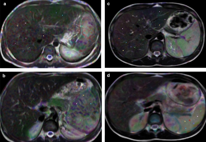
Purpose: To assess the role of diffusion tensor imaging in assessing liver and splenic parenchymal infiltration in Gaucher's disease (G.D.) type I and III before and after therapy.
Methods: A prospective study was conducted upon 28 consecutive patients with G.D. type I and III and 28 age and sex-matched controls. They underwent an MRI and DTI of the liver and spleen. Mean diffusivity (M.D.) and fractional anisotropy (F.A.) values of the liver and spleen were evaluated before and after treatment and compared with control.
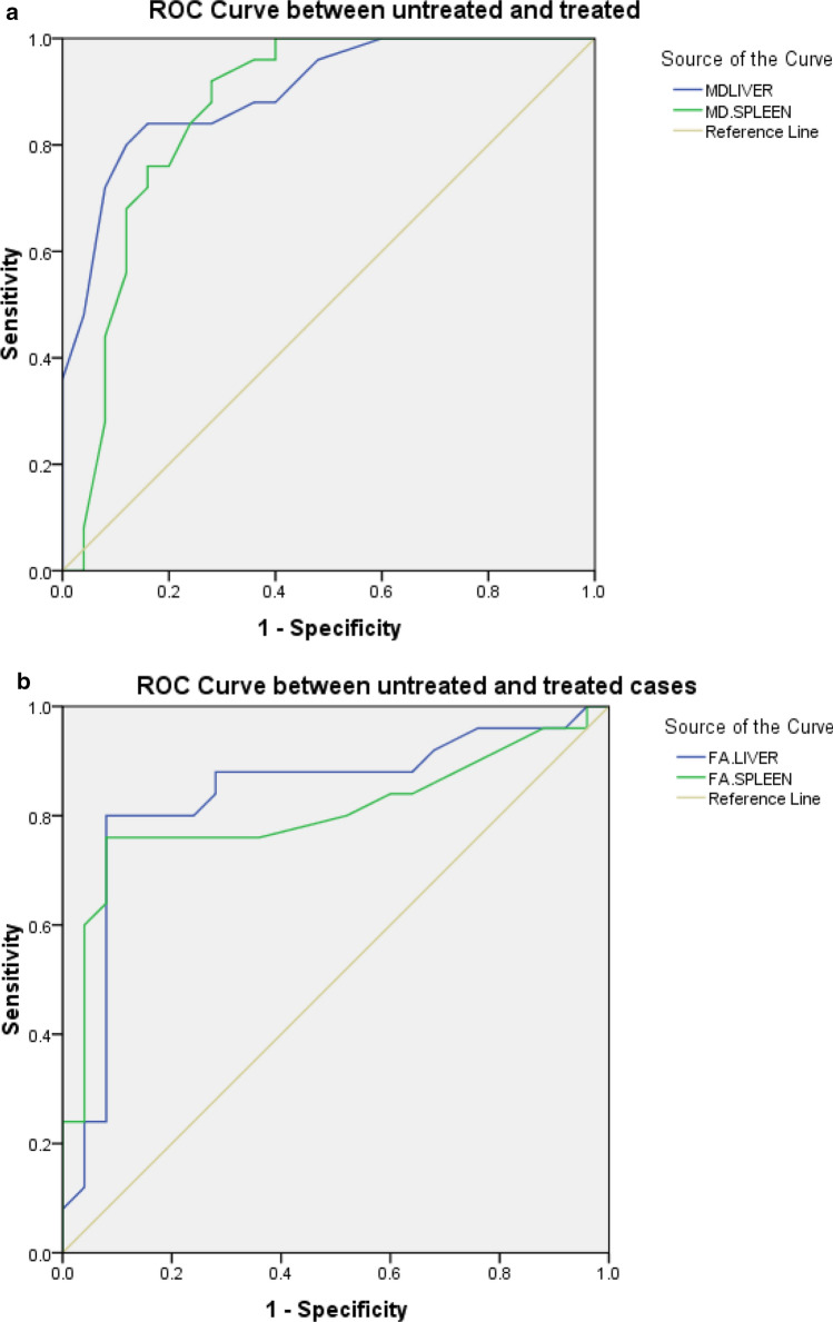
Results: There was a statistically significant difference in the M.D. value of the liver and spleen between untreated patients and controls and between control and treated patients and in the M.D. value of the liver and spleen between untreated and treated patients. There is a statistically significant difference in the F.A. value of the liver and spleen between untreated patients and controls and in the F.A. value of the liver and spleen between untreated and treated patients. Hemoglobin level was positively correlated with the M.D. value of the spleen. Clinical score was negatively correlated with M.D. value of the spleen and was positively correlated with F.A. values of the liver and F.A. values of the spleen. Spleen volume was negatively correlated with M.D. values of the spleen.
Conclusion: Significant difference in M.D. and F.A. values of liver and splenic parenchyma in p atients with type I and III G.D. and controls, and between untreated and treated patients. The M.D. and F.A. values were well correlated with some biomarkers of disease activity.
Keywords: Diffusion tensor imaging; Gaucher’s disease; Liver; M.R. imaging; Spleen.
© 2022. The Author(s).
Conflict of interest statement
The authors declare that they have no competing interests.
Figures


Similar articles
-
Diffusion tensor imaging of vertebral bone marrow in children with Gaucher's disease type I and III: Pre- and post-therapy.Clin Imaging. 2021 Nov;79:207-212. doi: 10.1016/j.clinimag.2021.06.002. Epub 2021 Jun 7. Clin Imaging. 2021. PMID: 34116297
-
Assessment of the liver and spleen in children with Gaucher disease type I with diffusion-weighted MR imaging.Blood Cells Mol Dis. 2018 Feb;68:139-142. doi: 10.1016/j.bcmd.2016.12.004. Epub 2016 Dec 20. Blood Cells Mol Dis. 2018. PMID: 28012701
-
Apparent diffusion coefficient of the vertebral bone marrow in children with Gaucher's disease type I and III.Skeletal Radiol. 2013 Feb;42(2):283-7. doi: 10.1007/s00256-012-1464-8. Epub 2012 Jun 21. Skeletal Radiol. 2013. PMID: 22718272
-
The role of diffusion tensor imaging and fractional anisotropy in the evaluation of patients with idiopathic normal pressure hydrocephalus: a literature review.Neurosurg Focus. 2016 Sep;41(3):E12. doi: 10.3171/2016.6.FOCUS16192. Neurosurg Focus. 2016. PMID: 27581308 Review.
-
Alglucerase. A review of its therapeutic use in Gaucher's disease.Drugs. 1992 Jul;44(1):72-93. doi: 10.2165/00003495-199244010-00007. Drugs. 1992. PMID: 1379912 Review.
Cited by
-
3.0 T diffusion tensor imaging and fiber tractography of the testes in nonobstructive azoospermia.Abdom Radiol (NY). 2024 Dec;49(12):4543-4555. doi: 10.1007/s00261-024-04457-8. Epub 2024 Jun 28. Abdom Radiol (NY). 2024. PMID: 38940912
-
Diffusion tensor imaging and fiber tractography of the normal epididymis.Abdom Radiol (NY). 2024 Aug;49(8):2932-2941. doi: 10.1007/s00261-024-04372-y. Epub 2024 Jun 5. Abdom Radiol (NY). 2024. PMID: 38836882
References
Publication types
MeSH terms
LinkOut - more resources
Full Text Sources
Medical
