. 2022 Oct;21(5):1339-1355.
doi: 10.1007/s10237-022-01593-2. Epub 2022 Jul 22.

Intracellular signaling control of mechanical homeostasis in the aorta

Affiliations

Intracellular signaling control of mechanical homeostasis in the aorta

Linda Irons et al. Biomech Model Mechanobiol. 2022 Oct.

Abstract

Mature arteries exhibit a preferred biomechanical state in health evidenced by a narrow range of intramural and wall shear stresses. When stresses are perturbed by changes in blood pressure or flow, homeostatic mechanisms tend to restore target values via altered contractility and/or cell and matrix turnover. In contrast, vascular disease associates with compromised homeostasis, hence we must understand mechanisms underlying mechanical homeostasis and its robustness. Here, we use a multiscale computational model wherein mechanosensitive intracellular signaling pathways drive arterial growth and remodeling. First, we identify an ensemble of cell-level parameterizations where tissue-level responses are well-regulated and adaptive to hemodynamic perturbations. The responsible mechanism is persistent multiscale negative feedback whereby mechanosensitive signaling drives mass turnover until homeostatic target stresses are reached. This demonstrates how robustness emerges despite inevitable cell and individual heterogeneity. Second, we investigate tissue-level effects of signaling node knockdowns (ATIR, ROCK, TGF[Formula: see text]RII, PDGFR, ERK1/2) and find general agreement with experimental reports of fault tolerance. Robustness against structural changes manifests via low engagement of the node under baseline stresses or compensatory multiscale feedback via upregulation of additional pathways. Third, we show how knockdowns affect collagen and smooth muscle turnover at baseline and with perturbed stresses. In several cases, basal production is not remarkably affected, but sensitivities to stress deviations, which influence feedback strength, are reduced. Such reductions can impair adaptive responses, consistent with previously reported aortic vulnerability despite grossly normal appearances. Reduced stress sensitivities thus form a candidate mechanism for how robustness is lost, enabling transitions from health towards disease.

Keywords: Growth and remodeling; Homeostasis; Mechanobiology; Multiscale; Robustness.


Conflict of interest statement

Statements and Declarations: The authors declare no conflicts of interest, financial or otherwise.

Figures

Figure 1:
Figure 1:
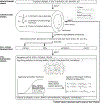
Schema of the coupled tissue-level to cell-signaling model. Blood pressure and flow are prescribed at the tissue-level. We use a constrained mixture model for three constituent-dominated behaviors (collagen, smooth muscle cells, and elastin), each contributing appropriate constituent-level stored energy functions. Equilibrium equations appropriate for an axisymmetric cylinder reveal the changes in intramural (Stress) and wall shear (Wss) stresses that become inputs to a logic-based cell signaling model as possible deviations from homeostatic values. The cell signaling model comprises 56 coupled ODEs and accounts for signaling through receptors including AT1R, AT2R, TGFβRI/II and PDGFR and pathways including MAPKs, Smads, PI3K and RhoA/ROCK. Cell-level parameters to be determined are initial values for four inputs: yStress,yWss,yIntegrins, and ySACs (stretch activated channels) as well as three parameters w,n, and EC50 for tuning sigmoidal activation functions; these three parameters can differ for each edge in the network but currently are prescribed uniformly as in previous models of this type. The outputs of the cell signaling model govern contractility and turnover rates of collagen and cells, which form a feedback loop to the tissue-level G&R model via an active stress contribution and constituent production and removal rates. Full details of the signaling network structure and model formulation are given in Supplementary Materials.
Figure 2:
Figure 2:
Distributions of cell-level parameters for 250 distinct sets {yStress(0),yWss(0),yIntegrins(0),ySACs(0),w,n,EC50 (dark grey bars) that led to apparent homeostatic tissue-level responses, characterized by an ability of the vessel to adapt to mild-to-moderate sustained changes in pressure and flow: wall thickness hγε1/3ho and inner radius aε1/3ao for prescribed changes in blood pressure PγPo and blood flow QεQo. Parameters were initially randomly sampled from near-uniform distributions within the bounds marked by asterisks, with the sampled parameter sets shown in light grey. Note, however, a skew in the distributions for n and EC50 which must additionally satisfy the constraint that EC50n<0.5 (see Eq S15) and were redrawn if this condition failed. Although visualized separately, these seven parameters are not independent and correlations between parameters can be identified for adaptive behavior (Supplementary Figs S2, S3). Selecting independently from these distributions therefore does not guarantee vessel adaptation.
Figure 3:
Figure 3:
Time-courses showing tissue- and cell-level responses to mild-to-modest sustained increases in pressure (+5, +10, and +15%). Asterisks indicate ideal tissue adaptation, with wall thickness hγε1/3ho and inner radius aε1/3ao for prescribed changes in blood pressure PγPo and blood flow QεQo. Solid lines and shaded regions indicate mean behavior ± one standard deviation of results generated from 250 homeostatic parameter sets (Fig 2), demonstrating well regulated tissue- and cell-level behavior for a range of network parameters, which reveals aortic robustness. Dependent variables are given as fold changes from basal values and time has been non-dimensionalized by scaling by the basal removal rate khc=khm.
Figure 4:
Figure 4:
Dimensionality reduction via t-distributed stochastic neighbor embedding (tSNE) showing localization of the 250 adaptive cell-level readouts (orange symbols) relative to 999 maladaptive (blue symbols) responses at 3 different snapshots in time for +15% perturbations of pressure (top row) and flow (bottom row). For further analysis of these groups see Figs S8–S13 and the associated description in Supplemental Materials; we find that both basal activity level and the sensitivity to deviations in stresses from homeostatic are important in further separating adaptive and maladaptive responses.
Figure 5:
Figure 5:
a. Tissue-level time-course responses to an ROCK node knockout in smooth muscle cells of a normal adult murine thoracic aorta showing mean (solid lines) ± one standard deviation (dashed lines) for all 250 model parameterizations, with the red dashed lines indicating the time of experimental measurement (Table 1). b. Predicted temporal cell-level responses for a representative parameter set.
Figure 6:
Figure 6:
a. (i) Response surface for the collagen production stimulus (Eq S26) generated by the cell-signaling network for simultaneous changes in intramural stress and wall shear stress. (ii) Best-fit response surface when using the linear approximation 1+KσαΔσKταΔτ used in previous studies as a phenomenological stimulus function. (iii) Differences between exact network response surfaces and the best-fit phenomenological approximation. Panels (iv–vi) are analogous to (i–iii) but for the cell production stimulus (Eq S29). All results are for one representative homeostatic parameterization. Note that model differences are greatest at extremes of perturbations in mechano-stimuli from homeostatic. b, c. Distributions of best-fit values of the gain parameters for collagen and cell production when fitting phenomenological approximations of the form 1+KσαΔσKταΔτ to network response surfaces, as demonstrated in A, for each of the 250 candidate adaptive parameterizations identified previously (Fig 2). Note the two-fold difference in scale on the abscissa. The example shown in a is marked in red.
Figure 7:
Figure 7:
Summary of qualitative effects of selected node knockouts on the tissue-level basal mass production, δα (Eq 9), the sensitivity of mass production to changes in intramural stress, Kσα, and the sensitivity of mass production to changes in wall shear stress, Kτα, where the superscripts α{c,m} indicate collagen and smooth muscle cells, respectively. Results were classified by taking the mean percent changes between knockout and baseline cases over the set of 250 candidate parameterizations (Fig 2) and then categorized as unremarkable (<3%) change), mild (between 3% and 20% change), or marked (≥20%) changes. The selection of the response threshold was optimized based on the percentage of parameter sets that agreed with the qualitative classification when all 52 knockouts were considered (Supplementary Figs S18–S20). The variability and agreement between the different parameterizations are shown in Supplementary Figs S19, S21.

References

    1. Bellini C, Bersi MR, Caulk AW, Ferruzzi J, Milewicz DM, Ramirez F, Rifkin DB, Tellides G, Yanagisawa H, and Humphrey JD. 2017. Comparison of 10 murine models reveals a distinct biomechanical phenotype in thoracic aortic aneurysms. Journal of The Royal Society Interface 14 (130): 20161036. - PMC - PubMed
    1. Bersi MR, Bellini C, Wu J, Montaniel KRC, Harrison DG, and Humphrey JD. 2016. Excessive adventitial remodeling leads to early aortic maladaptation in angiotensin-induced hypertension. Hypertension 67(5): 890–896. - PMC - PubMed
    1. Bersi MR, Khosravi R, Wujciak AJ, Harrison DG, and Humphrey JD. 2017. Differential cell-matrix mechanoadaptations and inflammation drive regional propensities to aortic fibrosis, aneurysm or dissection in hypertension. Journal of The Royal Society Interface 14 (136): 20170327. - PMC - PubMed
    1. Cao Z, Liao Q, Su M, Huang K, Jin J, and Cao D. 2019. Akt and erk dual inhibitors: The way forward? Cancer letters 459: 30–40. - PubMed
    1. Chien S 2007. Mechanotransduction and endothelial cell homeostasis: the wisdom of the cell. American Journal of Physiology-Heart and Circulatory Physiology 292 (3): H1209–H1224. - PubMed

LinkOut - more resources