Combinational Deletions of MGF360-9L and MGF505-7R Attenuated Highly Virulent African Swine Fever Virus and Conferred Protection against Homologous Challenge
- PMID: 35867564
- PMCID: PMC9327683
- DOI: 10.1128/jvi.00329-22
Combinational Deletions of MGF360-9L and MGF505-7R Attenuated Highly Virulent African Swine Fever Virus and Conferred Protection against Homologous Challenge
Abstract
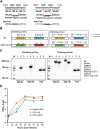
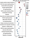
Multigene family (MGF) gene products are increasingly reported to be implicated in African swine fever virus (ASFV) virulence and attenuation of host defenses, among which the MGF360-9L and MGF505-7R gene products are characterized by convergent but distinct mechanisms of immune evasion. Herein, a recombinant ASFV mutant, ASFV-Δ9L/Δ7R, bearing combinational deletions of MGF360-9L and MGF505-7R, was constructed from the highly virulent ASFV strain CN/GS/2018 of genotype II that is currently circulating in China. Pigs inoculated intramuscularly with 104 50% hemadsorption doses (HAD50) of the mutant remained clinically healthy without any serious side effects. Importantly, in a virulence challenge, all four within-pen contact pigs demonstrated clinical signs and pathological findings consistent with ASF. In contrast, vaccinated pigs (5/6) were protected and clinical indicators tended to be normal, accompanied by extensive tissue repairs. Similar to most viral infections, innate immunity and both humoral and cellular immune responses appeared to be vital for protection. Notably, transcriptome sequencing (RNA-seq) and quantitative PCR (qPCR) analysis revealed a regulatory function of the mutant in dramatic and sustained expression of type I/III interferons and inflammatory and innate immune genes in vitro. Furthermore, infection with the mutant elicited an early and robust p30-specific IgG response, which coincided and was strongly correlated with the protective efficacy. Analysis of the cellular response revealed a strong ASFV-specific interferon gamma (IFN-γ) response and immunostaining of CD4+ T cells coupled with a high level of CD163+ macrophage infiltration in spleens of vaccinated pigs. Our study identifies a new mechanism of immunological regulation by ASFV MGFs that rationalizes the design of live attenuated vaccine for implementation of improved control strategies to eradicate ASFV. IMPORTANCE Currently, the deficiency in commercially available vaccines or therapeutic options against African swine fever constitutes a matter of major concern in the swine industry globally. Here, we report the design and construction of a recombinant ASFV mutant harboring combinational deletions of interferon inhibitors MGF360-9L and MGF505-7R based on a genotype II ASFV CN/GS/2018 strain currently circulating in China. The mutant was completely attenuated when inoculated at a high dose of 104 HAD50. In the virulence challenge with homologous virus, sterile immunity was achieved, demonstrating the mutant's potential as a promising vaccine candidate. This sufficiency of effectiveness supports the claim that this live attenuated virus may be a viable vaccine option with which to fight ASF.
Keywords: ASFV; MGF360-9L; MGF505-7R; attenuated phenotype; immune response; protective efficacy.
Conflict of interest statement
The authors declare no conflict of interest.
Figures


Similar articles
-
Sequential Deletions of Interferon Inhibitors MGF110-9L and MGF505-7R Result in Sterile Immunity against the Eurasia Strain of Africa Swine Fever.J Virol. 2022 Oct 26;96(20):e0119222. doi: 10.1128/jvi.01192-22. Epub 2022 Oct 5. J Virol. 2022. PMID: 36197109 Free PMC article.
-
African Swine Fever Virus Georgia Isolate Harboring Deletions of MGF360 and MGF505 Genes Is Attenuated in Swine and Confers Protection against Challenge with Virulent Parental Virus.J Virol. 2015 Jun;89(11):6048-56. doi: 10.1128/JVI.00554-15. Epub 2015 Mar 25. J Virol. 2015. PMID: 25810553 Free PMC article.
-
Combinational Deletions of MGF110-9L and MGF505-7R Genes from the African Swine Fever Virus Inhibit TBK1 Degradation by an Autophagy Activator PIK3C2B To Promote Type I Interferon Production.J Virol. 2023 May 31;97(5):e0022823. doi: 10.1128/jvi.00228-23. Epub 2023 May 10. J Virol. 2023. PMID: 37162350 Free PMC article.
-
How Does African Swine Fever Virus Evade the cGAS-STING Pathway?Pathogens. 2024 Nov 2;13(11):957. doi: 10.3390/pathogens13110957. Pathogens. 2024. PMID: 39599510 Free PMC article. Review.
-
Attenuated African swine fever viruses and the live vaccine candidates: a comprehensive review.Microbiol Spectr. 2024 Nov 5;12(11):e0319923. doi: 10.1128/spectrum.03199-23. Epub 2024 Oct 8. Microbiol Spectr. 2024. PMID: 39377589 Free PMC article. Review.
Cited by
-
Mucosal and cellular immune responses elicited by nasal and intramuscular inoculation with ASFV candidate immunogens.Front Immunol. 2023 Sep 1;14:1200297. doi: 10.3389/fimmu.2023.1200297. eCollection 2023. Front Immunol. 2023. PMID: 37720232 Free PMC article.
-
Exploring Virus-Host Interactions Through Combined Proteomic Approaches Identifies BANF1 as a New Essential Factor for African Swine Fever Virus.Mol Cell Proteomics. 2025 Jul 22;24(9):101038. doi: 10.1016/j.mcpro.2025.101038. Online ahead of print. Mol Cell Proteomics. 2025. PMID: 40707000 Free PMC article.
-
African Swine Fever Virus I267L Is a Hemorrhage-Related Gene Based on Transcriptome Analysis.Microorganisms. 2024 Feb 17;12(2):400. doi: 10.3390/microorganisms12020400. Microorganisms. 2024. PMID: 38399804 Free PMC article.
-
Generation and Genetic Stability of a PolX and 5' MGF-Deficient African Swine Fever Virus Mutant for Vaccine Development.Vaccines (Basel). 2024 Sep 30;12(10):1125. doi: 10.3390/vaccines12101125. Vaccines (Basel). 2024. PMID: 39460292 Free PMC article.
-
Development and application of a TaqMan-based real-time PCR method for the detection of the ASFV MGF505-7R gene.Front Vet Sci. 2023 Apr 4;10:1093733. doi: 10.3389/fvets.2023.1093733. eCollection 2023. Front Vet Sci. 2023. PMID: 37256000 Free PMC article.
References
-
- Laddomada A, Rolesu S, Loi F, Cappai S, Oggiano A, Madrau MP, Sanna ML, Pilo G, Bandino E, Brundu D, Cherchi S, Masala S, Marongiu D, Bitti G, Desini P, Floris V, Mundula L, Carboni G, Pittau M, Feliziani F, Sanchez-Vizcaino JM, Jurado C, Guberti V, Chessa M, Muzzeddu M, Sardo D, Borrello S, Mulas D, Salis G, Zinzula P, Piredda S, De Martini A, Sgarangella F. 2019. Surveillance and control of African swine fever in free-ranging pigs in Sardinia. Transbound Emerg Dis 66:1114–1119. 10.1111/tbed.13138. - DOI - PMC - PubMed
-
- Sun E, Huang L, Zhang X, Zhang J, Shen D, Zhang Z, Wang Z, Huo H, Wang W, Huangfu H, Wang W, Li F, Liu R, Sun J, Tian Z, Xia W, Guan Y, He X, Zhu Y, Zhao D, Bu Z. 2021. Genotype I African swine fever viruses emerged in domestic pigs in China and caused chronic infection. Emerg Microbes Infect 10:2183–2193. 10.1080/22221751.2021.1999779. - DOI - PMC - PubMed
Publication types
MeSH terms
Substances
LinkOut - more resources
Full Text Sources
Research Materials

