

The Acquisition and Retention of Lumpy Skin Disease Virus by Blood-Feeding Insects Is Influenced by the Source of Virus, the Insect Body Part, and the Time since Feeding
- PMID: 35867566
- PMCID: PMC9364806
- DOI: 10.1128/jvi.00751-22
The Acquisition and Retention of Lumpy Skin Disease Virus by Blood-Feeding Insects Is Influenced by the Source of Virus, the Insect Body Part, and the Time since Feeding
Abstract
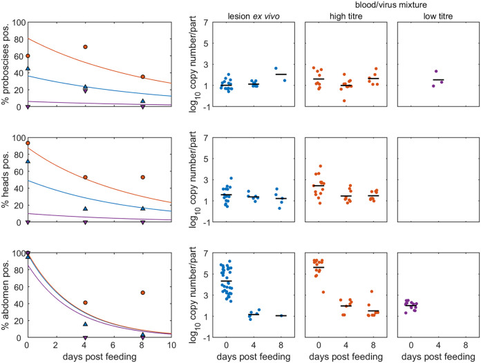
Lumpy skin disease virus (LSDV) is a poxvirus that causes severe systemic disease in cattle and is spread by mechanical arthropod-borne transmission. This study quantified the acquisition and retention of LSDV by four species of Diptera (Stomoxys calcitrans, Aedes aegypti, Culex quinquefasciatus, and Culicoides nubeculosus) from cutaneous lesions, normal skin, and blood from a clinically affected animal. The acquisition and retention of LSDV by Ae. aegypti from an artificial membrane feeding system was also examined. Mathematical models of the data were generated to identify the parameters which influence insect acquisition and retention of LSDV. For all four insect species, the probability of acquiring LSDV was substantially greater when feeding on a lesion compared with feeding on normal skin or blood from a clinically affected animal. After feeding on a skin lesion LSDV was retained on the proboscis for a similar length of time (around 9 days) for all four species and for a shorter time in the rest of the body, ranging from 2.2 to 6.4 days. Acquisition and retention of LSDV by Ae. aegypti after feeding on an artificial membrane feeding system that contained a high titer of LSDV was comparable to feeding on a skin lesion on a clinically affected animal, supporting the use of this laboratory model as a replacement for some animal studies. This work reveals that the cutaneous lesions of LSD provide the high-titer source required for acquisition of the virus by insects, thereby enabling the mechanical vector-borne transmission. IMPORTANCE Lumpy skin disease virus (LSDV) is a high consequence pathogen of cattle that is rapidly expanding its geographical boundaries into new regions such as Europe and Asia. This expansion is promoted by the mechanical transmission of the virus via hematogenous arthropods. This study quantifies the acquisition and retention of LSDV by four species of blood-feeding insects and reveals that the cutaneous lesions of LSD provide the high titer virus source necessary for virus acquisition by the insects. An artificial membrane feeding system containing a high titer of LSDV was shown to be comparable to a skin lesion on a clinically affected animal when used as a virus source. This promotes the use of these laboratory-based systems as replacements for some animal studies. Overall, this work advances our understanding of the mechanical vector-borne transmission of LSDV and provides evidence to support the design of more effective disease control programmes.
Keywords: Aedes aegypti; Culex quinquefasciatus; Culicoides nubeculosus; Stomoxys calcitrans; control; flies; lumpy skin disease; midges; mosquitoes; poxvirus; transmission; vector.
Conflict of interest statement
The authors declare no conflict of interest.
Figures
Similar articles
-
Quantifying and Modeling the Acquisition and Retention of Lumpy Skin Disease Virus by Hematophagus Insects Reveals Clinically but Not Subclinically Affected Cattle Are Promoters of Viral Transmission and Key Targets for Control of Disease Outbreaks.J Virol. 2021 Apr 12;95(9):e02239-20. doi: 10.1128/JVI.02239-20. Print 2021 Apr 12. J Virol. 2021. PMID: 33568514 Free PMC article.
-
Using the basic reproduction number to assess the risk of transmission of lumpy skin disease virus by biting insects.Transbound Emerg Dis. 2019 Sep;66(5):1873-1883. doi: 10.1111/tbed.13216. Epub 2019 May 15. Transbound Emerg Dis. 2019. PMID: 31038286 Free PMC article.
-
Infection, dissemination, and transmission of lumpy skin disease virus in Aedes aegypti (Linnaeus), Culex tritaeniorhynchus (Giles), and Culex quinquefasciatus (Say) mosquitoes.Acta Trop. 2024 Jun;254:107205. doi: 10.1016/j.actatropica.2024.107205. Epub 2024 Apr 3. Acta Trop. 2024. PMID: 38579960
-
Transmission of lumpy skin disease virus: A short review.Virus Res. 2019 Aug;269:197637. doi: 10.1016/j.virusres.2019.05.015. Epub 2019 May 29. Virus Res. 2019. PMID: 31152757 Review.
-
Epidemiological Risk Factors and Modelling Approaches for Risk Assessment of Lumpy Skin Disease Virus Introduction and Spread: Methodological Review and Implications for Risk-Based Surveillance in Australia.Transbound Emerg Dis. 2024 May 2;2024:3090226. doi: 10.1155/2024/3090226. eCollection 2024. Transbound Emerg Dis. 2024. PMID: 40303055 Free PMC article. Review.
Cited by
-
Development of a Multi-Locus Real-Time PCR with a High-Resolution Melting Assay to Differentiate Wild-Type, Asian Recombinant, and Vaccine Strains of Lumpy Skin Disease Virus.Vet Sci. 2025 Mar 1;12(3):213. doi: 10.3390/vetsci12030213. Vet Sci. 2025. PMID: 40266924 Free PMC article.
-
A new variant of lumpy skin disease virus circulating in Vietnam based on sequencing analysis of GPCR gene.Open Vet J. 2024 Jul;14(7):1701-1707. doi: 10.5455/OVJ.2024.v14.i7.19. Epub 2024 Jul 31. Open Vet J. 2024. PMID: 39175973 Free PMC article.
-
Lumpy Skin Disease: A Systematic Review of Mode of Transmission, Risk of Emergence and Risk Entry Pathway.Viruses. 2023 Jul 25;15(8):1622. doi: 10.3390/v15081622. Viruses. 2023. PMID: 37631965 Free PMC article.
-
Molecular detection of lumpy skin disease virus in naturally infected cattle and buffaloes: unveiling the role of tick vectors in disease spread.Vet Res Commun. 2024 Dec;48(6):3921-3939. doi: 10.1007/s11259-024-10541-7. Epub 2024 Oct 8. Vet Res Commun. 2024. PMID: 39377904 Free PMC article.
-
Detection of Culex tritaeniorhynchus Giles and Novel Recombinant Strain of Lumpy Skin Disease Virus Causes High Mortality in Yaks.Viruses. 2023 Mar 29;15(4):880. doi: 10.3390/v15040880. Viruses. 2023. PMID: 37112860 Free PMC article.
References
-
- Foil L, Gorham JR. 2000. Mechanical transmission of disease agents by arthropods, p 461–514. In Eldridge BF, Edman JD (ed), Medical entomology. Kluwer Academic Publishers, Alphen aan den Rijn.
-
- Brody AL. 1936. The transmission of fowl-pox. Memoir vol. 195. Cornell University Agricultural Experiment Station, Ithaca, NY.
MeSH terms
Substances
Grants and funding
- BB/R002606/UKRI | Biotechnology and Biological Sciences Research Council (BBSRC)
- BB/T005173/1/UKRI | Biotechnology and Biological Sciences Research Council (BBSRC)
- BBS/E/I/00007036/UKRI | Biotechnology and Biological Sciences Research Council (BBSRC)
- BBS/E/I/00007037/UKRI | Biotechnology and Biological Sciences Research Council (BBSRC)
- BBS/E/I/00007039/UKRI | Biotechnology and Biological Sciences Research Council (BBSRC)
LinkOut - more resources
Full Text Sources
Medical
