

Shared N417-Dependent Epitope on the SARS-CoV-2 Omicron, Beta, and Delta Plus Variants
- PMID: 35867572
- PMCID: PMC9364786
- DOI: 10.1128/jvi.00558-22
Shared N417-Dependent Epitope on the SARS-CoV-2 Omicron, Beta, and Delta Plus Variants
Abstract
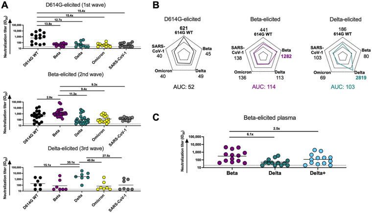
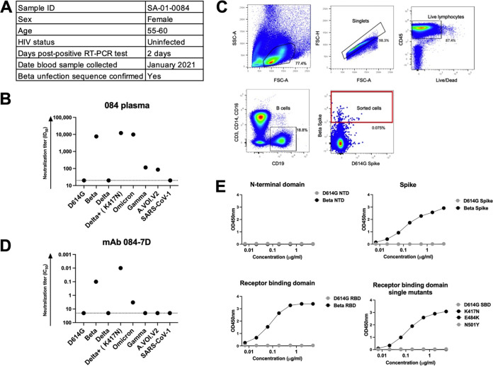
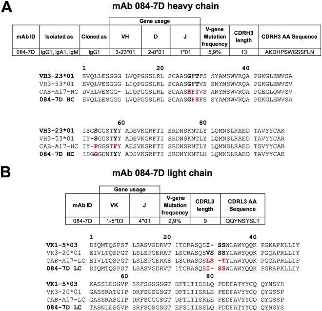
As severe acute respiratory syndrome coronavirus 2 (SARS-CoV-2) continues to evolve, several variants of concern (VOCs) have arisen which are defined by multiple mutations in their spike proteins. These VOCs have shown variable escape from antibody responses and have been shown to trigger qualitatively different antibody responses during infection. By studying plasma from individuals infected with either the original D614G, Beta, or Delta variants, we showed that the Beta and Delta variants elicit antibody responses that are overall more cross-reactive than those triggered by D614G. Patterns of cross-reactivity varied, and the Beta and Delta variants did not elicit cross-reactive responses to each other. However, Beta-elicited plasma was highly cross-reactive against Delta Plus (Delta+), which differs from Delta by a single K417N mutation in the receptor binding domain, suggesting that the plasma response targets the N417 residue. To probe this further, we isolated monoclonal antibodies from a Beta-infected individual with plasma responses against Beta, Delta+, and Omicron, which all possess the N417 residue. We isolated an N417-dependent antibody, 084-7D, which showed similar neutralization breadth to the plasma. The 084-7D MAb utilized the IGHV3-23*01 germ line gene and had somatic hypermutations similar to those of previously described public antibodies which target the 417 residue. Thus, we have identified a novel antibody which targets a shared epitope found on three distinct VOCs, enabling their cross-neutralization. Understanding antibodies targeting escape mutations, such as K417N, which repeatedly emerge through convergent evolution in SARS-CoV-2 variants, may aid in the development of next-generation antibody therapeutics and vaccines. IMPORTANCE The evolution of SARS-CoV-2 has resulted in variants of concern (VOCs) with distinct spike mutations conferring various immune escape profiles. These variable mutations also influence the cross-reactivity of the antibody response mounted by individuals infected with each of these variants. This study sought to understand the antibody responses elicited by different SARS-CoV-2 variants and to define shared epitopes. We show that Beta and Delta infections resulted in antibody responses that were more cross-reactive than the original D614G variant, but they had differing patterns of cross-reactivity. We further isolated an antibody from Beta infection which targeted the N417 site, enabling cross-neutralization of Beta, Delta+, and Omicron, all of which possess this residue. The discovery of antibodies which target escape mutations common to multiple variants highlights conserved epitopes to target in future vaccines and therapeutics.
Keywords: SARS-CoV-2; antibody cross-reactivity; antibody isolation; variants.
Conflict of interest statement
The authors declare no conflict of interest.
Figures

References
-
- Tegally H, Wilkinson E, Giovanetti M, Iranzadeh A, Fonseca V, Giandhari J, Doolabh D, Pillay S, San EJ, Msomi N, Mlisana K, von Gottberg A, Walaza S, Allam M, Ismail A, Mohale T, Glass AJ, Engelbrecht S, Van Zyl G, Preiser W, Petruccione F, Sigal A, Hardie D, Marais G, Hsiao N-Y, Korsman S, Davies M-A, Tyers L, Mudau I, York D, Maslo C, Goedhals D, Abrahams S, Laguda-Akingba O, Alisoltani-Dehkordi A, Godzik A, Wibmer CK, Sewell BT, Lourenço J, Alcantara LCJ, Kosakovsky Pond SL, Weaver S, Martin D, Lessells RJ, Bhiman JN, Williamson C, de Oliveira T. 2021. Detection of a SARS-CoV-2 variant of concern in South Africa. Nature 592:438–443. 10.1038/s41586-021-03402-9. - DOI - PubMed
-
- Wibmer CK, Ayres F, Hermanus T, Madzivhandila M, Kgagudi P, Oosthuysen B, Lambson BE, de Oliveira T, Vermeulen M, van der Berg K, Rossouw T, Boswell M, Ueckermann V, Meiring S, von Gottberg A, Cohen C, Morris L, Bhiman JN, Moore PL. 2021. SARS-CoV-2 501Y.V2 escapes neutralization by South African COVID-19 donor plasma. Nat Med 27:622–625. 10.1038/s41591-021-01285-x. - DOI - PubMed
-
- Scheepers C, Everatt J, Amoako DG, Tegally H, Wibmer CK, Mnguni A, Ismail A, Mahlangu B, Lambson BE, Richardson SI, Martin DP, Wilkinson E, San JE, Giandhari J, Manamela N, Ntuli N, Kgagudi P, Cele S, Pillay S, Mohale T, Ramphal U, Naidoo Y, Khumalo ZT, Kwatra G, Gray G, Bekker L-G, Madhi SA, Baillie V, Van Voorhis WC, Treurnicht FK, Venter M, Mlisana K, Wolter N, Sigal A, Williamson C, Hsiao N-Y, Msomi N, Maponga T, Preiser W, Makatini Z, Lessells R, Moore PL, de Oliveira T, von Gottberg A, Bhiman JN, NGS-SA. 2021. Emergence and phenotypic characterization of C.1.2, a globally detected lineage that rapidly accumulated mutations of concern. medRxiv. 10.1101/2021.08.20.21262342. - DOI
-
- Viana R, Moyo S, Amoako DG, Tegally H, Scheepers C, Althaus CL, Anyaneji UJ, Bester PA, Boni MF, Chand M, Choga WT, Colquhoun R, Davids M, Deforche K, Doolabh D, Du Plessis L, Engelbrecht S, Everatt J, Giandhari J, Giovanetti M, Hardie D, Hill V, Hsiao N-Y, Iranzadeh A, Ismail A, Joseph C, Joseph R, Koopile L, Kosakovsky Pond SL, Kraemer MUG, Kuate-Lere L, Laguda-Akingba O, Lesetedi-Mafoko O, Lessells RJ, Lockman S, Lucaci AG, Maharaj A, Mahlangu B, Maponga T, Mahlakwane K, Makatini Z, Marais G, Maruapula D, Masupu K, Matshaba M, Mayaphi S, Mbhele N, Mbulawa MB, Mendes A, Mlisana K, et al. 2022. Rapid epidemic expansion of the SARS-CoV-2 Omicron variant in southern Africa. Nature 603:679–686. 10.1038/d41586-021-03832-5. - DOI - PMC - PubMed
MeSH terms
Substances
Supplementary concepts
LinkOut - more resources
Full Text Sources
Research Materials
Miscellaneous
