. 2022 Jul 27;96(14):e0081622.
doi: 10.1128/jvi.00816-22. Epub 2022 Jul 11.

Tembusu Virus Nonstructural Protein 2B Antagonizes Type I Interferon Production by Targeting MAVS for Degradation

Affiliations

Tembusu Virus Nonstructural Protein 2B Antagonizes Type I Interferon Production by Targeting MAVS for Degradation

Peng Zhou et al. J Virol. .

Abstract

Tembusu virus (TMUV) is a newly emerged avian flavivirus that has caused severe egg-drop syndrome and fatal encephalitis in domestic ducks. It has spread widely throughout the main duck-producing areas in Asia, resulting in substantial economic losses to the duck industry. Previous studies have reported that TMUV has evolved several strategies to counteract the duck's innate immune responses to successfully establish infection in its host cells. However, the mechanisms underlying this phenomenon have not been elucidated. Here, we discovered that TMUV-encoded NS2B is a negative regulator of poly(I:C)-induced duck interferon-β (IFN-β) expression. Mechanistically, TMUV NS2B was found to interact specifically with the mitochondrial antiviral-signaling protein (duMAVS). Consequently, duMAVS was degraded through the K48-linked ubiquitination and proteasomal pathway, leading to the interruption of the RIG-I-like receptor (RLR) signaling. Further analyses also identified K321, K354, K398, and K411 as crucial residues for NS2B-mediated ubiquitination and degradation of duMAVS. Additionally, we demonstrated that NS2B functions by recruiting the E3 ubiquitin ligase duck membrane-associated RING-CH-type finger 5 (duMARCH5) to modify duMAVS via polyubiquitination, blocking the duMAVS-mediated innate immune response and promoting TMUV replication. Taken together, our findings revealed a novel mechanism by which TMUV evades the duck's antiviral innate immune responses. IMPORTANCE Tembusu virus (TMUV), an emerging pathogenic flavivirus, has spread to most duck farming areas in Asia since 2010, causing significant economic losses to the duck industry. Recently, TMUV has expanded its host range and may pose a potential threat to mammals, including humans. Understanding the interaction between TMUV and its host is essential for the development of effective vaccines and therapeutics. Here, we show that NS2B encoded by TMUV inhibits IFN production by interacting with duck MAVS (duMAVS) to mediate ubiquitination and proteasomal degradation. Further studies suggest that the E3 ubiquitin ligase duck membrane-associated RING-CH-type finger 5 (duMARCH5) is recruited by NS2B to mediate proteasomal degradation of duMAVS. As a result, the innate immune response triggered by the RIG-I-like receptor (RLR) is disrupted, facilitating viral replication. Overall, our results reveal a novel mechanism by which TMUV evades host innate immunity and provide new therapeutic strategies to prevent TMUV infection.

Keywords: MAVS; NS2B; Tembusu virus; degradation; interferon.


Conflict of interest statement

The authors declare no conflict of interest.

Figures
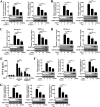
FIG 1
FIG 1
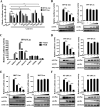
TMUV infection inhibited poly(I:C)- or SeV-induced expression of IFNs and ISGs. (A, B, F, G) DEFs were transfected with poly(I:C) or stimulated with SeV for 12 h and then infected with TMUV (0.5 or 5 MOI). At 12-h postinfection, cells were harvested, and mRNA levels of duck IFN-α (A), IFN-β (B), ZAP, PKR, and MX (F, G) were measured by RT-qPCR. All gene expression levels were normalized to duck GAPDH expression levels. (C, D) DEFs were transfected with 50 ng/well of duck IFN-β-Luc plasmid (C) or ISRE-Luc plasmid (D), along with 50 ng/well of the pRL-TK plasmid. At 24-h posttransfection, the cells were stimulated with poly(I:C) or SeV for 12 h and then infected with TMUV (0.5 or 5 MOI). At 12-h postinfection, cell lysates were harvested, and luciferase activities were measured using dual-luciferase reporter assays. (E) DEFs were transfected with poly(I:C) or stimulated with SeV for 12 h and then infected with TMUV (0.5 or 5 MOI). At 12-h postinfection, duck IFN-β levels in the supernatants were quantified by an IFN-β ELISA kit. All data are presented as mean ± SEM (n = 3). *, P < 0.05 and **, P < 0.01 (unpaired Student’s t test).
FIG 2
FIG 2
TMUV NS2B inhibits duMAVS signal transduction (A). DEFs were transfected with each of TMUV-derived expression plasmids (300 ng/well) or empty vector (300 ng/well), along with pRL-TK (50 ng/well), duck IFN-β-Luc (50 ng/well), or IRF7-Luc (50 ng/well). At 24-h posttransfection, cells were stimulated with poly(I:C), and the luciferase reporter assays were performed at 12 h after stimulation. (B) DEFs were transfected with increasing amounts of pCAGGS-HA-NS2B or pCAGGS-HA-NS3 (50, 100, 200 ng/well), along with IFN-β-Luc (50 ng/well) and pRL-TK (50 ng/well). At 24-h posttransfection, cells were stimulated with poly(I:C), and the luciferase activity was measured at 12-h poststimulation. (C) Each of the components (pCAGGS-Flag-duRIG-I, duMDA5, duMAVS, duTBK1, duIKKε, and duIRF7) (100 ng/well) or empty vector (100 ng/well) were cotransfected into DEFs with pCAGGS-HA-NS2B (200 ng/well), IFN-β-Luc (50 ng/well), and pRL-TK (50 ng/well). Luciferase assays were performed at 30 h after transfection. (D to F) DEFs were cotransfected with increasing amounts pCAGGS-HA-NS2B or pCAGGS-HA-NS3 (50, 100, 200 ng/well) and pCAGGS-Flag-duMAVS (100 ng/well), along with 50 ng/well of pRL-TK and 50 ng/well of IFN-β-Luc (D) or IRF7-Luc (E) or NF-κB-Luc (F). Luciferase assays were performed at 30-h posttransfection. Data are presented as mean ± SEM of three independent experiments. **, P < 0.01 (unpaired Student’s t test).
FIG 3
FIG 3
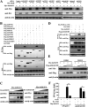
TMUV NS2B interacts with duMAVS. (A) HEK-293T cells were cotransfected with plasmids encoding HA-tagged NS2B and Flag-tagged duRIG-I, duMDA5, duMAVS, duTBK1, duIKKε, or duIRF7. Cell lysates were immunoprecipitated with anti-Flag antibodies, followed by immunoblot analysis with anti-HA and anti-Flag antibodies. Expression of transfected plasmids was detected by immunoblotting with the indicated antibodies (bottom). (B) HEK-293T cells were cotransfected with plasmids encoding Flag-tagged duMAVS and HA-tagged NS2B. Cell lysates were immunoprecipitated with anti-HA antibodies, and the immunocomplexes were detected by Western blotting with the indicated antibodies. (C) HeLa cells were cotransfected with plasmids expressing Flag-tagged duMAVS and HA-tagged NS2B. At 28-h posttransfection, the cells were fixed for immunofluorescence assays to detect duMAVS (green) and NS2B (blue) with anti-HA and anti-Flag antibodies, respectively. Mitochondria were stained with mito-Tracker (red). The fluorescence intensity profile of duMAVS (green), NS2B (blue), and mitochondria (red) was measured along the line drawn by Image J. Scale bar, 10 μm. (D) Coimmunoprecipitation analysis of the interaction of HA-tagged NS2B with Flag-tagged duMAVS or its truncation mutants in HEK-293T cells. (E) Coimmunoprecipitation analysis of the interaction of Flag-tagged duMAVS with HA-tagged NS2B or its truncation mutants in HEK-293T cells.
FIG 4
FIG 4
TMUV NS2B promotes the degradation of duMAVS. (A) DEFs were cotransfected with pCAGGS-HA-NS2B (2 μg/well) and pCAGGS-Flag-duRIG-I, -duMDA5, -duMAVS, -duTBK1, -duIKKε, or -duIRF7 (2 μg/well). After 28 h, cells were lysed and subjected to immunoblot analysis with anti-Flag, anti-HA, or anti-β-actin antibodies. (B) DEFs were transfected with increasing amounts of pCAGGS-HA-NS2B or pCAGGS-HA-NS3 (0.5, 1, 2 μg/well), along with pCAGGS-Flag-duMAVS (2 μg/well), followed by Western blotting with the indicated antibodies. (C) DEFs were transfected with increasing amounts of pCAGGS-HA-NS2B. At 24-h posttransfection, cells were collected to analyze mRNA levels of duMAVS by qRT-PCR. (D-G) DEFs were cotransfected with pCAGGS-Flag-duMAVS and pCAGGS-HA-NS2B or an empty vector. After 24 h, cells were treated with MG132 (10 μM) (D), 3-methyladenine (3-MA, 10 mM) (E), chloroquine (CQ, 20 μM) (F), NH4Cl (20 mM) (G) for 6 h. Cell lysates were then analyzed by Western blotting using the indicated antibodies. (H to I) DEFs were transfected with pCAGGS-Flag-duMAVS, together with pCAGGS- HA-NS2B or an empty vector. At 24-h posttransfection, cells were treated with MG132 (10 μM) (H) or 3-MA (10 mM) (I) for 6 h. Cell lysates were used for SDD-AGE analysis with the indicated antibodies. SDS-PAGE immunoblotting was used as a loading control.
FIG 5
FIG 5
NS2B catalyzes the K48-linked ubiquitination of duMAVS. (A, B) DEFs were cotransfected with Myc-tagged duMAVS and Flag-tagged NS2B, along with HA-tagged ubiquitin (Ub) (A) or HA-tagged K48/K63-Ub (B). At 24-h posttransfection, cells were treated with MG132 (10 μM) for 6 h. Cell lysates were subjected to coimmunoprecipitation with anti-Myc antibodies and subsequently detected by Western blotting with anti-HA and anti-Myc antibodies. Protein expression in cell lysates was detected by Western blotting with anti-HA, anti-Myc, anti-Flag, or anti-β-actin antibodies. (C) DEFs were cotransfected with Myc-tagged duMAVS or its truncated mutants and Flag-tagged NS2B, along with HA-tagged Ub. At 24-h posttransfection, the cells were treated with MG132 (10 μM) for 6 h. Cell lysates were subjected to coimmunoprecipitation and Western blotting with the indicated antibodies. (D, E) DEF were transiently cotransfected with Flag-tagged NS2B and HA-tagged Ub, along with wild-type Myc-tagged duMAVS or its point mutants K262R, K321R, K354R, K386R, K398R, K411R, K461R, and K619R (D) or K321R/K354R/K398R/K411R (4KR) (E). At 24-h posttransfection, the cells were treated with MG132 (10 μM) for 6 h. Cell lysates were subjected to coimmunoprecipitation and immunoblotting analysis with the indicated antibodies. (F) DEFs were cotransfected with Flag-tagged NS2B and Myc-tagged duMAVS or its point mutants. After 28 h, the cells were lysed and subjected to immunoblot analysis with the indicated antibodies. (G) DEFs were cotransfected with Myc-tagged duMAVS or duMAVS (4KR) and Flag-tagged NS2B or empty vector, together with pRL-TK and IFN-β or ISRE-Luc. At 30-h posttransfection, cells were harvested and lysed for luciferase assays. Data are presented as mean ± SEM of three independent experiments. **, P < 0.01 (unpaired Student’s t test).
FIG 6
FIG 6
DuMARCH5 is the E3 ubiquitin ligase involved in NS2B-induced duMAVS degradation. (A) DEFs were transfected with either a negative siRNA or specific siRNAs targeting duTAX1BP1, duTRIM25, duNDFIP1, duOTUD1, duPCBP2, duITCH, duMARCH5, duSMURF1, and duWWP1 for 12 h and subsequently transfected with Myc-tagged duMAVS and HA-tagged NS2B. At 24-h posttransfection, cell lysates were analyzed by immunoblotting with the indicated antibodies. (B) HEK-293T cells were cotransfected with HA-tagged NS2B and Flag-tagged duTAX1BP1, duTRIM25, duNDFIP1, duOTUD1, duPCBP2, duITCH, duMARCH5, duSMURF1, or duWWP1. At 28-h posttransfection, cell lysates were immunoprecipitated with anti-Flag antibodies, followed by Western blotting with the indicated antibodies. (C) DEFs were transfected with increasing amounts of pCAGGS-HA-duNDFIP or pCAGGS-HA-duMARCH5 (0.5, 1, or 2 μg/well), along with Myc-duMAVS (2 μg/well), followed by Western blotting with indicated antibodies. (D) DEFs were transiently cotransfected with Myc-tagged duMAVS and Flag-tagged duMARCH5, along with wild-type HA-tagged NS2B or its mutant NS2B (aa 1 to 50). At 24-h posttransfection, the cells were treated with MG132 (10 μM) for 6 h. Cell lysates were subjected to coimmunoprecipitation and immunoblotting analysis with the indicated antibodies. (E) DEFs were transfected with Myc-tagged duMAVS together with increasing amounts of Flag-tagged duMARCH5. At 24-h posttransfection, the cells were treated with DMSO or MG132 (10 μM) for 6 h. Cell lysates were subjected to Western blotting with the indicated antibodies. (F) DEFs were transfected with either negative siRNA or specific siRNA targeting duMARCH5 for 12 h and subsequently cotransfected with pRL-TK and duck IFN-β-Luc, along with Flag-tagged duMAVS or Flag empty vector and HA-tagged NS2B or HA empty vector. Luciferase assays were performed 24 h after transfection. Data are presented as mean ± SEM of three independent experiments. *, P < 0.05 and **, P < 0.01 (unpaired Student’s t test).
FIG 7
FIG 7
DuMARCH5 positively regulates the replication of TMUV. (A) DEFs were transfected with either negative siRNA or specific siRNA targeting duMARCH5 for 12 h and subsequently infected with TMUV at MOI of 0.5. Cells were harvested at 24-h postinfection, followed by Western blotting with anti-duMAVS, anti-TMUV E, or anti-β-actin antibodies. (B, C) DEFs were transfected with either negative siRNA or specific siRNA targeting duMARCH5, followed by TMUV infection (MOI = 0.1). At the indicated time points, viral titer and viral RNA were determined by TCID50 (B) and RT-qPCR (C). (D, E) DEFs were transfected with Flag-duMARCH5 or an empty vector, followed by TMUV infection (MOI = 0.1). At the indicated time points, viral titer and viral RNA were determined by TCID50 (D) and RT-qPCR (E).
FIG 8
FIG 8
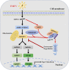
A hypothetical model explaining the role of TMUV NS2B in the disruption of duck RLR signaling.

Similar articles

Cited by

References

    1. Cao X. 2016. Self-regulation and cross-regulation of pattern-recognition receptor signalling in health and disease. Nat Rev Immunol 16:35–50. 10.1038/nri.2015.8. - DOI - PubMed
    1. Takeuchi O, Akira S. 2010. Pattern recognition receptors and inflammation. Cell 140:805–820. 10.1016/j.cell.2010.01.022. - DOI - PubMed
    1. Shi H, Wu J, Chen ZJ, Chen C. 2015. Molecular basis for the specific recognition of the metazoan cyclic GMP-AMP by the innate immune adaptor protein STING. Proc Natl Acad Sci USA 112:8947–8952. 10.1073/pnas.1507317112. - DOI - PMC - PubMed
    1. Kawai T, Akira S. 2006. Innate immune recognition of viral infection. Nat Immunol 7:131–137. 10.1038/ni1303. - DOI - PubMed
    1. Pichlmair A, Reis e Sousa C. 2007. Innate recognition of viruses. Immunity 27:370–383. 10.1016/j.immuni.2007.08.012. - DOI - PubMed

Publication types

MeSH terms

Substances

Supplementary concepts