. 2022 Jul 22;18(7):e1010723.
doi: 10.1371/journal.ppat.1010723. eCollection 2022 Jul.

The role of CD101-expressing CD4 T cells in HIV/SIV pathogenesis and persistence

Affiliations

The role of CD101-expressing CD4 T cells in HIV/SIV pathogenesis and persistence

Zachary Strongin et al. PLoS Pathog. .

Abstract

Despite the advent of effective antiretroviral therapy (ART), human immunodeficiency virus (HIV) continues to pose major challenges, with extensive pathogenesis during acute and chronic infection prior to ART initiation and continued persistence in a reservoir of infected CD4 T cells during long-term ART. CD101 has recently been characterized to play an important role in CD4 Treg potency. Using the simian immunodeficiency virus (SIV) model of HIV infection in rhesus macaques, we characterized the role and kinetics of CD101+ CD4 T cells in longitudinal SIV infection. Phenotypic analyses and single-cell RNAseq profiling revealed that CD101 marked CD4 Tregs with high immunosuppressive potential, distinct from CD101- Tregs, and these cells also were ideal target cells for HIV/SIV infection, with higher expression of CCR5 and α4β7 in the gut mucosa. Notably, during acute SIV infection, CD101+ CD4 T cells were preferentially depleted across all CD4 subsets when compared with their CD101- counterpart, with a pronounced reduction within the Treg compartment, as well as significant depletion in mucosal tissue. Depletion of CD101+ CD4 was associated with increased viral burden in plasma and gut and elevated levels of inflammatory cytokines. While restored during long-term ART, the reconstituted CD101+ CD4 T cells display a phenotypic profile with high expression of inhibitory receptors (including PD-1 and CTLA-4), immunsuppressive cytokine production, and high levels of Ki-67, consistent with potential for homeostatic proliferation. Both the depletion of CD101+ cells and phenotypic profile of these cells found in the SIV model were confirmed in people with HIV on ART. Overall, these data suggest an important role for CD101-expressing CD4 T cells at all stages of HIV/SIV infection and a potential rationale for targeting CD101 to limit HIV pathogenesis and persistence, particularly at mucosal sites.


Conflict of interest statement

The authors have declared that no competing interests exist

Figures

Fig 1
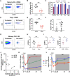
Fig 1. Phenotype of CD101-expressing CD4 T cells in healthy rhesus macaques.
A) Representative staining of CD101 on CD4 subsets in healthy RM PBMC. B) Representative histograms of CD101 on detailed CD4 subsets in healthy RM PBMC. C) Expression of CD101 within detailed CD4 subsets in PBMC and LN from healthy RM (top); total contribution of CD101+ cells to the overall CD4 pool from each CD4 subset in PBMC and LN. D) UMAP plot of flow cytometry data showing overall Phenograph cluster location and expression intensity of markers of interest. E) Hierarchical clustering of expression (z-score) for selected markers from flow cytometry data in each Phenograph cluster. F) Heatmap of the top 50 differentially expressed genes between CD101+ and CD101- Tregs (CD95+ CD25+ CD127-) by scRNA-seq analysis of healthy RM PBMC. G) Violin plots showing RNA expression levels of 4 genes of interest. H) Cytokine expression levels in CD101- and CD101+ memory CD4 after 3hr PMA/ionomycin stimulation of total PBMC from healthy individuals.
Fig 2
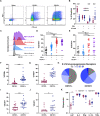
Fig 2. Dynamics of CD101-expressing CD4 T cells after SIV infection.
A) Representative staining of CD101 on PBMC memory CD4 pre- and post-SIV infection. B) Expression levels of CD101 on PBMC memory CD4 pre- and post-SIV infection. C) Total loss of CD101+ and CD101- cells within CD4 subsets calculated based on absolute counts in blood pre- and post-SIV infection (mean+SEM). D) Representative staining of CD101 on PBMC CD4 Tregs pre- and post-SIV infection. E) Expression levels of CD101 on PBMC CD4 Tregs pre- and post-SIV infection. F) Expression levels of CD101 on PBMC CD4 Tregs from HIV-infected individuals during active viremia and during suppressive antiretroviral therapy (ART). G) Representative staining of CD101 on rectal biopsy (RB) memory CD4 pre- and post-SIV infection. H) Expression levels of CD101 on RB memory CD4 pre- and post-SIV infection. I) Expression levels of α4β7 and CCR5 on RB memory CD4 CD101- and CD101+ cells from healthy RM. J) Association between depletion of CD101+ CD4 T cells in rectal biopsies at day 14 p.i. (calculated from baseline to acute infection as % of live lymphocytes) and SIV DNA levels in rectal biopsies at day 14 p.i. (spearman correlation, n = 9). K) CD101 expression within PBMC CD4 subsets during longitudinal SIV infection (mean±SEM). L) Overall contribution of CD101+ cells to the overall CD4 pool from each PBMC CD4 subset during longitudinal SIV infection (mean±SEM). Lines designate mean values.
Fig 3
Fig 3. Phenotypic profile of CD101+ CD4 during long-term antiretroviral therapy.
A) Representative staining of PD-1 and CTLA-4 within naïve, memory CD101- and CD101+ CD4 T cells in lymph node (LN) from ART-suppressed SIV-infected RM. B) Quadrant distribution of PD-1 and CTLA-4 expression within CD101- and CD101+ LN memory CD4 from ART-suppressed SIV-infected RM. C) Representative histogram of CD101 expression across PD-1 and CTLA-4 expression quadrants in LN memory CD4 from ART-suppressed SIV-infected RM. D) CD101 expression across PD-1 and CTLA-4 quadrants in LN memory CD4 from ART-suppressed SIV-infected RM. E) CD101 expression across PD-1 and CTLA-4 quadrants in PBMC memory CD4 from ART-suppressed HIV-infected individuals. F,G) Expression levels of CD39 (F) and TIGIT (G) within CD101- and CD101+ LN memory CD4. H) Pie chart representing the frequency of cells expressing PD-1 and/or CTLA-4 and/or CD39 and/or TIGIT within CD101- and CD101+ LN memory CD4. I,J) Expression levels of Ki-67 within CD101- and CD101+ memory CD4 from LN from ART-suppressed SIV-infected RM (I) and PBMC from ART-suppressed HIV-infected individuals (J). K) Cytokine expression levels in CD101- and CD101+ memory CD4 after 3hr PMA/ionomycin stimulation of total PBMC from ART-suppressed PLWH. Lines designate mean values.
Fig 4
Fig 4. HIV DNA levels in sorted memory CD4 T cells.
A) Sorting strategy for memory CD101- and CD101+ memory CD4 T cells. B-D) Total HIV proviruses (B), defective HIV DNA copies (C) and intact HIV DNA copies (D) measured by IPDA in sorted CD101- and CD101+ memory CD4 T cells from PBMC from ART-suppressed individuals.

References

    1. Bhaskaran K, Hamouda O, Sannes M, Boufassa F, Johnson AM, Lambert PC, et al.. Changes in the risk of death after HIV seroconversion compared with mortality in the general population. JAMA. 2008;300(1):51–9. Epub 2008/07/03. doi: 10.1001/jama.300.1.51 . - DOI - PubMed
    1. Antiretroviral Therapy Cohort C. Life expectancy of individuals on combination antiretroviral therapy in high-income countries: a collaborative analysis of 14 cohort studies. Lancet. 2008;372(9635):293–9. Epub 2008/07/29. doi: 10.1016/S0140-6736(08)61113-7 ; PubMed Central PMCID: PMC3130543. - DOI - PMC - PubMed
    1. Finzi D, Blankson J, Siliciano JD, Margolick JB, Chadwick K, Pierson T, et al.. Latent infection of CD4+ T cells provides a mechanism for lifelong persistence of HIV-1, even in patients on effective combination therapy. Nat Med. 1999;5(5):512–7. Epub 1999/05/06. doi: 10.1038/8394 . - DOI - PubMed
    1. Wong JK, Hezareh M, Gunthard HF, Havlir DV, Ignacio CC, Spina CA, et al.. Recovery of replication-competent HIV despite prolonged suppression of plasma viremia. Science. 1997;278(5341):1291–5. Epub 1997/11/21. doi: 10.1126/science.278.5341.1291 . - DOI - PubMed
    1. Hiener B, Horsburgh BA, Eden JS, Barton K, Schlub TE, Lee E, et al.. Identification of Genetically Intact HIV-1 Proviruses in Specific CD4(+) T Cells from Effectively Treated Participants. Cell Rep. 2017;21(3):813–22. doi: 10.1016/j.celrep.2017.09.081 ; PubMed Central PMCID: PMC5960642. - DOI - PMC - PubMed

Publication types