. 2022 Jul 22;8(29):eabo0155.
doi: 10.1126/sciadv.abo0155. Epub 2022 Jul 22.

The ER protein Creld regulates ER-mitochondria contact dynamics and respiratory complex 1 activity

Affiliations

The ER protein Creld regulates ER-mitochondria contact dynamics and respiratory complex 1 activity

Marie Paradis et al. Sci Adv. .

Abstract

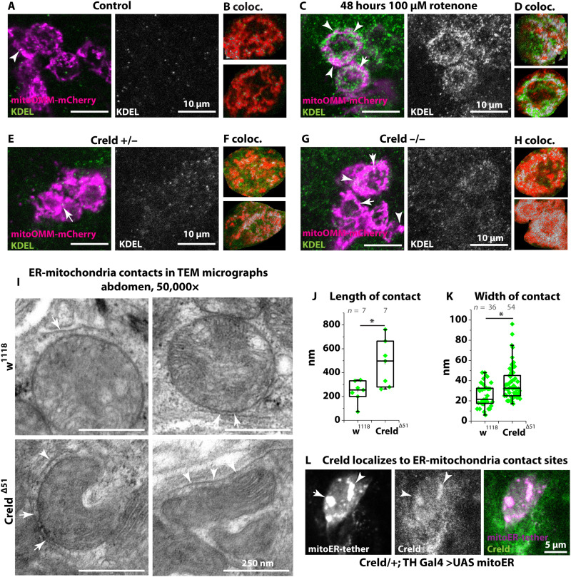
Dynamic contacts are formed between endoplasmic reticulum (ER) and mitochondria that enable the exchange of calcium and phospholipids. Disturbed contacts between ER and mitochondria impair mitochondrial dynamics and are a molecular hallmark of Parkinson's disease, which is also characterized by impaired complex I activity and dopaminergic neuron degeneration. Here, we analyzed the role of cysteine-rich with EGF-like domain (Creld), a poorly characterized risk gene for Parkinson's disease, in the regulation of mitochondrial dynamics and function. We found that loss of Creld leads to mitochondrial hyperfusion and reduced ROS signaling in Drosophila melanogaster, Xenopus tropicalis, and human cells. Creld fly mutants show differences in ER-mitochondria contacts and reduced respiratory complex I activity. The resulting low-hydrogen peroxide levels are linked to disturbed neuronal activity and lead to impaired locomotion, but not neurodegeneration, in Creld mutants. We conclude that Creld regulates ER-mitochondria communication and thereby hydrogen peroxide formation, which is required for normal neuron function.


Figures

Fig. 1.
Fig. 1.. Impaired locomotion, increased diastole, and mitochondrial accumulation in Creld mutants.
(A) Domain structure of human CRELD1 and CRELD2 and Drosophila Creld indicating protein identity of domains. (B) Single images from videos of w1118 and Creld mutants, showing their walking behavior. (C) Startle-induced negative geotaxis (SING) assay of 2-day-old w1118 and Creld mutants. (D) M-mode tracing of pixel movement of the adult heart. (E) Quantification of diastole, systole [not significant (ns), P = 0.43], and heart period. (F) Heart rate of w1118 and Creld mutant flies. (G) Transmission electron micrographs of abdominal tissue of w1118 and Creld mutant adults. Sections show myocardial cells. (H) Transmission electron micrographs of thorax tissue of w1118 and Creld mutant adults. Sections show indirect flight muscles (IFMs). Boxes represent the interquartile range and median; whiskers represent minimum and maximum. ***P < 0.001.
Fig. 2.
Fig. 2.. Creld interaction with Pink1 and Parkin.
(A) X. tropicalis stage 40 bodywall muscle cells labeled with mito-eGFP and close-ups. (B) Quantification of the mitochondrial fragmentation index as the number of mitochondria divided by the total mitochondrial area of cells from animals injected with control and Creld1 morpholino (MO). (C) Untreated (control) or transfected with dsRNA against CRELD1 HeLa cells and close-ups. Mitochondria are stained with anti-TOMM20. (D) Quantification of the mitochondrial fragmentation index based on TOMM20 (C). (E) SING assay of w1118 and Creld mutants untreated or fed with 20 μM CsA in 10% sucrose solution. Analysis of variance (ANOVA) with F = 34.662 and 77 degrees of freedom. (F) SING assay of flies expressing a constitutively active form of Pink1. Genotypes are Creld−/−; tubulin Gal4/+, Creld/+ or −/−; and tubulin Gal4/UAS Pink1.C. ANOVA with F = 8.023 and 59 degrees of freedom. (G) SING assay of Pink1 mutants expressing UAS-Creld-HA. Genotypes are as indicated. ANOVA with F = 39.562 and 122 degrees of freedom. ns, P = 0.86. (H) SING assay of Creld-Parkin double mutants. Genotypes are as indicated. ANOVA with F = 56.584 and 204 degrees of freedom. ns, P = 0.29. Boxes represent the interquartile range and median; whiskers represent minimum and maximum. *P < 0.05, **P < 0.01, and *** P < 0.001.
Fig. 3.
Fig. 3.. Creld is required in neurons for normal locomotion.
(A) SING assay of animals expressing UAS-Creld-HA in specific tissues. Climbed distance in millimeters was measured after 6 s. Genotypes are (from left to right) Creld−/−; UAS-Creld-HA/+, Creld/+, or −/− as indicated; tubulin-Gal4/UAS Creld-HA, Creld+/−, or −/−; mef-Gal4; UAS-Creld-HA, Creld/+, or −/−; handC-Gal4; UAS-Creld-HA, Creld/+, or −/−; elav-Gal4; and UAS-Creld-HA. ANOVA with F = 46.1 and 195 degrees of freedom. (B) SING assay of animals expressing UAS-Creld-HA in specific neurons. Climbed distance in millimeters was measured after 6 s. Genotypes are Creld−/−; UAS-Creld-HA/+, Creld/+, or −/− as indicated; elav-Gal4/UAS Creld-HA, Creld/+, or −/−; dilp2,3-Gal4; UAS-Creld-HA, Creld/+, or −/−; D42-Gal4; UAS-Creld-HA, Creld/+, or −/−; TH-Gal4; and UAS-Creld-HA. ANOVA with F = 38.167 and 168 degrees of freedom. (C) Airyscan confocal imaging of ER in PPL1 cluster of dopaminergic neurons labeled by GFP KDEL (left) and mitochondria labeled by mCherry-mitoOMM (right) under the control of TH-Gal4. Genotypes are Creld/+; TH Gal4/UAS GFP KDEL or mCherry-mitoOMM and Creld−/−; and TH Gal4/UAS GFP KDEL or mCherry-mitoOMM. mCherry was detected with a DsRed antibody. (D) Mitochondrial fragmentation index in somata of dopaminergic neurons. (E and F) Calcium imaging in PPL2 clusters of dopaminergic neurons with UAS CaMPARI2. Images were taken after 30-s ultraviolet light induction and 4-min photoconversion. Genotypes are Creld/+ or −/− and TH Gal4/UAS CaMPARI2. Boxes represent the interquartile range and median; whiskers represent minimum and maximum. *P < 0.05, **P < 0.01, and ***P < 0.001.
Fig. 4.
Fig. 4.. Low H2O2 impairs dopaminergic neuron function.
(A and B) Adult gut tissue was stained with DHE to label superoxide anions. Adult gut was stained with MitoSOX to label mitochondrial superoxide anions. a.u., arbitrary units. (C) Hydrogen peroxide was measured in extracts of whole adults with Amplex Red (n = 8). (D) Transcript levels of ROS-scavenging enzymes in Creld mutants. Fold change was normalized to w1118 (n = 8). (E) SING assay of Creld mutants ubiquitously expressing UAS Sod1. Genotypes are Creld−/−; armadillo Gal4/+, Creld/+, or −/−; and armadillo Gal4/UAS Sod1. (F) Hydrogen peroxide was measured in extracts of whole adults with Amplex Red. Genotypes are Creld−/−; armadillo Gal4/+, Creld/+ or −/−; armadillo Gal4/UAS Pink1.C, Creld/+ or −/−; armadillo Gal4/UAS Sod1, Creld/+ or −/−; armadillo Gal4/UAS catalase-RNAi, Creld/+ or −/−; and armadillo Gal4/UAS Duox RNAi. (G) SING assay of Creld mutants expressing PINK1, Sod1, catalase-RNAi, or Duox-RNAi in dopaminergic neurons. Genotypes are Creld−/−; TH-Gal4;+; Creld/+ or −/−; TH-Gal4/UAS Pink1.C, Creld/+, or −/−; TH Gal4/UAS Sod1, Creld/+, or −/−; TH-Gal4/UAS catalase-RNAi, Creld/+, or −/−; and TH-Gal4/UAS Duox-RNAi. ANOVA with F = 33.627 and 145 degrees of freedom. (H) Oxygen consumption of larval bodywall preparations of w1118 and Creld mutants treated with pyruvate and malate and rotenone. (I) SING assay of Creld mutants expressing ND-39 in dopaminergic neurons. Genotypes are Creld−/−, TH-Gal4;+, Creld/+ or −/−, and TH-Gal4/UAS ND-39. Boxes represent the interquartile range and median; whiskers represent minimum and maximum. ***P < 0.001.
Fig. 5.
Fig. 5.. Creld and complex I activity modulate ER-mitochondria contact.
(A) PPL1 cluster of dopaminergic neurons expressing mCherry-mitoOMM, stained with anti-DsRed and anti-KDEL in +/+; TH-Gal4/UAS mitoOMM. (B, D, F, and H) Regions of interest of single soma of dopaminergic neurons after colocalization analysis with ImageJ colocalization threshold tool. Colocalization is shown in gray. (C) PPL1 cluster of dopaminergic neurons expressing mCherry-mitoOMM, stained with anti-DsRed and anti-KDEL in +/+; TH-Gal4/UAS mitoOMM after treatment with rotenone. (E and G) PPL1 cluster of dopaminergic neurons expressing mCherry-mitoOMM, stained with anti-DsRed and anti-KDEL in Creld/+; TH-Gal4/UAS mitoOMM (A) and Creld−/−; TH-Gal4/UAS mitoOMM. Arrowheads show colocalization of ER and mitochondria. (I) TEM micrographs of adult abdomen showing ER-mitochondria contact sites. Scale bars are as indicated. (J) Quantification of ER-mitochondria contact length. (K) Quantification of ER-mitochondria contact width. (L) PPL1 cluster of dopaminergic neurons expressing an artificial tether construct (mitoER-tether-RFP) stained with α-Creld. Boxes represent the interquartile range and median; whiskers represent minimum and maximum. *P < 0.05 and ***P < 0.001.
Fig. 6.
Fig. 6.. Phospholipid transfer at ER-mitochondria contacts is impaired in Creld mutants.
(A) Total lipid, phospholipid, PE, and PC content of fractions containing the cytoplasm, ER, plasma membrane, and lysosomes. Fold change of extracts of Creld mutant flies normalized to w111. (B) Lipids as indicated in fractions containing mitochondria and MAM (crude mitochondria). (C) Lipids as indicated in fractions containing MAM. (D to F) Model of Creld action under (D) wild-type, (E) complex I–inhibited, and (F) Creld loss-of-function conditions. Bar charts show means and SD, and dots represent single data points. *P < 0.05. ns, not significant or P as indicated.

References

    1. Lapointe J., Hekimi S., When a theory of aging ages badly. Cell. Mol. Life Sci. 67, 1–8 (2010). - PMC - PubMed
    1. Ohashi M., Hirano T., Watanabe K., Shoji H., Ohashi N., Baba H., Endo N., Kohno T., Hydrogen peroxide modulates neuronal excitability and membrane properties in ventral horn neurons of the rat spinal cord. Neuroscience 331, 206–220 (2016). - PubMed
    1. Scialò F., Sriram A., Fernández-Ayala D., Gubina N., Lõhmus M., Nelson G., Logan A., Cooper H. M., Navas P., Enríquez J. A., Murphy M. P., Sanz A., Mitochondrial ROS produced via reverse electron transport extend animal lifespan. Cell Metab. 23, 725–734 (2016). - PMC - PubMed
    1. Morán M., Moreno-Lastres D., Marín-Buera L., Arenas J., Martín M. A., Ugalde C., Mitochondrial respiratory chain dysfunction: Implications in neurodegeneration. Free Radic. Biol. Med. 53, 595–609 (2012). - PubMed
    1. Trist B. G., Hare D. J., Double K. L., Oxidative stress in the aging substantia nigra and the etiology of Parkinson’s disease. Aging Cell 18, e13031 (2019). - PMC - PubMed

MeSH terms

Substances