. 2022 Jul 26;119(30):e2120489119.
doi: 10.1073/pnas.2120489119. Epub 2022 Jul 22.

A mechanism of self-lipid endocytosis mediated by the receptor Mincle

Affiliations

A mechanism of self-lipid endocytosis mediated by the receptor Mincle

Alexey V Kostarnoy et al. Proc Natl Acad Sci U S A. .

Abstract

Cellular lipid uptake (through endocytosis) is a basic physiological process. Dysregulation of this process underlies the pathogenesis of diseases such as atherosclerosis, obesity, diabetes, and cancer. However, to date, only some mechanisms of lipid endocytosis have been discovered. Here, we show a previously unknown mechanism of lipid cargo uptake into cells mediated by the receptor Mincle. We found that the receptor Mincle, previously shown to be a pattern recognition receptor of the innate immune system, tightly binds a range of self-lipids. Moreover, we revealed the minimal molecular motif in lipids that is sufficient for Mincle recognition. Superresolution microscopy showed that Mincle forms vesicles in cytoplasm and colocalizes with added fluorescent lipids in endothelial cells but does not colocalize with either clathrin or caveolin-1, and the added lipids were predominantly incorporated in vesicles that expressed Mincle. Using a model of ganglioside GM3 uptake in brain vessel endothelial cells, we show that the knockout of Mincle led to a dramatic decrease in lipid endocytosis. Taken together, our results have revealed a fundamental lipid endocytosis pathway, which we call Mincle-mediated endocytosis (MiME), and indicate a prospective target for the treatment of disorders of lipid metabolism, which are rapidly increasing in prevalence.

Keywords: C-type lectin receptors; Mincle; endocytosis; endothelial cells; ganglioside.


Conflict of interest statement

The authors declare no competing interest.

Figures
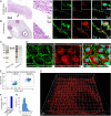
Fig. 1.
Fig. 1.
Human endothelial cells express Mincle. (A) Hematoxylin/eosin and immunohistochemical staining using antibodies against Mincle (red) and CD31 (green) in human carotid plaque specimens collected immediately after endarterectomy. Nuclei were costained with DAPI (cyan); n = 6 samples. Representative images are shown. (B) Hematoxylin/eosin and immunohistochemical staining of human umbilical cord specimens for Mincle (red) and CD31 (green). Nuclei were costained with DAPI (cyan); n = 6 samples. Representative images are shown. (C) HUVEC lysates were evaluated for Mincle expression by Western blotting using two clones of monoclonal antibodies. (D) HUVECs were costained for Mincle (red) and CD31 (green) and imaged by confocal microscopy. (E and F) Flow cytometry was performed on HUVECs stained for surface and intracellular Mincle. Flow cytometry plots (E) and quantification of Mincle+CD31+ cell percentages (F) are depicted. (G) SIM of HUVECs stained for Mincle revealed the presence of this receptor in the cytoplasm in small, uniformly sized bodies with an average diameter of 115.2 ± 33.2 nm (mean ± SD); the histogram is presented in H; in total, 14,768 Mincle-expressing bodies were measured in n = 5 cells with an Imaris 7.2. The results are presented as the mean ± SD values.
Fig. 2.
Fig. 2.
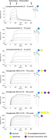
Mincle recognizes various GSLs through direct binding. SPR sensorgrams of glycolipids binding to chip-immobilized Mincle are expressed in response units (RU) vs. time after double referencing (blank surface and blank buffer referencing). Recombinant human Mincle was produced in human cells. The concentrations of lysoglucosylceramide, glucosylceramide, and lactosylceramide were 2, 4, 6, 8, and 10 µM (from bottom to top), and the concentrations of gangliosides GM3, GD3, and GM1 were 0.78, 1.56, 3.13, 6.25, 12.5, 25, 50, 75, and 100 µM (from bottom to top). All data shown are representative of at least two independent experiments.
Fig. 3.
Fig. 3.
Mincle colocalizes with added fluorescent GSLs in human endothelial cells. (Left) Three-dimensional SIM in HUVECs. HUVECs were incubated for 1 h with exogenously added fluorescent GSLs, washed, fixed, labeled with antibodies against Mincle, and analyzed by SIM. Glucosylceramide and lactosylceramide fluorescently labeled with NBD and ganglioside GM3 fluorescently labeled with TopFluor were used. Fluorescent glycoshingolipids are pseudocolored in green and Mincle in red; colocalization is indicated by yellow pseudocoloring. Representative cells are shown. Two independent experiments were performed. (Right) Statistical analyses of GSL/Mincle colocalization using the Pearson correlation coefficient; n = 15 independent fields per quantification. The results are presented as the mean ± SD values.
Fig. 4.
Fig. 4.
Mincle is necessary for rapid uptake of ganglioside GM3 by brain endothelial cells. (A) Brain endothelial cells isolated from Mincle-deficient mice and WT mice using MACS were incubated with fluorescent TopFluor-labeled ganglioside GM3 for 2 h, stained with phalloidin to delineate their boundaries, and evaluated by confocal microscopy. Phalloidin is pseudocolored in red and GM3 in green. (B) Quantification of fluorescent GM3 uptake by endothelial cells isolated from Mincle-deficient mice and WT mice using MACS by confocal microscopy. (C) Brain endothelial cells isolated from Mincle-deficient (Mincle-KO) mice and WT mice via puromycin selection were incubated with TopFluor-labeled ganglioside GM3 for 2 h, stained using anti-CD31 antibodies to delineate cell boundaries, and evaluated by confocal microscopy. CD31 is pseudocolored in red and GM3 in green. Statistics were calculated with an unpaired two-tailed Student’s t test *P < 0.0001. The results are presented as the mean ± SD values.

References

    1. Johannes L., Wunder C., Shafaq-Zadah M., . Glycolipids and Lectins in Endocytic Uptake Processes. Journal of Molecular Biology 428, 4792–4818 (2016). - PubMed
    1. Ferreira A. P. A., Boucrot E., Mechanisms of carrier formation during clathrin-independent endocytosis. Trends Cell Biol. 28, 188–200 (2018). - PubMed
    1. Kaksonen M., Roux A., Mechanisms of clathrin-mediated endocytosis. Nat. Rev. Mol. Cell Biol. 19, 313–326 (2018). - PubMed
    1. Kumano-Kuramochi M., et al. , Lectin-like oxidized LDL receptor-1 is palmitoylated and internalizes ligands via caveolae/raft-dependent endocytosis. Biochem. Biophys. Res. Commun. 434, 594–599 (2013). - PubMed
    1. Conway E. M., Boffa M. C., Nowakowski B., Steiner-Mosonyi M., An ultrastructural study of thrombomodulin endocytosis: Internalization occurs via clathrin-coated and non-coated pits. J. Cell. Physiol. 151, 604–612 (1992). - PubMed

Publication types

LinkOut - more resources