. 2023 Feb 13;146(2):629-644.
doi: 10.1093/brain/awac271.

Shh activation restores interneurons and cognitive function in newborns with intraventricular haemorrhage

Affiliations

Shh activation restores interneurons and cognitive function in newborns with intraventricular haemorrhage

Bokun Cheng et al. Brain. .

Abstract

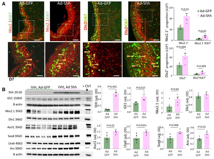
Premature infants with germinal matrix haemorrhage-intraventricular haemorrhage (GMH-IVH) suffer from neurobehavioural deficits as they enter childhood and adolescence. Yet the underlying mechanisms remain unclear. Impaired development and function of interneurons contribute to neuropsychiatric disorders. Therefore, we hypothesized that the occurrence of IVH would reduce interneuron neurogenesis in the medial ganglionic eminence and diminish the population of parvalbumin+ and somatostatin+ cortical interneurons. Because Sonic Hedgehog promotes the production of cortical interneurons, we also postulated that the activation of Sonic Hedgehog signalling might restore neurogenesis, cortical interneuron population, and neurobehavioural function in premature newborns with IVH. These hypotheses were tested in a preterm rabbit model of IVH and autopsy samples from human preterm infants. We compared premature newborns with and without IVH for intraneuronal progenitors, cortical interneurons, transcription factors regulating neurogenesis, single-cell transcriptome of medial ganglionic eminence and neurobehavioural functions. We treated premature rabbit kits with adenovirus expressing Sonic Hedgehog (Ad-Shh) or green fluorescence protein gene to determine the effect of Sonic Hedgehog activation on the interneuron production, cortical interneuron population and neurobehaviour. We discovered that IVH reduced the number of Nkx2.1+ and Dlx2+ progenitors in the medial ganglionic eminence of both humans and rabbits by attenuating their proliferation and inducing apoptosis. Moreover, IVH decreased the population of parvalbumin+ and somatostatin+ neurons in the frontal cortex of both preterm infants and kits relative to controls. Sonic Hedgehog expression and the downstream transcription factors, including Nkx2.1, Mash1, Lhx6 and Sox6, were also reduced in kits with IVH. Consistent with these findings, single-cell transcriptomic analyses of medial ganglionic eminence identified a distinct subpopulation of cells exhibiting perturbation in genes regulating neurogenesis, ciliogenesis, mitochondrial function and MAPK signalling in rabbits with IVH. More importantly, restoration of Sonic Hedgehog level by Ad-Shh treatment ameliorated neurogenesis, cortical interneuron population and neurobehavioural function in kits with IVH. Additionally, Sonic Hedgehog activation alleviated IVH-induced inflammation and several transcriptomic changes in the medial ganglionic eminence. Taken together, IVH reduced intraneuronal production and cortical interneuron population by downregulating Sonic Hedgehog signalling in both preterm rabbits and humans. Notably, activation of Sonic Hedgehog signalling restored interneuron neurogenesis, cortical interneurons and cognitive function in rabbit kits with IVH. These findings highlight disruption in cortical interneurons in IVH and identify a novel therapeutic strategy to restore cortical interneurons and cognitive function in infants with IVH. These studies can accelerate the development of new therapies to enhance the neurodevelopmental outcome of survivors with IVH.

Keywords: Sonic Hedgehog; cortical interneuron; germinal matrix haemorrhage; neurogenesis; premature newborns.


Conflict of interest statement

The authors report no competing interests.

Figures

Figure 1
Figure 1
IVH reduces interneuron production and the number of cortical PV+ and SST+ neurons in human infants. (A) Representative immunofluorescence of coronal sections from the MGE of a 24-week premature infant labeled with Ki67 and Nkx2.1/Dlx2 antibodies, as indicated. Bottom panel: High-power view of the boxed area in the top panel. Note that the number of both proliferating Nkx2.1+ and Dlx2+ cells (arrowhead) in the ventricular (VZ) and subventricular zone (SVZ) are fewer in the infants with IVH relative to controls. Scale bars 50 µM (top), 20 µM (bottom). Bar charts represent mean ± SEM (n = 5 each group). Student’s t-test used. Dlx2+Ki67+ and Dlx2+ cells were fewer in number in premature infants with IVH relative to controls without IVH, but not Nkx2.1+ cells. Data points are shown as dots within or above the bar. (B) Coronal sections from the frontoparietal cortex of a 29-week infant was stained with TLE4 (lower layer marker) and PV/SST antibodies. Bottom panel: High-power view of the boxed area in the top panel. The number of PV+ (arrow) and SST+ (arrowhead) neurons was reduced in the lower layer of the cortex of infants with IVH relative to controls without IVH. Scale bars 50 µM (top), 20 µM (bottom). Bar charts represent mean ± SEM (n = 5 each group). Student’s t-test used. PV+ and SST+ neurons were decreased in infants with IVH compared to infants without IVH in the lower layers. Data points are shown as dots within or above the bar.
Figure 2
Figure 2
IVH inhibits neurogenesis in rabbits. (A) Coronal sections at the level of mid-septal nucleus from D3 kits with and without IVH were labelled with Nkx2.1- and Ki67-specific antibodies. The number of both Nkx2.1+ and Nkx2.1+Ki67+ cells (arrowhead) was reduced in the kits with IVH. Scale bars 20 µM. Bar charts represent mean ± SEM (n = 5 each group). Two-way ANOVA used. Data points are shown as dots within or above the bar. Nkx2.1+ cells were diminished in number in premature kits with IVH relative to healthy (no glycerol) and glycerol-treated controls without IVH. Nkx2.1+Ki67+ cells were reduced in kits with IVH compared to healthy controls at D3. (B) Representative immunofluorescence of coronal section from D3 kits with and without IVH were labelled with Dlx2 and Ki67 antibodies. The number of both Dlx2 and Dlx2+Ki67+ cells (arrowhead) were reduced in the kits with IVH. Scale bars = 20 µM. Bar charts represent mean ± SEM (n = 5 each group). Two-way ANOVA used. Data points are shown as dots within or above the bar. Dlx2+ cells were few in premature kits with IVH relative to healthy (no glycerol) and glycerol-treated controls without IVH. Dlx2+Ki67+ cells were reduced in kits with IVH compared to both controls. (C) Representative western blot analyses of homogenates made from MGE of D3 kits with and without IVH, as indicated. Rat brain was used as a positive control. Data represent mean ± SEM (n = 5 each group). Student’s t-test used. Values are normalized to β-actin levels. Protein levels of Nkx2.1, Dlx1, Ascl1, Lhx6 and Sox6 were reduced in the MGE of kits with IVH compared to controls without IVH. (D) Western blot analyses from D7 kits with and without IVH, as indicated. Rat brain was used as a positive control. Data represent mean ± SEM (n = 5 each group). Student’s t-test used. Data points are shown as dots within or above the bar. Values are normalized to β-actin levels. Protein levels of Dlx1, Ascl1, Lhx6 and Sox6 were diminished in the MGE of kits with IVH relative to controls without IVH.
Figure 3
Figure 3
IVH reduced PV+ and SST+ neurons in the cerebral cortex. (A) Representative immunofluorescence of coronal section at the level of mid-septal nucleus from D14 kits with and without IVH were labelled with TLE4- (lower cortical layer marker), PV- and SST-specific antibodies. Bottom panel: High-power view of the boxed area in the top panel. Scale bars 50 µM (top), 20 µM (bottom). Bar charts represent mean ± SEM (n = 5 each group). Two-way ANOVA used. PV+ cells were fewer in number in the upper layer of premature kits with IVH relative to both glycerol-treated and untreated controls without IVH, a difference noted for IVH versus glycerol untreated kits with IVH in the lower layer. The number of SST+ neurons was reduced in both upper and lower layer of kits with IVH compared to both glycerol-treated and untreated controls without IVH. (B) Western blot analyses from D14 kits with and without IVH, as indicated. Rat brain was used as a positive control. Data represent mean ± SEM (n = 5 each group). Student’s t-test used. Values are normalized to β-actin levels. Protein levels were diminished for SST and elevated for SP8 in the cerebral cortex of kits with IVH relative to controls without IVH. Protein levels of PV were comparable between groups.
Figure 4
Figure 4
Transcriptional profiling of MGE cells by scRNA-seq. (A) Coronal slice taken at the mid-septal nucleus level shows slit like normal ventricle (arrow) in a kit without IVH and blood-filled ventricle (arrowhead) in kit with IVH. The dotted red line in each slice depicts the MGE that was dissected for RNA sequencing. Cells dissociated from these regions were processed for RNA seq. library using 10× chromium technologies. We next performed RNAseq. (B) UMAP (uniform manifold approximation) visualization depicting 10 categories of cells including radial glial, intermediate progenitors, immature interneurons, oligodendrocyte and others, as indicated. (C) Dot plot for select genes differentially expressed in the radial glia, intermediate progenitors and immature interneurons. Genes for a cell type compared with the remaining two cell types to determine the uniqueness of each category. The graph depicts unique gene sets that identify each of the three categories of cells. Dot size and colour represent percent and average expression, respectively. (D) Heat map of select genes differential expressed in radial glia, intermediate progenitors and immature neurons in the MGE that depict the gene sets unique to each category. The red, white and blue colours represent high, average and lower expression of a particular genes, respectively, as measured by row-standardized Z-scores.
Figure 5
Figure 5
Single-cell transcriptome-wide profiling of radial glia, intermediate progenitors and immature interneurons from MGE of kits with and without IVH. (A) UMAP (uniform manifold approximation) visualization of radial glial cells (top), intermediate progenitors (middle) and immature interneurons (bottom) showing their unbiased clustering into multiple subclusters (represented by distinct colours) in kits with and without IVH, as indicated. Top: Note subcluster 0 in ‘IVH’ and subclusters 3 and 9 (highlighted by broken line) in ‘no IVH’ show greater abundance of radial glia cells. Middle: Subcluster 3 in ‘no IVH’ and subcluster 0 in IVH category is dominating. Bottom: 13 subclusters of immature interneurons, and cluster 6 in kits with IVH and clusters 3 as well as 8 in kits without IVH were the prevailing clusters of cells. (B) Dot plot for selected genes differentially expressed in radial glia, intermediate progenitors, and immature interneurons. Size of the dot and the colour represent percent and average expression, respectively. Annotations on the y-axis describe the functional role of the gene. (C) Violin plot of canonical genes for interneuron neurogenesis differentially expressed in the radial glia, intermediate progenitors and immature neurons of the MGE of kits with and without IVH. The expression of SOX2, CCND2, DLX1, DLX5, GAD1, GAD5 and ARX genes are reduced in immature interneurons of kits with IVH relative to controls without IVH.
Figure 6
Figure 6
Activation of Shh signalling promotes neurogenesis in kits with IVH. (A) Coronal sections from Ad-Shh- and Ad-GFP-treated kit with IVH (D7) were labelled with Ki67 and Nkx2.1 or Dlx2 specific antibodies. Bottom panel: High-power view of the boxed area in the top panel. Note that both Nkx2.1+ and Nkx2.1+Ki67+ cells (arrowhead) as well as Dlx2+ and Dlx2+Ki67+ (arrowhead) neurons are increased in Ad-Shh-treated kits relative to controls. Scale bars 50 µM (top), 20 µM (bottom). Bar charts represent mean ± SEM. n = 5 each group. Two-way ANOVA used. Ad-Shh treatment increased the number of total and cycling Nkx2.1+ and Dlx2+ cells. (B) Western blot analyses were performed from homogenates made of dissected MGE (D7 kits) treated with Ad-Shh and Ad-GFP, as indicated. Rat brain was used as a positive control. Data represent mean ± SEM (n = 5 each group). Student’s t-test used. Values are normalized to β-actin levels. Protein levels of Shh, Gli1, Nkx2.1, Ascl1, Lhx6, Sox6 and Arx were elevated in the MGE of Ad-Shh-treated kits relative to Ad-GFP controls.
Figure 7
Figure 7
Ad-Shh treatment enhances cognitive function in kits with IVH. (A) Representative track plots of Ad-Shh- and Ad-GFP-treated kits with IVH in 3-chamber socialization test at D21. Bar charts show mean ± SEM, n = 8–10 each group, Two-way ANOVA used. Note that Ad-Shh-treated kits spent more time with stranger relative to Ad-GFP controls. (B) Typical track plots of Ad-Shh- and Ad-GFP-treated kits with IVH in elevated plus maze test at D21. Bar charts show mean ± SEM (n = 8–10 each group). Two-way ANOVA used. Note that Ad-Shh-treated kits spent more time in open arm relative Ad-GFP controls. (C) Representative track plots of Ad-Shh- and Ad-GFP-treated kits with IVH in object placement test at D21. Bar charts show mean ± SEM (n = 8–10 each group), Student’s t-test. Note that Ad-Shh-treated kits spent more time with moved object compared to Ad-GFP controls. (D) Bar charts show mean ± SEM (n = 8–10 each group). Two-way ANOVA used. Ad-Shh- and Ad-GFP-treated kits spent comparable time with novel object in Novel Object recognition test. (E) Bar charts show mean ± SEM (n = 8–10 each group). Two-way ANOVA used. Ad-Shh- and Ad-GFP-treated kits spent similar time in both centre and periphery of the arena in open field test.
Figure 8
Figure 8
Single-cell transcriptome profiling of radial glia, intermediate progenitors and immature interneurons from MGE of Ad-Shh- and Ad-GFP-treated kits with IVH. (A) UMAP plot of radial glial cells (top), intermediate progenitors (middle) and immature interneuron (bottom) showing their unbiased clustering into multiple subclusters (represented by distinct colours and a number) in Ad-Shh- and Ad-GFP-treated kits with IVH, as indicated. Top: Subclusters of radial glial cells. Of the six subclusters (top), subclusters 5 and 6 in Ad-Shh-treated and subclusters 2 and 3 in Ad-GFP-treated kits exhibit a higher abundance of cells. Middle: Eight subclusters of intermediate progenitors. Of these, clusters 0 and 1 display fewer cells in Ad-Shh-treated kits relative to Ad-GFP controls. Bottom: Clusters of immature interneurons. Note that clusters 0 and 3 show less abundance of cells in Ad-Shh-treated kits compared to controls. (B) Gene expression of selected genes from subclusters 2 and 3 of Ad-GFP-treated kits and subclusters 5 and 6 of Ad-Shh-treated kits shown as a dot plot. The selected signature genes from subclusters 2, 3, 5 and 6 represent differential expression profile between the radial glia of Ad-GFP- and Ad-Shh-treated kits at the subcluster level. The x-axis shows subcluster number and the y-axis shows gene names and their functional annotations. (C) Heat map of select genes differentially expressed in radial glia, intermediate progenitors and immature neurons of the MGE from Ad-GFP- and Ad-Shh-treated kits with IVH. Annotations on the y-axis describe the functional role of the genes. The red, white and blue colours represent high, average and lower expression of a particular genes, respectively, as measured by row-standardized Z-scores.

Similar articles

Cited by

References

    1. Ballabh P, de Vries LS. White matter injury in infants with intraventricular haemorrhage: Mechanisms and therapies. Nat Rev Neurol. 2021;17:199–214. - PMC - PubMed
    1. Indredavik MS, Skranes JS, Vik T, et al. . Low-birth-weight adolescents: Psychiatric symptoms and cerebral MRI abnormalities. Pediatr Neurol. 2005;33:259–266. - PubMed
    1. Whitaker AH, Feldman JF, Lorenz JM, et al. . Neonatal head ultrasound abnormalities in preterm infants and adolescent psychiatric disorders. Arch Gen Psychiatry. 2011;68:742–752. - PubMed
    1. Kepecs A, Fishell G. Interneuron cell types are fit to function. Nature. 2014;505:318–326. - PMC - PubMed
    1. Marín O. Interneuron dysfunction in psychiatric disorders. Nat Rev Neurosci. 2012;13:107–120. - PubMed

Publication types