

Covid-19 associated shoulder girdle calcific myositis: a novel entity
- PMID: 35867893
- PMCID: PMC10996957
- DOI: 10.1259/bjr.20220411
Covid-19 associated shoulder girdle calcific myositis: a novel entity
Abstract
Objective: To investigate the prevalence, describe the radiological features, and consider the clinical sequelae of COVID-19- associated shoulder girdle calcific myositis.
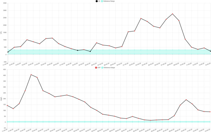
Methods: All patients who underwent a CT pulmonary angiogram study at our institution (Queen Alexandra Hospital, Portsmouth Hospitals University NHS Trust, Portsmouth, United Kingdom) in April and May 2020, January 2021, and July 2021 were included. A total of 1239 CT pulmonary angiogram studies for 1201 patients were reviewed. Patients with COVID-19 and associated shoulder girdle calcific myositis were identified. Their electronic patient records were reviewed. The patients' demographics, serum inflammatory markers, and proning history were recorded.
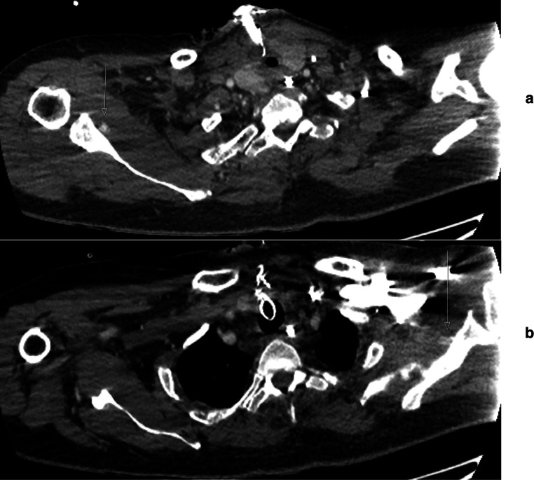
Results: Of the 364 patients in Wave 1, 71 patients (19.5%) had COVID-19, and of those, 2 patients (2.8%) had shoulder girdle calcific myositis. Of the 521 patients in Wave 2, 354 patients (67.9%) had COVID-19, and of those, 3 patients (0.8%) had shoulder girdle calcific myositis. Of the 316 patients in Wave 3, 37 patients (11.7%) had COVID-19, and of those, 1 patient (2.7%) had shoulder girdle calcific myositis. The overall prevalence was 1.3%. The most common site of calcific myositis was within the subscapularis muscle.
Conclusion: COVID-19-associated shoulder girdle calcific myositis is a rare extrapulmonary musculoskeletal manifestation of COVID-19. Early recognition and increased awareness of this disease entity, in our experience, aids in reducing patient morbidity and improving long-term functional outcome.
Advances in knowledge: We have reported a novel disease entity associated with COVID-19, in the form of shoulder girdle calcific myositis. We have described the common imaging features and discussed our experience of management and clinical sequelae.
Figures


Similar articles
-
Effectiveness of Extracorporeal Shock Wave Therapy and kinesio taping in calcific tendinopathy of the shoulder: a randomized controlled trial.Eur J Phys Rehabil Med. 2018 Jun;54(3):333-340. doi: 10.23736/S1973-9087.17.04749-9. Epub 2017 Nov 29. Eur J Phys Rehabil Med. 2018. PMID: 29185674 Clinical Trial.
-
Incidence of rotator cuff tears in the setting of calcific tendinopathy on MRI: a case controlled comparison.Skeletal Radiol. 2019 Feb;48(2):245-250. doi: 10.1007/s00256-018-3018-1. Epub 2018 Jul 7. Skeletal Radiol. 2019. PMID: 29982853
-
Prevalence of calcific deposits within the rotator cuff tendons in adults with and without subacromial pain syndrome: clinical and radiologic analysis of 1219 patients.J Shoulder Elbow Surg. 2015 Oct;24(10):1588-93. doi: 10.1016/j.jse.2015.02.024. Epub 2015 Apr 11. J Shoulder Elbow Surg. 2015. PMID: 25870115
-
Intramuscular migration of calcific tendinopathy in the rotator cuff: ultrasound appearance and a review of the literature.J Ultrasound. 2016 Mar 25;19(3):175-81. doi: 10.1007/s40477-016-0202-9. eCollection 2016 Sep. J Ultrasound. 2016. PMID: 27635162 Free PMC article. Review.
-
Calcific tendinopathy of the shoulder with intraosseous extension: experience with arthroscopic treatment and review of the literature.Rev Esp Cir Ortop Traumatol (Engl Ed). 2020 Jan-Feb;64(1):13-21. doi: 10.1016/j.recot.2019.09.009. Epub 2019 Nov 14. Rev Esp Cir Ortop Traumatol (Engl Ed). 2020. PMID: 31734180 Review. English, Spanish.
References
-
- Johns Hopkins . University & Medicine Coronavirus Resource Center . Available from : https://coronavirus.jhu.edu/map.html
-
- NHS England . Consultant-led Referral to Treatment Waiting Times Data 2021-22 . 2021. . Available from : https://www.england.nhs.uk/statistics/statistical-work-areas/rtt-waiting...
MeSH terms
LinkOut - more resources
Full Text Sources
Medical
Miscellaneous
