. 2022 Aug:88:106096.
doi: 10.1016/j.ultsonch.2022.106096. Epub 2022 Jul 18.

Lysozyme crystallization in hydrogel media under ultrasound irradiation

Affiliations

Lysozyme crystallization in hydrogel media under ultrasound irradiation

Mariia Savchenko et al. Ultrason Sonochem. 2022 Aug.

Abstract

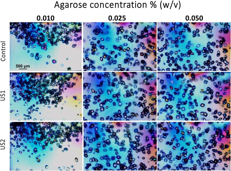
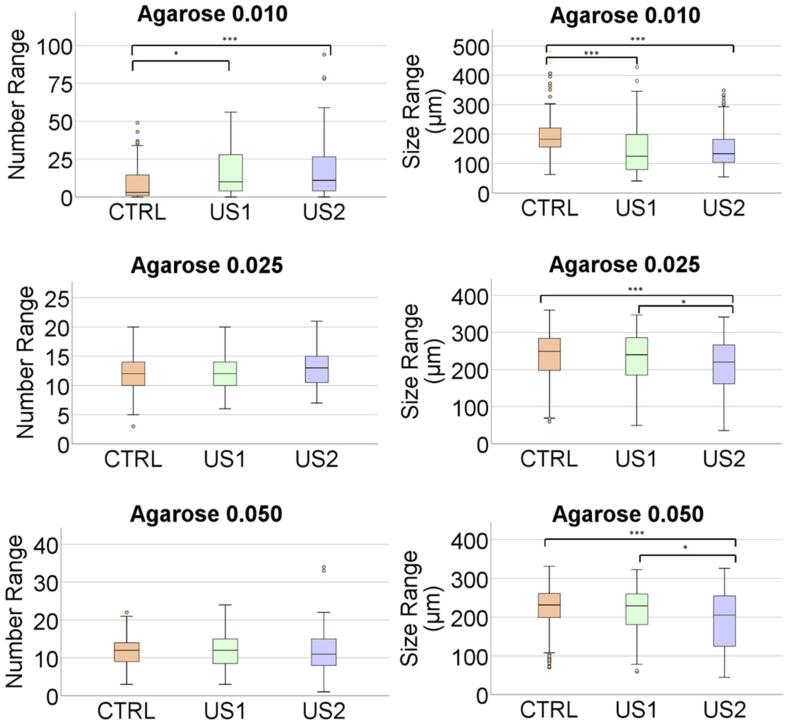
Sonocrystallization implies the application of ultrasound radiation to control the nucleation and crystal growth depending on the actuation time and intensity. Its application allows to induce nucleation at lower supersaturations than required under standard conditions. Although extended in inorganic and organic crystallization, it has been scarcely explored in protein crystallization. Now, that industrial protein crystallization is gaining momentum, the interest on new ways to control protein nucleation and crystal growth is advancing. In this work we present the development of a novel ultrasound bioreactor to study its influence on protein crystallization in agarose gel. Gel media minimize convention currents and sedimentation, favoring a more homogeneous and stable conditions to study the effect of an externally generated low energy ultrasonic irradiation on protein crystallization avoiding other undesired effects such as temperature increase, introduction of surfaces which induce nucleation, destructive cavitation phenomena, etc. In-depth statistical analysis of the results has shown that the impact of ultrasound in gel media on crystal size populations are statistically significant and reproducible.

Keywords: Hydrogels; Lysozyme; Nucleation; Protein crystallization; Ultrasound.


Conflict of interest statement

The authors declare that they have no known competing financial interests or personal relationships that could have appeared to influence the work reported in this paper.

Figures

Fig. 1
Fig. 1
The experimental scheme: A) Preparation of the samples A1: Experiments in solution, A2: Experiments in gels made with agarose; B) US-Bioreactor design and sonification procedure and illustration of the set-up; C) Observation of the samples and data analysis.
Fig. 2
Fig. 2
A and B) Shows the lateral and top 1–2 transversal section views, respectively. The chamber Type 1 was designed to hold the cuvette and it is located at 3.5 mm in front of the block. The dimensions of this chamber had 15 mm width, 12 mm of effective high and 91.4 mm of thickness (approximated volume of 16.4 cm3). The chambers Type 2 and Type 3 were introduced to hold several cuvettes in the future considering different pressures changing the main configuration. C) Exhibits the 3D printed support to maintain the ultrasonic transducer (100 KHz) in a correct position to ensure the alignment of the propagating waves. D) 3D schematic full configuration of the US-Bioreactor. E) Picture of the US-Bioreactor prepared for the experimental irradiation of gels.
Fig. 3
Fig. 3
3D-CAD illustration of the prototyped of US-Bioreactor with a recreation of multilayer media propagation through vaseline, PMMA, water, polystyrene and gel sample.
Fig. 4
Fig. 4
Row A) Shows lysozyme crystals obtained in solution under silent condition (Control) and ultrasonic irradiation for 30 min immediately after preparation of the experiment (US1) and 30 min after preparation (US2). The statistical analysis of the number (B) and size (C) of the crystals are shown for the three replicates. The scale bar in the optical microscopy images is 500 µm in all the pictures.
Fig. 5
Fig. 5
Gelation kinetic of 0.100 % (A) and 0.200 % (B) solutions of agarose. Darker lines represent the mean values, whereas the lighter bands around them represent the standard deviations. The bottom part showing the viscoelastic moduli as a function of frequency at a constant stress of 1 Pa for 0.100 % (C) and 0.200 % (D) agarose gels before and after applying the protocol US1. Darker lines represent the mean values, whereas the lighter bands around them represent the standard deviations. Note that the nonsymmetric appearance of standard deviations is due to the logarithmic scale.
Fig. 6
Fig. 6
Lysozyme crystals obtained in solution under silent condition (Control) and ultrasonic irradiation for 30 min immediately after preparing the experiment (US1) and 30 min after preparation, irradiated for 30 min (US2). From left to right it is shown the result in solution and increasing concentration of agarose. The scale bar in the optical microscopy images is 500 µm in all the pictures.
Fig. 7
Fig. 7
Represents the crystal numbers distribution and average size range as a function of agarose concentration. There are significant differences in all groups irradiated.

References

    1. McPherson A., Gavira J.A. Introduction to protein crystallization. Acta Crystallogr. Sect. F Struct. Biol. Commun. 2014;70:2–20. doi: 10.1107/S2053230X13033141. - DOI - PMC - PubMed
    1. Simone E., Zhang W., Nagy Z.K. Application of process analytical technology-based feedback control strategies to improve purity and size distribution in biopharmaceutical crystallization. Cryst. Growth Des. 2015;15:2908–2919. doi: 10.1021/acs.cgd.5b00337. - DOI
    1. Simone E., Zhang W., Nagy Z.K. Analysis of the crystallization process of a biopharmaceutical compound in the presence of impurities using process analytical technology (PAT) tools. J. Chem. Technol. Biotechnol. 2016;91:1461–1470. doi: 10.1002/jctb.4743. - DOI
    1. García-Ruiz J.M. Nucleation of protein crystals. J. Struct. Biol. 2003;142:22–31. doi: 10.1016/S1047-8477(03)00035-2. - DOI - PubMed
    1. Galkin O., Vekilov P.G. Direct determination of the nucleation rates of protein crystals. J. Phys. Chem. B. 1999;103:10965–10971. doi: 10.1021/jp992786x. - DOI