Review
. 2022 Jul 21;185(15):2739-2755.
doi: 10.1016/j.cell.2022.06.031.

What is a cell type and how to define it?

Affiliations
Review

What is a cell type and how to define it?

Hongkui Zeng. Cell. .

Abstract

Cell types are the basic functional units of an organism. Cell types exhibit diverse phenotypic properties at multiple levels, making them challenging to define, categorize, and understand. This review provides an overview of the basic principles of cell types rooted in evolution and development and discusses approaches to characterize and classify cell types and investigate how they contribute to the organism's function, using the mammalian brain as a primary example. I propose a roadmap toward a conceptual framework and knowledge base of cell types that will enable a deeper understanding of the dynamic changes of cellular function under healthy and diseased conditions.


Conflict of interest statement

Declaration of interests The author declares no competing interests.

Figures
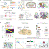
Figure 1.
Figure 1.. Approaches to characterize cell types.
(A) Molecular and anatomical approaches as primary ways of single-cell characterization include single-cell transcriptomics by sc/snRNA-seq, single-cell epigenomics exampled by snATAC-seq, spatially resolved transcriptomics exampled by MERFISH, full morphology reconstruction exampled by MouseLight (image adopted from Winnubst et al., 2019), EM connectomics (image adopted from Hulse et al., 2021), and barcoded connectomics exampled by BARseq (image adopted from Chen et al., 2019). (B) Cross-modality integrated approaches include Patch-seq (image adopted from Lee et al., 2021), retrograde tracing followed by molecular profiling, functional imaging followed by spatially resolved transcriptomics, using Patch-seq data as a Rosetta stone to assign molecular identities to neurons reconstructed from EM dataset (image adopted from Turner et al., 2022), and generation of enhancer based viral vectors (image adopted from Mich et al., 2021).
Figure 2.
Figure 2.. Hierarchical organization of cell types.
(A) A transcriptomic cell atlas for the mouse nervous system (image adopted from Zeisel et al., 2018). (B) A transcriptomic cell type taxonomy for the mouse primary motor cortex, with annotation (image adopted from Brain Initiative Cell Census Network, 2021). (C) UMAP representation of a transcriptomic cell type taxonomy for the glutamatergic neuron types in mouse isocortex and hippocampal formation, revealing discrete and continuous variations (image adopted from Yao et al., 2021b).
Figure 3.
Figure 3.. Multimodal correspondence of cell type phenotypic properties.
(A) MERFISH data from mouse motor cortex shows that continuous variation of glutamatergic IT transcriptomic types is correlated with their continuous spatial distribution along the cortical depth from L2/3 to L6 (image adopted from Zhang et al., 2021a). (B) Patch-seq data on GABAergic interneurons from mouse visual cortex shows correspondence between transcriptomic (T) types and morpho-electrical (ME) types (image adopted from Gouwens et al., 2020). (C) Brain-wide complete morphology reconstruction of cortical glutamatergic neurons shows distinct axon projection patterns between major transcriptomic types and further heterogeneity within each type (image adopted from Peng et al., 2021). Each color outlines the projection of one neuron within the type in each panel.
Figure 4.
Figure 4.. Dynamic changes of cell types and states during development, aging and various physiological or pathological contexts.
Major neuronal and non-neuronal classes are shown along the life stages of development, adulthood and aging. Neural progenitors generate different neuronal types, astrocytes and oligodendrocytes at different developmental timepoints, whereas microglia have a separate developmental origin. Major developmental events, various physiological states in adulthood, and different diseased states throughout the lifespan are shown below the timeline.

Similar articles

Cited by

References

    1. Abbott LF, Bock DD, Callaway EM, Denk W, Dulac C, Fairhall AL, Fiete I, Harris KM, Helmstaedter M, Jain V, et al. (2020). The Mind of a Mouse. Cell 182, 1372–1376. - PubMed
    1. Adamson B, Norman TM, Jost M, Cho MY, Nunez JK, Chen Y, Villalta JE, Gilbert LA, Horlbeck MA, Hein MY, et al. (2016). A Multiplexed Single-Cell CRISPR Screening Platform Enables Systematic Dis of the Unfolded Protein Response. Cell 167, 1867–1882 e1821. - PMC - PubMed
    1. Agathocleous M, and Harris WA (2009). From progenitors to differentiated cells in the vertebrate retina. Annu Rev Cell Dev Biol 25, 45–69. - PubMed
    1. Allaway KC, Gabitto MI, Wapinski O, Saldi G, Wang CY, Bandler RC, Wu SJ, Bonneau R, and Fishell G (2021). Genetic and epigenetic coordination of cortical interneuron development. Nature 597, 693–697. - PMC - PubMed
    1. Arendt D (2008). The evolution of cell types in animals: emerging principles from molecular studies. Nature reviews Genetics 9, 868–882. - PubMed

Publication types

LinkOut - more resources