. 2022 Oct 1:172:105822.
doi: 10.1016/j.nbd.2022.105822. Epub 2022 Jul 20.

Increased GABA transmission to GnRH neurons after intrahippocampal kainic acid injection in mice is sex-specific and associated with estrous cycle disruption

Affiliations

Increased GABA transmission to GnRH neurons after intrahippocampal kainic acid injection in mice is sex-specific and associated with estrous cycle disruption

Robbie J Ingram et al. Neurobiol Dis. .

Abstract

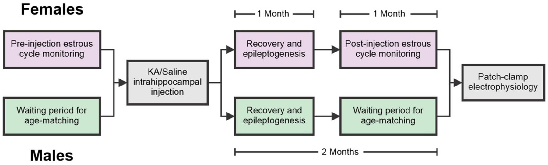
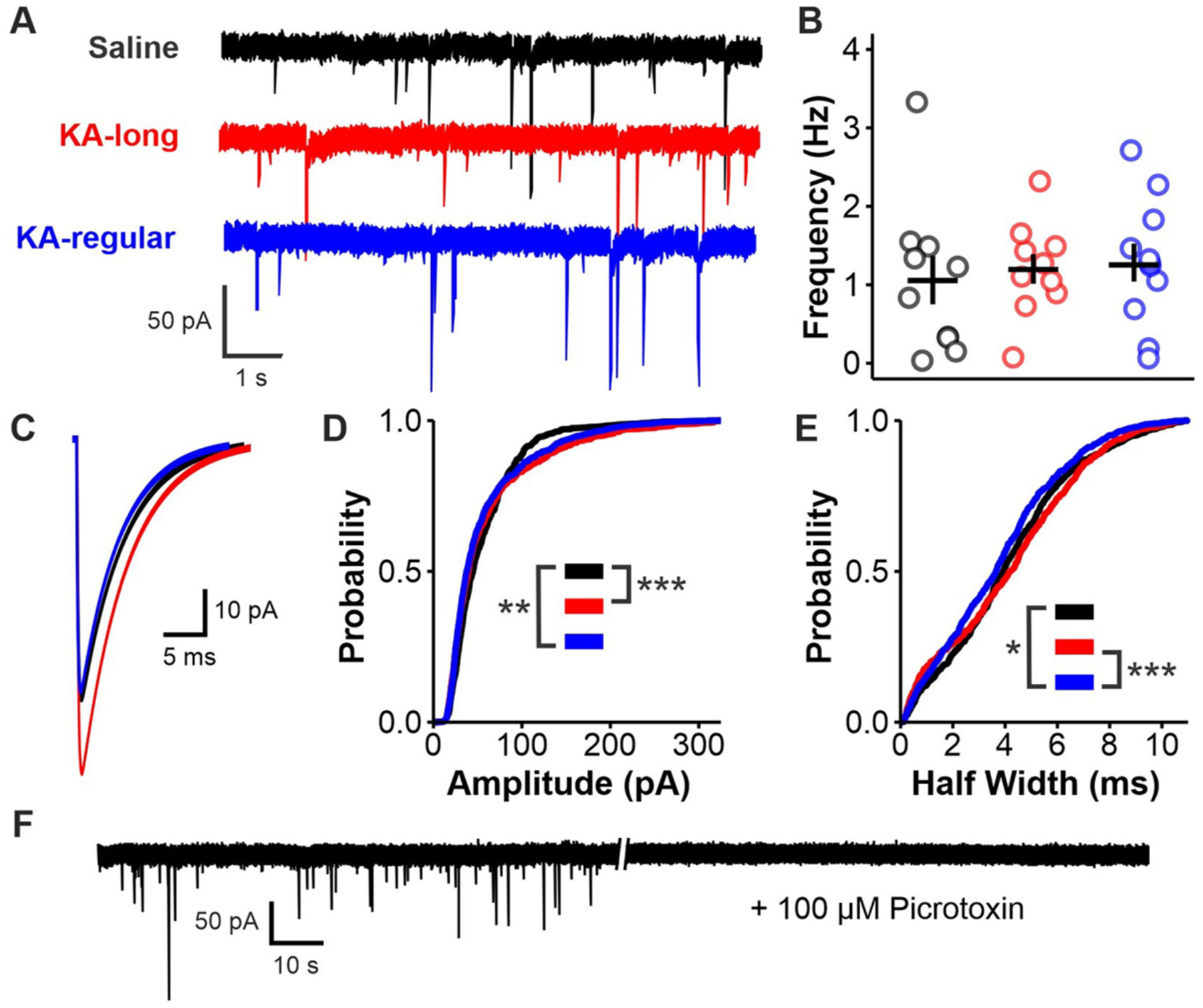
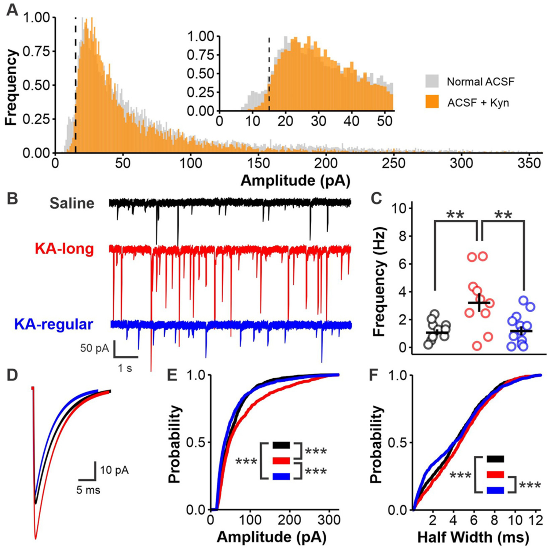
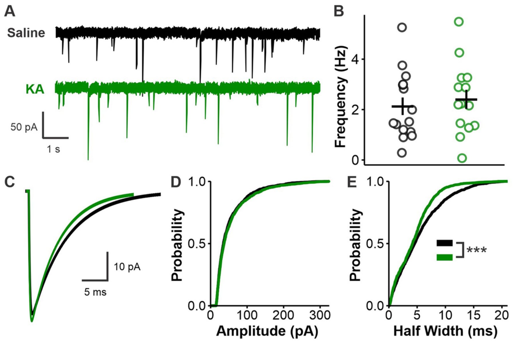
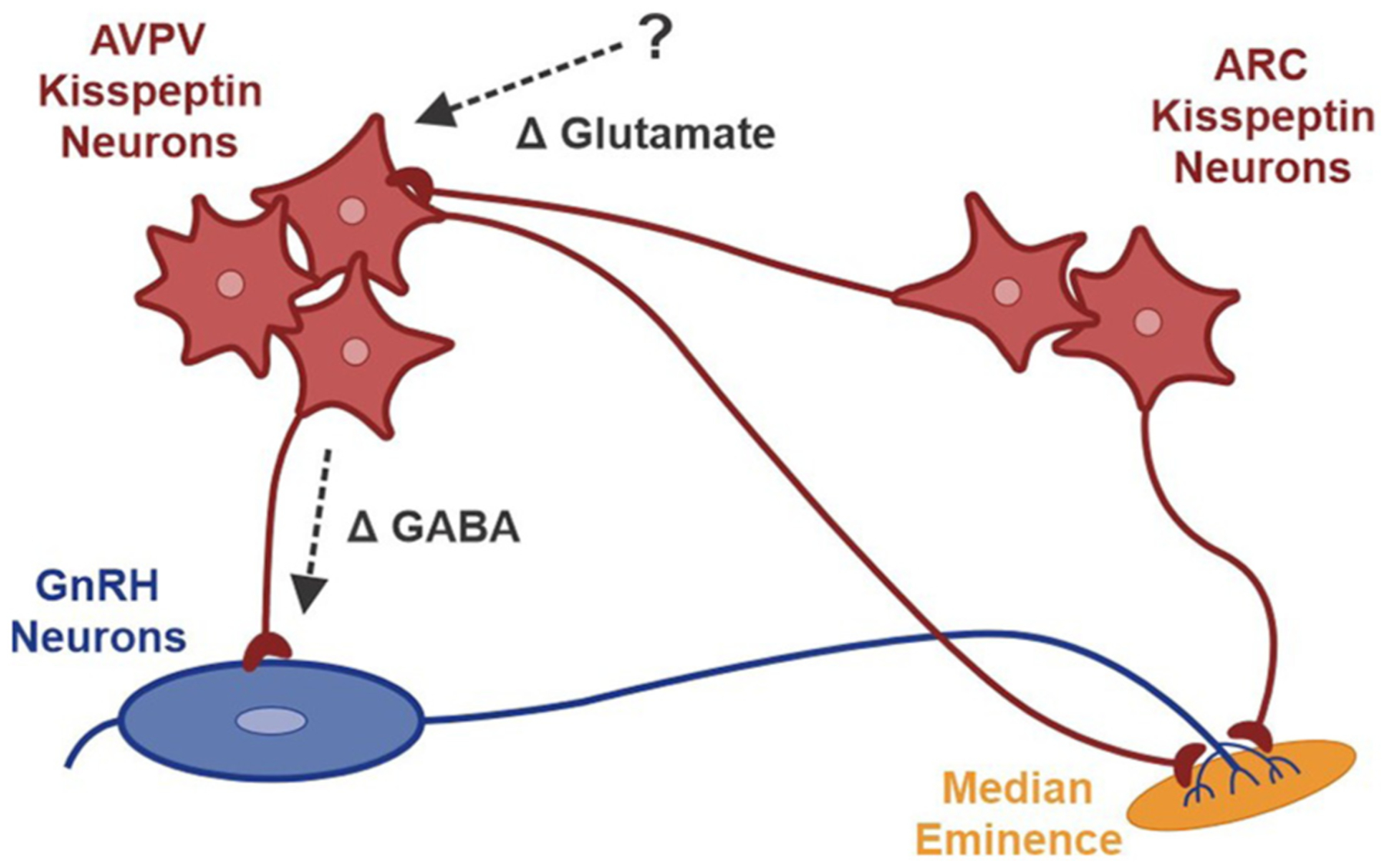
Patients with epilepsy develop reproductive endocrine comorbidities at a rate higher than that of the general population. Clinical studies have identified disrupted luteinizing hormone (LH) release patterns in patients of both sexes, suggesting potential epilepsy-associated changes in hypothalamic gonadotropin-releasing hormone (GnRH) neuron function. In previous work, we found that GnRH neuron firing is increased in diestrous females and males in the intrahippocampal kainic acid (IHKA) mouse model of temporal lobe epilepsy. Notably, GABAA receptor activation is depolarizing in adult GnRH neurons. Therefore, here we tested the hypothesis that increased GnRH neuron firing in IHKA mice is associated with increased GABAergic drive to GnRH neurons. When ionotropic glutamate receptors (iGluRs) were blocked to isolate GABAergic postsynaptic currents (PSCs), no differences in PSC frequency were seen between GnRH neurons from control and IHKA diestrous females. In the absence of iGluR blockade, however, GABA PSC frequency was increased in GnRH neurons from IHKA females with disrupted estrous cycles, but not saline-injected controls nor IHKA females without estrous cycle disruption. GABA PSC amplitude was also increased in IHKA females with disrupted estrous cycles. These findings suggest the presence of an iGluR-dependent increase in feed-forward GABAergic transmission to GnRH neurons specific to IHKA females with comorbid cycle disruption. In males, GABA PSC frequency and amplitude were unchanged but PSC duration was reduced. Together, these findings suggest that increased GABA transmission helps drive elevated firing in IHKA females on diestrus and indicate the presence of a sex-specific hypothalamic mechanism underlying reproductive endocrine dysfunction in IHKA mice.

Keywords: Electrophysiology; Epilepsy; GABA; Gonadotropin-releasing hormone; Hypothalamus; Neuroendocrinology.


Conflict of interest statement

Declaration of Competing Interest

The authors declare no competing financial interests.

Figures

Fig. 1.
Fig. 1.
Flowchart illustrating similarities and differences between the experimental paradigms used for male and female mice. Note that all intrahippocampal injections and patch clamp electrophysiology experiments in females were performed using diestrous mice.
Fig. 2.
Fig. 2.
Histological verification of successful IHKA injection targeting. A, Example cresyl violet-stained sections showing presence (top) or absence (bottom) of sclerosis in ipsilateral hippocampus. B, Example GFAP (green) and DAPI (blue) staining showing presence (top) and absence (bottom) of gliosis in sections from IHKA mice. The example section exhibiting no gliosis is provided for visual comparison; other sections from the same mouse did exhibit signs of gliosis, and this mouse was thus included in the final data analyses. Note that gliosis was only examined in sections from IHKA mice that did not show signs of sclerosis in cresyl violet staining after screening for video-recorded seizures. Scale bars = 1 mm.
Fig. 3.
Fig. 3.
Increased GABA sPSC amplitude in GnRH neurons from diestrous female IHKA mice in the presence of iGluR blockade. A, Example traces. B, Scatter plot showing individual cell values (circles) and mean ± SEM for GABA PSC frequency in GnRH neurons from saline (black), KA-long (red), and KA-regular (blue) mice. C, Average PSC traces from all events obtained in each group. D-E, Cumulative probability distributions for PSC amplitude (D) and half width (E). F, Representative trace showing that the addition of 100 μM picrotoxin eliminates all remaining PSCs in this recording configuration; *p < 0.017, **p < 0.003, ***p < 0.0003 by Kolmogorov-Smirnov (K-S) tests corrected for multiple comparisons with the Bonferroni method.
Fig. 4.
Fig. 4.
GABAergic transmission to GnRH neurons in female IHKA mice is increased in an iGluR-dependent manner. A, Histogram showing normalized distributions of unitary GABA PSC amplitude values from all recordings made in the presence (orange) or absence (grey) of 1 mM kynurenic acid. Dotted line indicates 15 pA minimum cutoff for putative GABAergic currents. B, Example traces recorded in control ACSF. C, Scatter plot showing individual cell values (circles) and mean ± SEM for GABA PSC frequency in GnRH neurons from saline (black), KA-long (red), and KA-regular (blue) mice; **p < 0.01 by Kruskal-Wallis with Dunn’s post hoc tests. D, Average PSC traces from all events obtained in each group. E-F, Cumulative probability distributions for PSC amplitude (E) and half width (F); ***p < 0.0003 by Kolmogorov-Smirnov (K-S) tests corrected for multiple comparisons with the Bonferroni method.
Fig. 5.
Fig. 5.
Reduced GABAergic PSC duration but no changes in frequency or amplitude in male IHKA mice. A, Example traces. B, Scatter plot showing individual cell values (circles) and mean ± SEM for GABA PSC frequency in GnRH neurons from saline (black) and KA (green) mice. C, Average PSC traces from all events obtained in each group. D-E, Cumulative probability distributions for PSC amplitude (D) and half width (E); ***p < 0.001 by Kolmogorov-Smirnov (K-S) tests.
Fig. 6.
Fig. 6.
A working model of epilepsy-associated dysfunction in the hypothalamic kisspeptin-GnRH neural circuit in KA-long females. Altered glutamatergic transmission, perhaps arising at least in part from ARC kisspeptin neurons, reaches AVPV kisspeptin neurons, which then release increased amounts of GABA at synapses on GnRH neurons. Note that GnRH neurons send extended projections to the median eminence, which is the site at which GnRH is released into the bloodstream.

Comment in

Similar articles

Cited by

References

    1. Adams C, Chen X, Moenter SM, 2018. Changes in GABAergic transmission to and intrinsic excitability of gonadotropin-releasing hormone (GnRH) neurons during the estrous cycle in mice. eNeuro 5. 10.1523/ENEURO.0171-18.2018. - DOI - PMC - PubMed
    1. Auger C, Marty A, 2000. Quantal currents at single-site central synapses. J. Physiol 526 (Pt 1), 3–11. 10.1111/j.1469-7793.2000.t01-3-00003.x. - DOI - PMC - PubMed
    1. Bauer J, Cooper-Mahkorn D, 2008. Reproductive dysfunction in women with epilepsy: menstrual cycle abnormalities, fertility, and polycystic ovary syndrome. Int. Rev. Neurobiol 83, 135–155. 10.1016/S0074-7742(08)00007-X. - DOI - PubMed
    1. Bauer J, Jarre A, Klingmüller D, Elger CE, 2000. Polycystic ovary syndrome in patients with focal epilepsy: a study in 93 women. Epilepsy Res 41, 163–167. 10.1016/s0920-1211(00)00139-x. - DOI - PubMed
    1. Bauer J, Blumenthal S, Reuber M, Stoffel-Wagner B, 2004. Epilepsy syndrome, focus location, and treatment choice affect testicular function in men with epilepsy. Neurology 62, 243–246. 10.1212/01.wnl.0000091866.48962.79. - DOI - PubMed

Publication types