. 2022 Oct;81(10):1453-1464.
doi: 10.1136/annrheumdis-2022-222629. Epub 2022 Jul 22.

Gain-of-function mutations in ALPK1 cause an NF-κB-mediated autoinflammatory disease: functional assessment, clinical phenotyping and disease course of patients with ROSAH syndrome

Christina Torres Kozycki  1   2 Shilpa Kodati  3 Laryssa Huryn  3 Hongying Wang  4 Blake M Warner  5 Priyam Jani  5 Dima Hammoud  6 Mones S Abu-Asab  7 Yingyos Jittayasothorn  3 Mary J Mattapallil  3 Wanxia Li Tsai  8 Ehsan Ullah  9 Ping Zhou  10 Xiaoying Tian  10 Ariane Soldatos  11 Niki Moutsopoulos  5 Marie Kao-Hsieh  5 Theo Heller  12 Edward W Cowen  13 Chyi-Chia Richard Lee  14 Camilo Toro  15   16 Shelley Kalsi  17 Zohreh Khavandgar  5 Alan Baer  5 Margaret Beach  5 Debra Long Priel  18 Michele Nehrebecky  4 Sofia Rosenzweig  4 Tina Romeo  4 Natalie Deuitch  4   19 Laurie Brenchley  5 Eileen Pelayo  5 Wadih Zein  3 Nida Sen  3 Alexander H Yang  12 Gary Farley  20 David A Sweetser  21   22 Lauren Briere  21 Janine Yang  23 Fabiano de Oliveira Poswar  24   25 Ida Vanessa D Schwartz  24   25 Tamires Silva Alves  24 Perrine Dusser  26 Isabelle Koné-Paut  27 Isabelle Touitou  28 Salah Mohamed Titah  29 Petrus Martin van Hagen  30 Rogier T A van Wijck  31 Peter J van der Spek  31 Hiromi Yano  32 Andreas Benneche  33 Ellen M Apalset  34 Ragnhild Wivestad Jansson  35 Rachel R Caspi  36 Douglas Byron Kuhns  18 Massimo Gadina  8 Hidetoshi Takada  37 Hiroaki Ida  38 Ryuta Nishikomori  39 Elena Verrecchia  40   41 Eugenio Sangiorgi  42 Raffaele Manna  40 Brian P Brooks  9 Lucia Sobrin  23 Robert B Hufnagel  9 David Beck  43 Feng Shao  10 Amanda K Ombrello  4 Ivona Aksentijevich  4 Daniel L Kastner  1 Undiagnosed Diseases Network
Collaborators, Affiliations

Gain-of-function mutations in ALPK1 cause an NF-κB-mediated autoinflammatory disease: functional assessment, clinical phenotyping and disease course of patients with ROSAH syndrome

Christina Torres Kozycki et al. Ann Rheum Dis. 2022 Oct.

Abstract

Objectives: To test the hypothesis that ROSAH (retinal dystrophy, optic nerve oedema, splenomegaly, anhidrosis and headache) syndrome, caused by dominant mutation in ALPK1, is an autoinflammatory disease.

Methods: This cohort study systematically evaluated 27 patients with ROSAH syndrome for inflammatory features and investigated the effect of ALPK1 mutations on immune signalling. Clinical, immunologic and radiographical examinations were performed, and 10 patients were empirically initiated on anticytokine therapy and monitored. Exome sequencing was used to identify a new pathogenic variant. Cytokine profiling, transcriptomics, immunoblotting and knock-in mice were used to assess the impact of ALPK1 mutations on protein function and immune signalling.

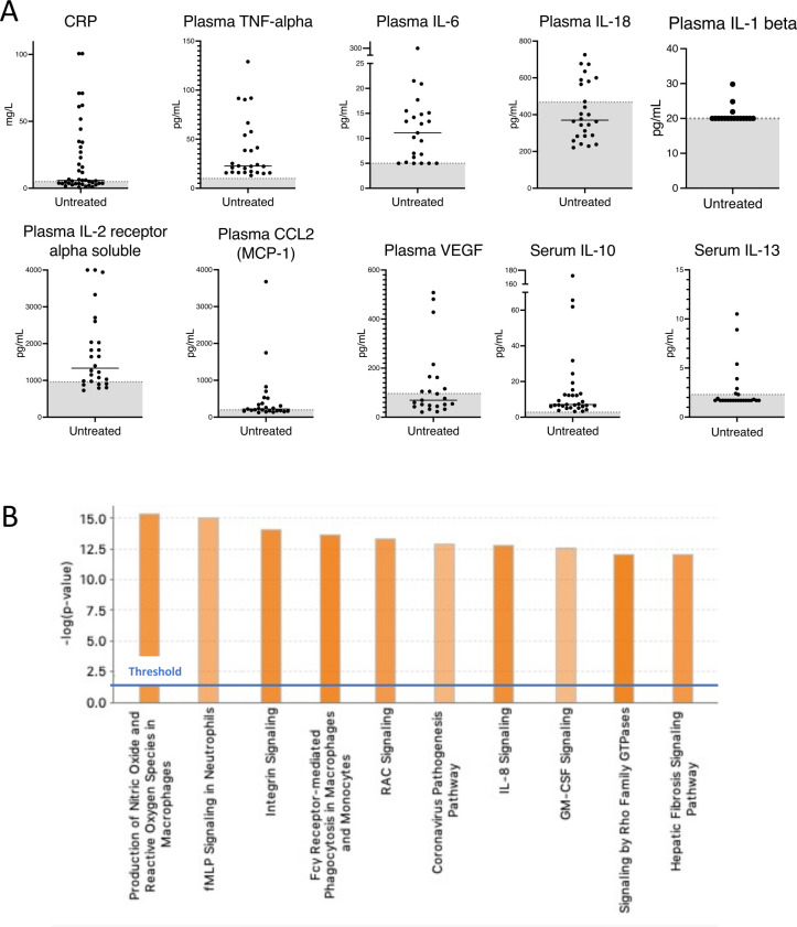
Results: The majority of the cohort carried the p.Thr237Met mutation but we also identified a new ROSAH-associated mutation, p.Tyr254Cys.Nearly all patients exhibited at least one feature consistent with inflammation including recurrent fever, headaches with meningeal enhancement and premature basal ganglia/brainstem mineralisation on MRI, deforming arthritis and AA amyloidosis. However, there was significant phenotypic variation, even within families and some adults lacked functional visual deficits. While anti-TNF and anti-IL-1 therapies suppressed systemic inflammation and improved quality of life, anti-IL-6 (tocilizumab) was the only anticytokine therapy that improved intraocular inflammation (two of two patients).Patients' primary samples and in vitro assays with mutated ALPK1 constructs showed immune activation with increased NF-κB signalling, STAT1 phosphorylation and interferon gene expression signature. Knock-in mice with the Alpk1 T237M mutation exhibited subclinical inflammation.Clinical features not conventionally attributed to inflammation were also common in the cohort and included short dental roots, enamel defects and decreased salivary flow.

Conclusion: ROSAH syndrome is an autoinflammatory disease caused by gain-of-function mutations in ALPK1 and some features of disease are amenable to immunomodulatory therapy.

Keywords: Amyloidosis; Arthritis; Immune System Diseases; Inflammation; Therapeutics.


Conflict of interest statement

Competing interests: FS is a cofounder and stockholder of Pyrotech therapeutics, a company that aims to develop agonist/inhibitor drugs for ALPK1. RM has received honorary fees for lectures from SHIRE-TAKEDA- SANOFI- NOVARTIS-SOBI and Nida Sen is employed by Janssen.

Figures

Figure 1
Figure 1
Heterozygous missense mutations of ALPK1 in the cohort and overview of clinical manifestations observed in patients with ROSAH syndrome. (A) Electropherogram of the previously unreported Y254C mutation for patients F13.1. (B) Domain structure of ALPK1 protein, indicating the location of the observed ROSAH-associated mutations (T237M and Y254C). (C) Schematic showing cross-species conservation of ALPK1 in the regions flanking the T237M and Y254C mutations. Sequences were obtained from Uniprot and multiple sequence alignments were created on Clustal Omega. (D) Bar chart indicating the prevalence of clinical manifestations reported in our ROSAH syndrome cohort. Patient F2.4 has cerebral palsy and was unable to provide any information about subjective clinical features. Blue shading indicates yes, and grey shading indicates no. Splenomegaly as determined by ultrasounds and arthritis as demonstrated by X-ray. Arthritis was present in all individuals evaluated by X-ray. ROSAH, retinal dystrophy, optic nerve oedema, splenomegaly, anhidrosis and headache.
Figure 2
Figure 2
Clinical manifestations associated with ROSAH syndrome. (A) Optic disc elevation. Fundus photographs demonstrating flagrant optic disc oedema (black arrow) in patient F9.2 (left) and more subtle changes in patient F9.3 (right). (B) Inflammatory arthritis with erosive changes in patient F3.3 (top left) and patient F13.1 (top right). X-ray demonstrating advanced diffuse changes of inflammatory arthritis involving wrist, metacarpophalangeal and interphalangeal joints with evolving joint deformities for patient F13.1 (bottom). (C) Gastrointestinal inflammation. Patient F2.2’s endoscopy for dysphagia revealed oesophageal linear furrows (left, arrows) and erythematous duodenal mucosa (right, arrow). (D) Massive splenomegaly. Splenic histology showing red pulp expansion with mild histiocytic hyperplasia from patient F2.2 (resected at age 13, 26×15×6 cm, weighing 1320 grams) (left). Abdominal MRI from patient F1.1 at age 13 demonstrating hepatosplenomegaly with spleen craniocaudal diameter of 22.5 cm and liver craniocaudal diameter of 18.6 cm (right) in the coronal plane (normal range for age: spleen 8–12 cm, liver 8.5–14 cm). (E) Premature basal ganglia and brainstem mineralisation. Susceptibility weighted imaging from brain MRIs showing decreased signal intensity consistent with premature mineralisation of the globus pallidi (small black arrow), substantia nigra (open white arrows) and red nuclei (red arrows). The mineralisation worsens with age eventually involving the caudate nuclei and putamina (white arrow heads) (top row: patient F1.1, bottom row: patient F7.1). (F) Dental caries. Sjögren’s disease-like pattern of dental caries in patient F4.1. (G) Short dental roots. Three-dimensional rendering and two-dimensional slice of the maxillary central incisor from F5.3 (left) and an age matched healthy control (right). The crown length is similar between the teeth (green line to blue line). However, the root length is one third in F5.3 (blue line to red line). ROSAH, retinal dystrophy, optic nerve oedema, splenomegaly, anhidrosis and headache.
Figure 3
Figure 3
ROSAH patient salivary glands demonstrate salivary hypofunction, altered echoarchitecture and histopathological evidence of inflammation, atrophy and fibrosis. (A) Whole unstimulated saliva flow and total stimulated saliva flow (TSSF, collected while stimulating with 2% citric acid every 30 s) were measured in 4 patients with ROSAH and compared with patients with Sjögren’s disease (SjD) and healthy volunteers (HV). Like SjD, unstimulated and stimulated salivary flow rates were reduced in patients with ROSAH as compared with HV. Statistical significance was only reached for TSSF. (B) The parotid (PAR) and submandibular (SMG) salivary gland ultrasound (SGUS) of ROSAH patients exhibited abnormal echogenicity and homogeneity compared with HV. The most striking finding were isolated (<25% total surface area) to scattered (>50% total surface area) round, hypoechoic lesions which ranged in size from 1.5 mm to 6.5 mm (average of 3–3.5 mm; red arrows). These differ from hypoechoic lesions seen in SjD in shape, size, and distribution and are most likely attributable to pockets of trapped saliva (ie, sialectasias). (C) Labial minor salivary glands (LSG) were inspected using light microscopy. HV LSG are typified by mixed seromucous and mucous acinar cells, and associated ducts, with minimal atrophy or fibrosis and only minimal scattered, typically plasmacytic, inflammation. Alternately, SjD LSG exhibit overall architectural distortion with decreased proportions of seromucous >mucous acinar cells and increased proportion of immune infiltrates (eg, periductal focal lymphocytic sialadenitis (dashed ellipsis) with enhanced diffuse non-sialadenitis). Additional features included: atrophy (eg, decreased lobular size, decreased acinar size), fibrosis (eg, increased interlobular and intralobular collage deposition), adipocyte infiltration (‘fatty infiltration’; black arrow). SMG from patients with ROSAH syndrome exhibit features similar to SjD including periductal focal lymphocytic sialadentis (two of three cases; dashed elipsis), decreased seromucous acinar cells (three of three), prominent increased periductal fibrosis (three of three) and atrophy and increased fatty infiltration (three of three). Additional features included perivascular inflammation, and damage to ducts with mucous extravasation reaction was observed in two cases. ROSAH, retinal dystrophy, optic nerve oedema, splenomegaly, anhidrosis and headache.
Figure 4
Figure 4
Inflammatory signature in untreated patients with ROSAH syndrome. (A) CRP, cytokine and chemokine levels in serum (n=7) and plasma (n=5) of untreated patients with ROSAH syndrome. Grey zone indicates normal range. (B) Top 10 activated canonical pathways predicted based on differentially expressed genes from whole blood RNA of untreated adults with ROSAH syndrome (n=4) based on Ingenuity Pathway Analysis. Bars denote the different pathways based on Z-scores. CRP, C reactive protein; GM-CSF: granulocyte-macrophage colony-stimulating factor; ROSAH, retinal dystrophy, optic nerve oedema, splenomegaly, anhidrosis and headache.
Figure 5
Figure 5
Gain-of-function mutations in ALPK1 are associated with enhanced NF-κB activation in transfected cells and fibroblasts from patients with ROSAH syndrome. (A) 293 T cells were transiently cotransfected with an NF-B–responsive luciferase reporter gene and Flag-ALPK1 (wild-type or disease-associated mutant [T237M or Y254C]). Luciferase assay of NF-κB activation is shown as mean±SD. From three technical replicates (two-tailed unpaired Student’s t-test, ***p<0.001). (-) reflects transfection with empty vector. (B) Fibroblasts derived from patients with ROSAH syndrome were stimulated with ADP-heptose and whole cell lysates were immunoblotted against respective target proteins. Patient derived fibroblast showed increased levels of phospho-IκBα, increased degradation of IκBα, increased phospho-IKKα/β and increased MAPK activity (p38 and JNK). (C). Whole blood RNASeq data demonstrating ALPK1 mRNA expression was higher in untreated patients with ROSAH syndrome (red dots, n=4) as compared with controls (blue dots, n=3). ROSAH, retinal dystrophy, optic nerve oedema, splenomegaly, anhidrosis and headache.
Figure 6
Figure 6
ALPK1 mutations affect STAT1 phosphorylation, plasma levels of interferon-induced cytokines and transcription of interferon-regulated genes. (A, B) 293 T cells transiently transfected with ALPK1 variants (A) and ROSAH patient derived fibroblasts (B) were stimulated with ADP-heptose (5 uM) and whole cell lysates from both experiments were subjected to Western blotting for indicated proteins. Constitutive STAT1 phosphorylation (pSTAT) was observed in both transfected cells and patient fibroblasts. (-) reflects transfection with empty vector. (C) CD14-labelled monocytes from an untreated ROSAH patient (F2.4, middle of panel) showed constitutively phosphorylated STAT1 (pSTAT1) as compared with healthy control (top) and ROSAH patient treated with TNF-inhibitor (F2.2, bottom of panel). (D) Plasma CXCL10 (interferon-inducible protein 10 (IP-10)) as measured in patients F1.1, F2.2, F2.3, F2.4 and F7.1. Grey shaded area represents the mean plus or minus 2 SD from 114 healthy controls. (E) Heat map showing increased expression of interferon-regulated genes (type I: top (GO:0060337) and type II: bottom (GO:0034341)) in four untreated patients with ROSAH syndrome as compared with three healthy controls. Upregulated genes are shown in red and down-regulated genes in blue. ROSAH, retinal dystrophy, optic nerve oedema, splenomegaly, anhidrosis and headache; STAT1, signal transducer and activator of transcription.
Figure 7
Figure 7
Response to anticytokine therapy. (A) Pretreatment and post-treatment CRPs for patients initiated on anticytokine therapy (n=6). Shaded zone represents normal range. (B) Pretreatment and post-treatment cytokines from patients F1.1 (plasma), F2.2 (serum), F2.3 (serum). Shaded area indicates time on anti-cytokine therapy. Specific therapies are as indicated in the figures. (C) Heatmap showing differentially expressed inflammatory response genes (GO: 0006954) in whole blood of pre-treatment (n=4) and post-adalimumab (n=2) patients with ROSAH syndrome. Patient F2.2 had post-treatment samples collected on three separate visits. Upregulated genes are shown in red, and downregulated genes in blue. Complete list of genes in online supplemental table 9. (D) Fluorescein angiography from patient F2.3 demonstrating retinal vasculitis (dotted white arrows) and disc leakage (solid red arrow) that improved after initiation of tocilizumab. (E) Optical coherence tomography from patient F2.3 demonstrating cystoid macular oedema (white arrows) that improved after initiation of tocilizumab. CRP, C reactive protein; ROSAH, retinal dystrophy, optic nerve oedema, splenomegaly, anhidrosis and headache.

References

    1. Williams LB, Javed A, Sabri A, et al. . ALPK1 missense pathogenic variant in five families leads to ROSAH syndrome, an ocular multisystem autosomal dominant disorder. Genet Med 2019;21:2103–15. 10.1038/s41436-019-0476-3 - DOI - PMC - PubMed
    1. Chen HY, Welby E, Li T, et al. . Retinal disease in ciliopathies: recent advances with a focus on stem cell-based therapies. Transl Sci Rare Dis 2019;4:97–115. 10.3233/TRD-190038 - DOI - PMC - PubMed
    1. Zhou P, She Y, Dong N, et al. . Alpha-Kinase 1 is a cytosolic innate immune receptor for bacterial ADP-heptose. Nature 2018;561:122–6. 10.1038/s41586-018-0433-3 - DOI - PubMed
    1. Ryzhakov G, West NR, Franchini F, et al. . Alpha kinase 1 controls intestinal inflammation by suppressing the IL-12/Th1 axis. Nat Commun 2018;9:3797. 10.1038/s41467-018-06085-5 - DOI - PMC - PubMed
    1. García-Weber D, Dangeard A-S, Cornil J, et al. . ADP-heptose is a newly identified pathogen-associated molecular pattern of Shigella flexneri. EMBO Rep 2018;19. 10.15252/embr.201846943. [Epub ahead of print: 19 11 2018]. - DOI - PMC - PubMed

Publication types

Supplementary concepts