. 2022 Jul 22;12(1):12523.
doi: 10.1038/s41598-022-15969-y.

Anti-cardiolipin IgG autoantibodies associate with circulating extracellular DNA in severe COVID-19

Affiliations

Anti-cardiolipin IgG autoantibodies associate with circulating extracellular DNA in severe COVID-19

Daniel Bertin et al. Sci Rep. .

Abstract

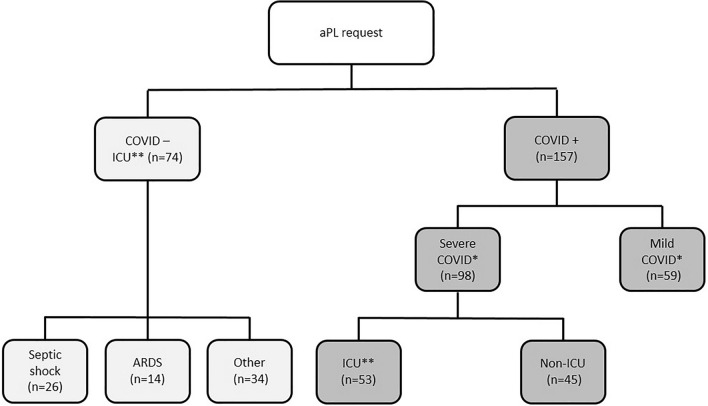
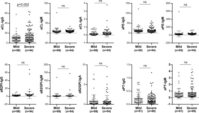
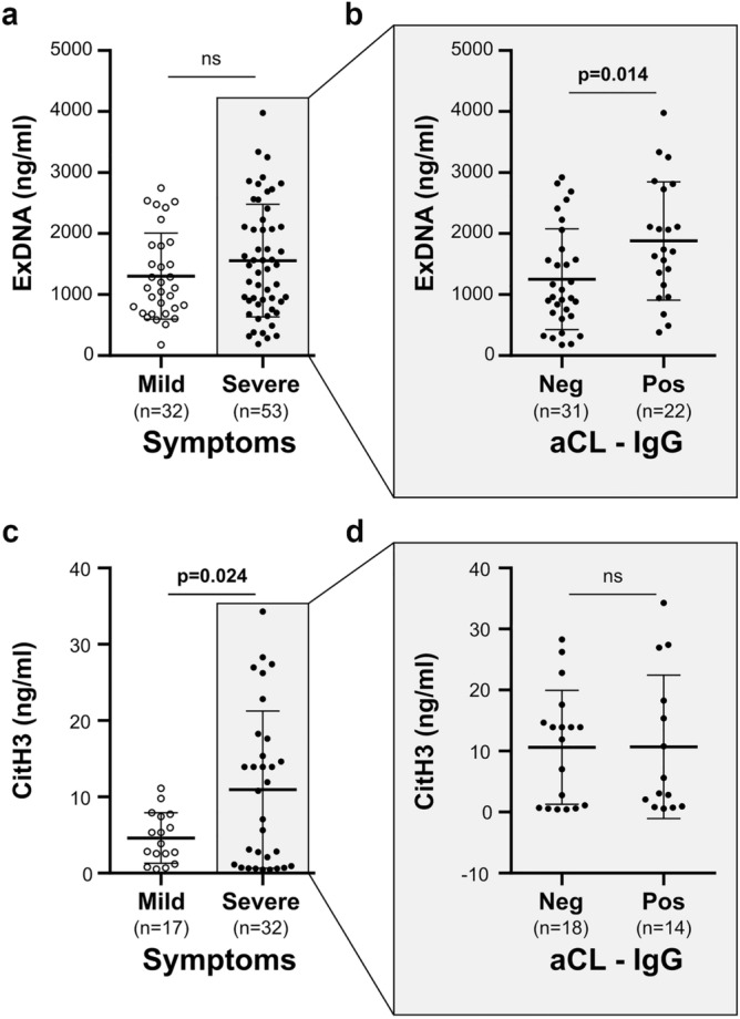
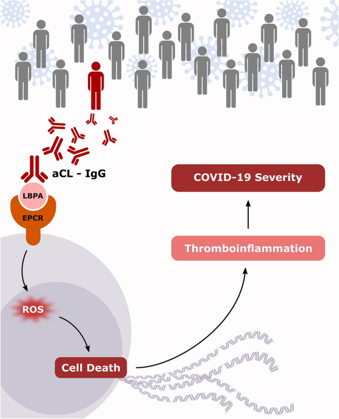
Whereas the detection of antiphospholipid autoantibodies (aPL) in COVID-19 is of increasing interest, their role is still unclear. We analyzed a large aPL panel in 157 patients with COVID-19 according to the disease severity. We also investigated a potential association between aPL and extracellular DNA (exDNA, n = 85) or circulating markers of neutrophil extracellular traps (NET) such as citrullinated histones H3 (CitH3, n = 49). A total of 157 sera of patients infected by SARS-CoV-2 were collected. A large aPL panel including lupus anticoagulant, anti-cardiolipin and anti-beta-2 glycoprotein I (IgG, IgM and IgA), anti-phosphatidylethanolamine IgA, anti-prothrombin (IgG and IgM) was retrospectively analyzed according to the disease severity. We found a total aPL prevalence of 54.8% with almost half of the cases having aCL IgG. Within an extended panel of aPL, only aCL IgG were associated with COVID-19 severity. Additionally, severe patients displayed higher CitH3 levels than mild patients. Interestingly, we highlighted a significant association between the levels of aCL IgG and exDNA only in aCL positive patients with severe disease. In conclusion, we showed a significant link between aPL, namely aCL IgG, and circulating exDNA in patients with severe form of COVID-19, that could exacerbate the thrombo-inflammatory state related to disease severity.


Conflict of interest statement

The authors declare no competing interests.

Figures

Figure 1
Figure 1
Flow Chart of patients. ARDS acute respiratory distress syndrome, ICU Intensive Care Unit. Two comparison were performed in this study: one between severe and mild COVID groups (*) and another between COVID ICU and non COVID ICU groups (**).
Figure 2
Figure 2
Antiphospholipid autoantibody levels in mild and severe patients with COVID-19. aCL anti-cardiolipin, aB2GPI anti-beta-2 glycoprotein I, aPE anti-phosphatidylethanolamine, aPT anti-prothrombin autoantibodies. Statistical analysis performed with Student's t test. ns not significant.
Figure 3
Figure 3
Extracellular DNA and citrullinated histones H3 levels in patients with COVID-19. Extracellular DNA levels in mild/severe COVID-19 patients (a) and in aCL IgG positive (Pos)/negative (Neg) patients with severe COVID-19 (b). Citrullinated histones H3 levels in mild /severe COVID-19 patients (c) and in aCL IgG positive (Pos)/negative (Neg) with severe COVID-19 (d). Statistical analysis performed with Student's t test. aCL anti-cardiolipin, ns not significant.
Figure 4
Figure 4
Putative mechanism of anticardiolipin autoantibodies in COVID-19 severity. We hypothesize that SARS-CoV-2 infection of genetically-prone patients results in autoimmunity. Anticardiolipin autoantibodies (aCL) can bind a cell surface complex composed of lysobiphosphatidic acid (LBPA) and endothelial protein C receptor (EPCR). This interaction promotes cell death via reactive oxygen species (ROS). Then, releases extracellular DNA contributes to a heightened thrombo-inflammatory state associated with COVID-19 severity. The figure has been drawn by AB using InkScape 0.92, http://www.inkscape.org.

Similar articles

Cited by

References

    1. Novelli L, Motta F, De Santis M, Ansari AA, Gershwin ME, Selmi C. The JANUS of chronic inflammatory and autoimmune diseases onset during COVID-19—A systematic review of the literature. J. Autoimmun. 2021;117:102592. doi: 10.1016/j.jaut.2020.102592. - DOI - PMC - PubMed
    1. Lin YS, Lin CF, Fang YT, Kuo YM, Liao PC, Yeh TM, et al. Antibody to severe acute respiratory syndrome (SARS)-associated coronavirus spike protein domain 2 cross-reacts with lung epithelial cells and causes cytotoxicity. Clin. Exp. Immunol. 2005;141(3):500–508. doi: 10.1111/j.1365-2249.2005.02864.x. - DOI - PMC - PubMed
    1. Yang Y-H, Huang Y-H, Chuang Y-H, Peng C-M, Wang L-C, Lin Y-T, et al. Autoantibodies against human epithelial cells and endothelial cells after severe acute respiratory syndrome (SARS)-associated coronavirus infection. J. Med. Virol. 2005;77(1):1–7. doi: 10.1002/jmv.20407. - DOI - PMC - PubMed
    1. Angileri F, Legare S, Marino Gammazza A, Conway de Macario E, JlMacario A, Cappello F. Molecular mimicry may explain multi-organ damage in COVID-19. Autoimmun. Rev. 2020;19(8):102591. doi: 10.1016/j.autrev.2020.102591. - DOI - PMC - PubMed
    1. Beaudoin CA, Jamasb AR, Alsulami AF, Copoiu L, van Tonder AJ, Hala S, et al. Predicted structural mimicry of spike receptor-binding motifs from highly pathogenic human coronaviruses. Comput. Struct. Biotechnol. J. 2021;19:3938–3953. doi: 10.1016/j.csbj.2021.06.041. - DOI - PMC - PubMed