Review
. 2022 Jul 22;5(1):737.
doi: 10.1038/s42003-022-03593-5.

Recent trends in bioartificial muscle engineering and their applications in cultured meat, biorobotic systems and biohybrid implants

Affiliations
Review

Recent trends in bioartificial muscle engineering and their applications in cultured meat, biorobotic systems and biohybrid implants

Eva Schätzlein et al. Commun Biol. .

Abstract

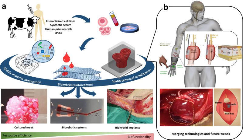
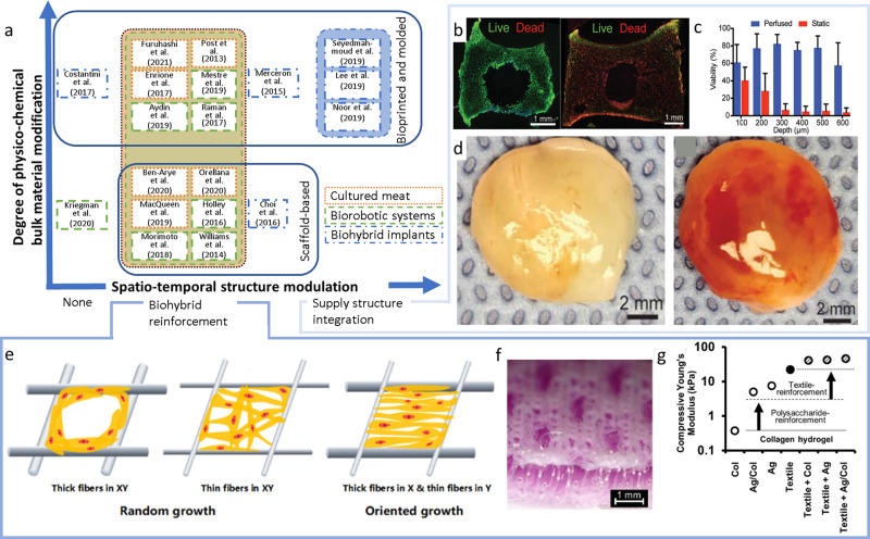
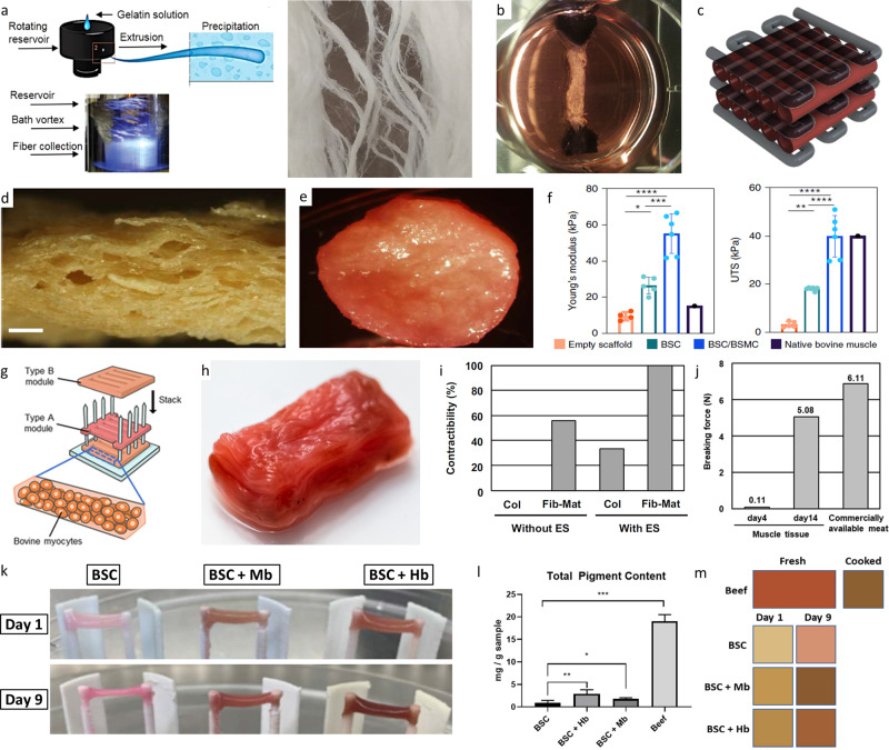
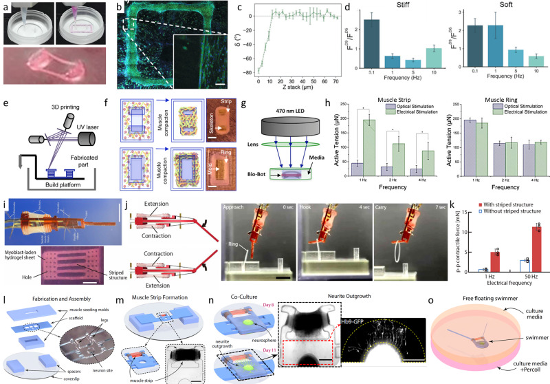
Recent advances in tissue engineering and biofabrication technology have yielded a plethora of biological tissues. Among these, engineering of bioartificial muscle stands out for its exceptional versatility and its wide range of applications. From the food industry to the technology sector and medicine, the development of this tissue has the potential to affect many different industries at once. However, to date, the biofabrication of cultured meat, biorobotic systems, and bioartificial muscle implants are still considered in isolation by individual peer groups. To establish common ground and share advances, this review outlines application-specific requirements for muscle tissue generation and provides a comprehensive overview of commonly used biofabrication strategies and current application trends. By solving the individual challenges and merging various expertise, synergetic leaps of innovation that inspire each other can be expected in all three industries in the future.


Conflict of interest statement

The authors declare no competing interests.

Figures

Fig. 1
Fig. 1. Biofabrication of muscle tissue and merging the expertise of the different fields of applications.
Biofabrication of muscle tissue enables multiple applications (a) ranging from cultured meat (assembly of fibrous muscle, fat, and vascular tissues to cultured steak by Kang et al (CC BY 4.0)), over biorobotic systems (from ref. . Reprinted with permission from AAAS.) to biohybrid implants (the pectoral branch of the thoracoacromial artery was identified beneath the pectoralis major by Liu et al (CC BY 4.0)). This review provides a comprehensive overview on the most important cellular and material-specific requirements as well as dedicated biofabrication strategies(adapted from refs. , (Schematic illustration of the concept, experimental procedure, goal, and outlook of the study by Schäfer et al. (CC BY 4.0))) for each of the three fields of application. While biofabrication of cultured meat, biorobotic systems, and bioartificial muscle implants has mostly been studied in isolation so far, the technological fusion will unleash unexpected innovations and determine future trends. The recently published combination of biorobotic systems and biohybrid implants is a path-breaking pointer to what lies ahead (b, adapted from Srinivasan and co-workers (reprinted with permission from Springer Nature Limited: Nature Biomedical Engineering, A cutaneous mechanoneural interface for neuroprosthetic feedback, Srinivasan et al., Copyright 2021).
Fig. 2
Fig. 2. Categorization and examples of strategies for biofabrication of muscle tissue.
Application-specific modi operandi for muscle tissue fabrication (a). According to the application, different degrees of physico-chemical bulk material modification and spatio-temporal structure modulation are applied for different examples found in the literature. The latter can be subdivided into biohybrid reinforcement (eg), and supply structure integration (bd). Live-Dead staining of actively perfused (b, left) vs. non-perfused (b, right) tissues as well as the measured cell viability as a function of the distance to the nutritional channel (c), exemplarily outline the importance of supply channel integration in thick tissues. The self-assembling capacity of vascular structures in bulk materials demonstrates the different potencies of compact (d, left) and highly porous scaffolds (d, right).(From ref. Reprinted with permission from AAAS.) Biohybrid reinforcement was shown to strengthen cell alignment (eg) and promote the mechanical properties of hydrogels (f, g) (difference in the morphological characteristics of a scaffold with different fiber diameters. by Xie et al (CC BY 4.0)). For instance, spacer fabric integration increased Young’s modulus of low concentrated collagen (Col) and alginate (Alg) hydrogels by several orders of magnitude (g) (Warp-knitted spacer fabric design and Morphological and mechanical analysis of warp-knitted spacer fabrics by Schäfer et al. (CC BY 4.0)).
Fig. 3
Fig. 3. Recent trends in cultured meat fabrication.
Fibrous anisotropic gelatin scaffolds could be produced by immersion rotary jet spinning (a) (fibrous gelatin production by immersion rotary jet spinning (iRJS) by MacQueen et al. (CC BY 4.0)). Picture of a muscle strip with anchoring system which was further developed for the first burger from cultured meat (b) (Reprinted from Principles of Tissue Engineering, Fourth Edition, M. Post, C. van der Weele, Principles of Tissue Engineering for Food, Pages 1647–1662, Copyright (2014), with permission from Elsevier). Schematic depiction of a possible cultured meat scaffold design (c) (Reprinted with permission from Springer Science Business Media, LLC, part of Springer Nature: Food Engineering Reviews, Cultured Meat: Meat Industry Hand in Hand with Biomedical Production Methods, Zidarič et al., Copyright (2020)). Textured soy protein scaffold (d) seeded with bovine satellite cells (BSC) and bovine aortic smooth muscle cells (BSMC) co-culture (e). Comparison of Young’s modulus and ultimate tensile strength (UTS) of the various seeded textured soy scaffold types of native bovine muscle from the literature (f).(Reprinted with permission from Springer Nature Limited: Nature Food, Textured soy protein scaffolds enable the generation of three-dimensional bovine skeletal muscle tissue for cell-based meat, Ben-Arye et al, Copyright (2020)) Construction process of a thick bovine muscle tissue (g) and image of the resulting product colored using red food dye (h). The rate of fiber-shaped bovine muscle tissues capable of contracting in response to applied electrical stimulation (ES), formed within the collagen (Col) or Fibrin-Matrigel (Fib-Mat) based tissue cultures (i) and mechanical characterization of the produced tissue (j)(Construction process of millimetre-thick bovine muscle tissue, Morphological and functional analysis of bovine muscle tissue, Morphological analysis of the millimetre-thick bovine muscle tissue and Food feature analysis of the large bovine muscle tissue by Furuhashi et al (CC BY 4.0)). Representative images of cultured meat strips of bovine muscle satellite cells (BSCs) grown in the presence of hemoglobin (Hb) or myoglobin (Mb) in the cell culture media for up to nine days in a fibrin hydrogel (k). Spectroscopic quantification of total pigment content (l) and average tissue coloration (m) of homogenized cultured meat strips after incubation in heme-protein-containing media in comparison to beef. (properties of skeletal muscle tissue formation and Pigment content and tissue coloration by Simsa et al. (CC BY 4.0)).
Fig. 4
Fig. 4. Examples of bioprinted and molded biorobotic systems.
Extrusion printing of muscle cell laden bioink and PDMS pillars for cell alignment (a). Microscopic image of the print (b). Myotube alignment in the printing direction and in parallel to the passive force from the pillars (c). Fold increment of the achieved force of the cells before (FD6) and after 4 days of stimulation (FD9) with differing stiffness of the pillars (d) (© 2018 WILEY-VCH Verlag GmbH & Co. KGaA, Weinheim) Schematic manufacturing process of muscle strips and rings with stereolithographic 3D printed molds (e), injection of the bioink and following muscle compaction to achieve muscle strips and rings for bioreactors (f). Actuation of the genetic modified muscle cells via optical stimulation (g) and electrical stimulation (h). Active tension changes with the stimulation method and the muscle shape h). Biohybrid robot powered by an antagonistic pair of skeletal muscle tissues (i). Example movement of the biorobot grabbing a ring (j). Contractile force is increased with a sufficient maturation and therefore striped structure of the muscle cells (k) (from ref. . Reprinted with permission from AAAS.) Biofabrication of a free floating biorobotic swimmer driven by on-board neuromuscular unit (o) with the steps of fabrication and assembly (l), muscle strip formation (m), co-culture of muscle cells and the neurosphere in a continuous ECM-gel (n) (from ref. Biofabrication timeline and free swimming driven by neuromuscular units by Aydin et al. (CC BY NC ND 4.0)).
Fig. 5
Fig. 5. Recent trends in the development of cell seeded biorobotic systems.
Concept design of a self-propelled biohybrid flagellum (right) with a similar motion to a spermatozoa (left) (a). Map of the predicted velocity as a function of head and tail dimensions of the biohybrid flagellum (b). Schematic of a two-tailed swimming biorobot (c). A sequence of images of the actuation of the two tailed swimmer (d) and the traveled distance and calculated velocity (e) (reprinted with permission from Nature Publishing Group, a division of Macmillan Publishers Limited: Nature Communications, A self-propelled biohybrid swimmer at low Reynolds number, Williams et al, Copyright 2014) Schematic diagram of the movement of a different biorobot, where the contraction of the cardiomyocyte bends the thin PDMS cantilever (f) of the floating or stationary biorobots (g). Immunostaining of cardiomyocyte marker, troponin-I (left) and actin cytoskeleton (right) show the growth of the cells without alignment (h). A tissue-engineered medusoid (i) with biomimetic jellyfish propulsion (j). Time lapse of a stroke cycle of a jellyfish and the medusoid (k) (reprinted with permission from Nature Publishing Group, a division of Macmillan Publishers Limited.: Springer Nature, Nature Biotechnology, A tissue-engineered jellyfish with biomimetic propulsion, Nawroth et al., Copyright 2012)) Artificial intelligence assisted design process of biorobots with predictable motion paths employing contractile (red) and passive (cyan) cell-based building blocks (l, left) as well as their in vivo realization using cardiomyocyte and epidermal cell progenitors (l, right). Predicted and in vivo movement of the designed models (m) (designing and manufacturing reconfigurable organisms and Transferal from silico to vivo by Kriegman et al. (CC BY 4.0)).
Fig. 6
Fig. 6. Examples of biofabricated muscle tissue for regenerative medicine.
Coaxial extruder nozzle (a) used for the printing of high shape fidelity structures utilizing ionic and UV-based bioink crosslinking (b). Fluorescence image of the printed multicellular result comprising C2C12 muscle cells (green) and BALB/3T3 fibroblasts (red) (c) (3D bioprinting set-up and Multi-cellular 3D bioprinting through a microfluidic printing head by Costantini et al. (CC BY NC ND 4.0)) Bioprinted multimaterial muscle-tendon unit (d) showing different elastic moduli of the integrated scaffold materials of polyurethane (PU) and polycaprolactone (PCL) (e). The C2C12 muscle cells of the muscle-tendon unit show morphological changes in the fluorescence microscopic image into an elongated shape while NIH/3T3 fibroblasts keep their morphology (f). Fluorescently-labeled dual-cell printed constructs (green: DiO-labeled C2C12 cells; red: DiI-labeled NIH/ 3T3 cells; imaged at 7 d in culture) shows cell–cell interactions and cell migration (g) (© IOP Publishing. Reproduced with permission. All rights reserved.) Printing path of a fiber bundle design for muscle organization with PCL pillars and sacrificial Pluronic F-127 channels for nutrition and cell alignment (h). The cell alignment can be seen in the immunofluorescent staining for myosin heavy chain of the 3D printed muscle after 7 days differentiation (i). Subcutaneous implantation of the bioprinted muscle fiber bundle with host nerve integration (j). Assessment of the function of the bioprinted muscle construct after 4 weeks of implantation (positive control: the normal gastrocnemius muscle; negative control: the gluteus muscle after dissected common peroneal nerve) (k) (reprinted with permission from Nature Publishing Group, a division of Macmillan Publishers Limited: Nature Biotechnology, A 3D bioprinting system to produce human-scale tissue constructs with structural integrity, Kang et al, Copyright 2016) Printing of cardiac patches (ln). Model of the cardiac patch (l) and a side view of the printing concept showing the sacrificial bioink with endothelial cells (EC) and the bioink made from decellularized omentum tissue (OM) with cardiomyocytes (CM) (m). Printed cardiac patch with cardiac tissue (actinin stained in pink) and blood vessels (CD31 stained in green) (n). Small scale human heart (o, p) printed in suspension to create hollow ventricles. The ventricles were filled with red and blue dye for visualization (o). The fluorescence image shows printed vasculature in the small-scale heart (CMs in pink, ECs in orange) (p) (© 2018 WILEY-VCH Verlag GmbH & Co. KGaA, Weinheim).

Similar articles

Cited by

References

    1. Dolgin E. Will cell-based meat ever be a dinner staple? Nature. 2020;588:S64–S67. doi: 10.1038/d41586-020-03448-1. - DOI - PubMed
    1. Morimoto Y, Onoe H, Takeuchi S. Biohybrid robot powered by an antagonistic pair of skeletal muscle tissues. Sci. Robot. 2018;3:1–11. doi: 10.1126/scirobotics.aat4440. - DOI - PubMed
    1. Liu M, Liu W, Yang X, Guo H, Peng H. Pectoralis major myocutaneous flap for head and neck defects in the era of free flaps: harvesting technique and indications. Sci. Rep. 2017;7:1–9. doi: 10.1038/s41598-016-0028-x. - DOI - PMC - PubMed
    1. Santoso, J. W. & McCain, M. L. Neuromuscular disease modeling on a chip. DMM Dis. Model. Mech. 13, dmm044867 (2020). - PMC - PubMed
    1. Ben-Arye T, Levenberg S. Tissue engineering for clean meat production. Front. Sustain. Food Syst. 2019;3:1–19. doi: 10.3389/fsufs.2019.00046. - DOI