. 2022 Jul 22;38(1):23.
doi: 10.1186/s42826-022-00133-4.

Ventilator-induced lung injury results in oxidative stress response and mitochondrial swelling in a mouse model

Affiliations

Ventilator-induced lung injury results in oxidative stress response and mitochondrial swelling in a mouse model

Jon Petur Joelsson et al. Lab Anim Res. .

Abstract

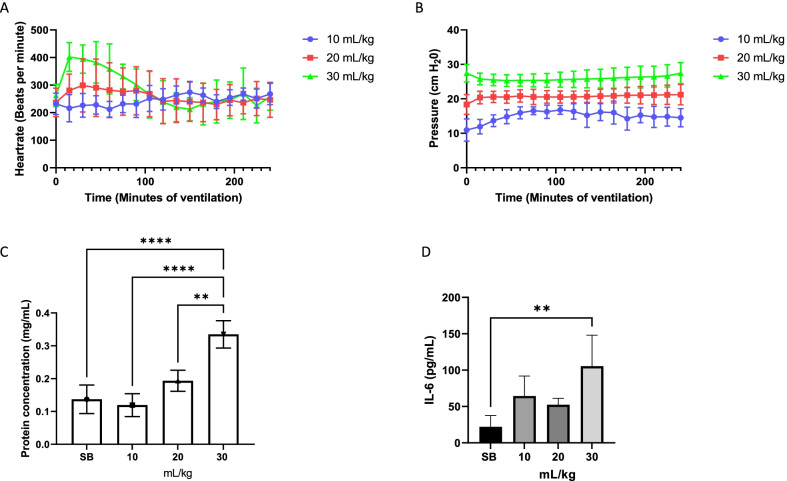
Background: Mechanical ventilation is a life-saving therapy for critically ill patients, providing rest to the respiratory muscles and facilitating gas exchange in the lungs. Ventilator-induced lung injury (VILI) is an unfortunate side effect of mechanical ventilation that may lead to serious consequences for the patient and increase mortality. The four main injury mechanisms associated with VILI are: baro/volutrauma caused by overstretching the lung tissues; atelectrauma, caused by repeated opening and closing of the alveoli resulting in shear stress; oxygen toxicity due to use of high ratio of oxygen in inspired air, causing formation of free radicals; and biotrauma, the resulting biological response to tissue injury, that leads to a cascade of events due to excessive inflammatory reactions and may cause multi-organ failure. An often-overlooked part of the inflammatory reaction is oxidative stress. In this research, a mouse model of VILI was set up with three tidal volume settings (10, 20 and 30 mL/kg) at atmospheric oxygen level. Airway pressures and heart rate were monitored and bronchoalveolar lavage fluid (BALF) and lung tissue samples were taken.

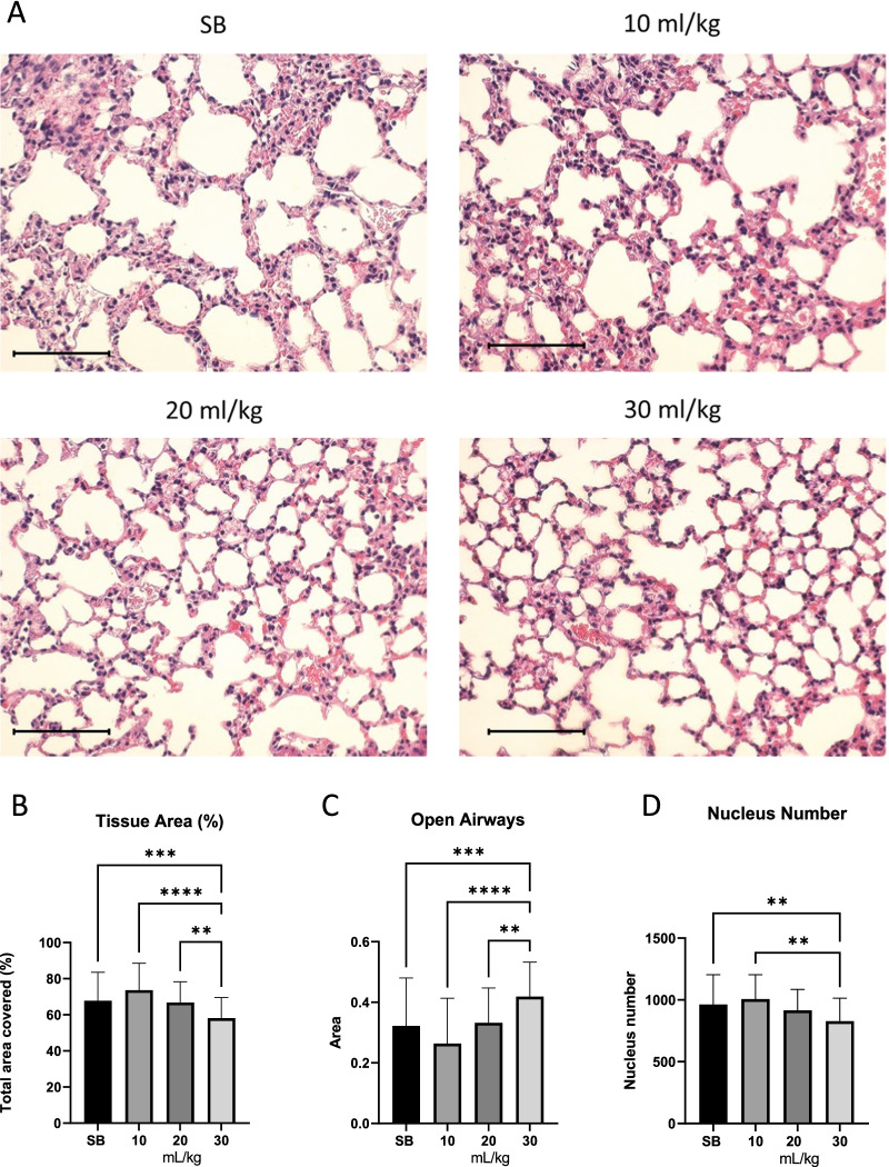
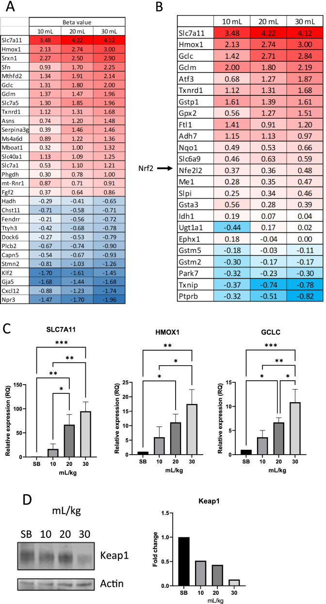
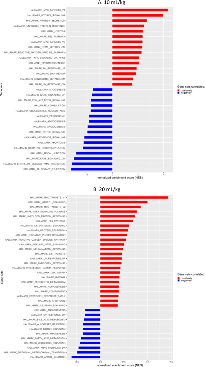
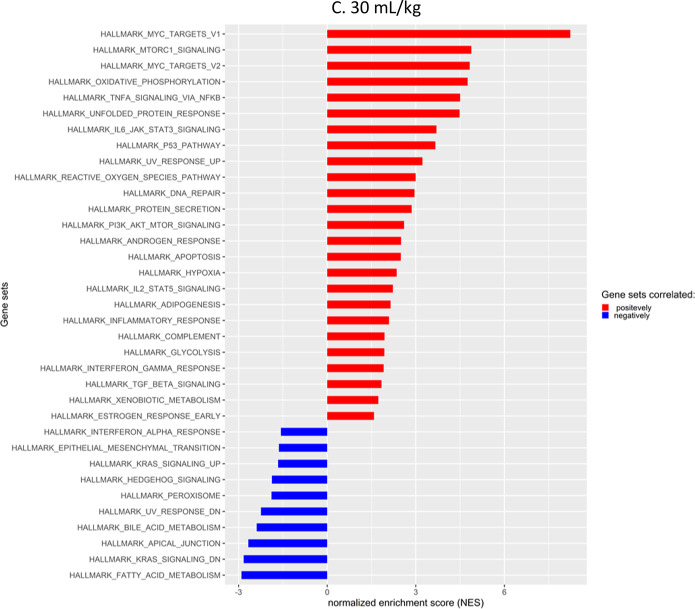
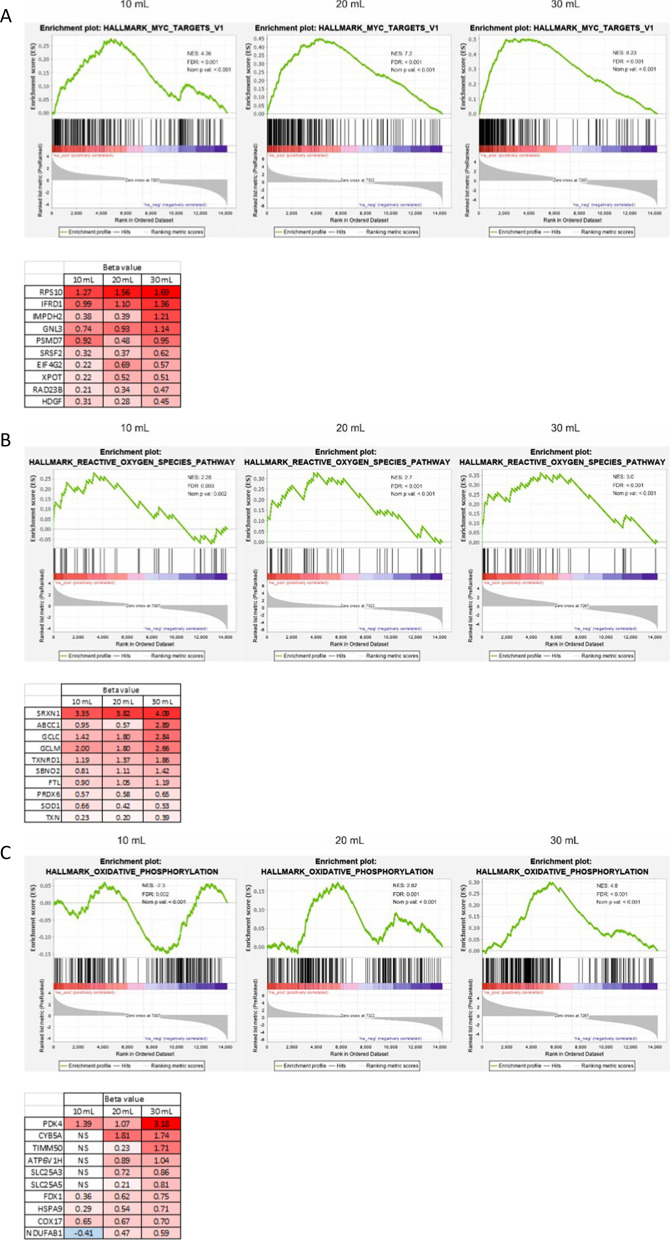
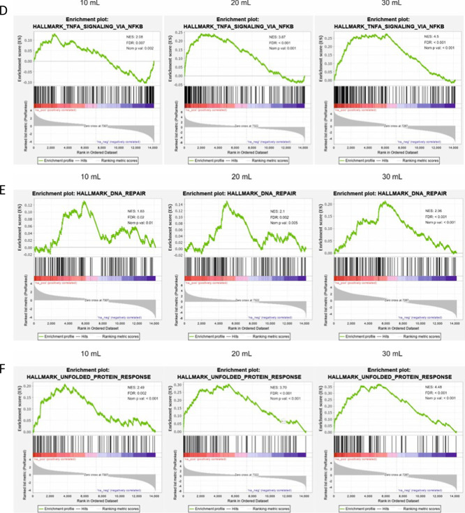
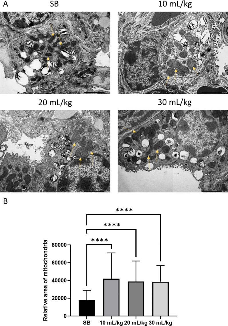
Results: We show a correlation between increased inflammation and barrier failure, and higher tidal volumes, evidenced by increased IL-6 expression, high concentration of proteins in BALF along with changes in expression of adhesion molecules. Furthermore, swelling of mitochondria in alveolar type II cells was seen indicating their dysfunction and senescence-like state. RNA sequencing data present clear increases in inflammation, mitochondrial biogenesis and oxidative stress as tidal volume is increased, supported by degradation of Keap1, a redox-regulated substrate adaptor protein.

Conclusions: Oxidative stress seems to be a more prominent mechanism of VILI than previously considered, indicating that possible treatment methods against VILI might be identified by impeding oxidative pathways.

Keywords: Acute lung injury; Mouse studies; Oxidative stress; Ventilator-induced lung injury.


Conflict of interest statement

Authors declare no competing interests.

Figures

Fig. 1
Fig. 1
High tidal volume results in decreased barrier protection and increased inflammatory response. When ventilating the mice at different tidal volumes, heart rate remained stable for the two lower tidal volumes. However, in the initial first hour of ventilation with tidal volume of 30 mL/kg heart rate increased but then remained stable (A). Pressure needed to maintain the applied tidal volumes remained relatively stable throughout the ventilation period (B). Increased protein concentration in BALF was detected in the 30 mL/kg tidal volume range (C). Pro-inflammatory cytokine, IL-6, expression increased significantly in the mice that were ventilated at 30 mL/kg (D). Standard deviation of the means is shown. Significant difference from the SB controls are shown (P ≤ 0.01 = **; P ≤ 0.0001 = ****)
Fig. 2
Fig. 2
Tissue area decreases as tidal volume increases. Shown are representative images of lung alveoli in SB and ventilated mice (A). Tissue area decreased significantly in mice ventilated at 30 mL/kg (B), while open airways increased (C). Total nucleus number decreased in mice ventilated at 30 mL/kg (D). Standard deviation of the means is shown. Significant difference from the SB controls are shown (P ≤ 0.01 = **; P ≤ 0.001 = ***; P ≤ 0.0001 = ****). Scale bar is 100 µm
Fig. 3
Fig. 3
Top 30 differentially expressed genes. Increased tidal volume leads to increased differential expression. Shown are the top 30 genes with the highest beta value from RNA sequencing of mouse lung tissues. Up regulated genes showed increased expression with increased tidal volume, while down regulated genes decreased in expression with increased tidal volume (A). Genes involved in glutathione production were significantly upregulated. The expression was shown to be tidal volume dependent (B). Real time PCR was done using lung tissue from ventilated mice and compared to SB controls (C). The top three genes from sequencing analysis were confirmed to be expressed in a tidal volume dependent manner. Immunoblotting from SB control, 10, 20 and 30 mL/kg tidal volume ventilated mice was performed for Keap1 and Actin. Actin used as a loading control and quantification carried out using FIJI (D). Significant difference from the SB controls are shown (P ≤ 0.05 = *; P ≤ 0.01 = **; P ≤ 0.001 = ***)
Fig. 4
Fig. 4
Gene set enrichment scores. Increased NES due to tidal volume increase. Gene set enrichment analysis from mouse tissue RNA sequencing, comparing SB to increased tidal volumes
Fig. 4
Fig. 4
Gene set enrichment scores. Increased NES due to tidal volume increase. Gene set enrichment analysis from mouse tissue RNA sequencing, comparing SB to increased tidal volumes
Fig. 5
Fig. 5
Genes set pertaining to mitochondrial biogenesis and respiration are enriched as a result of higher tidal volume. HALLMARK_MYC_TARGET_V1, REACTIVE_OXYGEN_SPECIES_PATHWAY and OXIDATIVE_PHOSPHORYLATION gene sets were enriched dependent on tidal volume increases (AC). The top 10 genes with highest beta value from the appropriate gene sets are displayed beneath each graph. TNFA_SIGNALING_VIA_NFKB, DNA_REPAIR and UNFOLDED_PROTEIN_RESPONSE gene sets increase their NES scores with increased tidal volumes (DF)
Fig. 5
Fig. 5
Genes set pertaining to mitochondrial biogenesis and respiration are enriched as a result of higher tidal volume. HALLMARK_MYC_TARGET_V1, REACTIVE_OXYGEN_SPECIES_PATHWAY and OXIDATIVE_PHOSPHORYLATION gene sets were enriched dependent on tidal volume increases (AC). The top 10 genes with highest beta value from the appropriate gene sets are displayed beneath each graph. TNFA_SIGNALING_VIA_NFKB, DNA_REPAIR and UNFOLDED_PROTEIN_RESPONSE gene sets increase their NES scores with increased tidal volumes (DF)
Fig. 6
Fig. 6
Increased area of mitochondria in ventilated mice. Mitochondrial swelling was observed in ventilated mice (A). Mitochondria were traced in images, using FIJI image processing (B). Mitochondria in ventilated mice were significantly larger. Standard deviation of the means is shown. A range of 41–84 measurements of mitochondria at the same magnifications were performed. (P ≤ 0.0001 = ****)

References

    1. Network TARDS. Ventilation with lower tidal volumes as compared with traditional tidal volumes for acute lung injury and the acute respiratory distress syndrome. N Engl J Med. 2000;342(18):1301–1308. doi: 10.1056/NEJM200005043421801. - DOI - PubMed
    1. Beitler JR, Malhotra A, Thompson BT. Ventilator-induced lung injury. Clin Chest Med. 2016;37(4):633–646. doi: 10.1016/j.ccm.2016.07.004. - DOI - PMC - PubMed
    1. Joelsson JP, Ingthorsson S, Kricker J, Gudjonsson T, Karason S. Ventilator-induced lung-injury in mouse models: Is there a trap? Lab Anim Res. 2021;37(1):30. doi: 10.1186/s42826-021-00108-x. - DOI - PMC - PubMed
    1. Amarelle L, Quintela L, Hurtado J, Malacrida L. Hyperoxia and lungs: what we have learned from animal models. Front Med. 2021;8:606678. doi: 10.3389/fmed.2021.606678. - DOI - PMC - PubMed
    1. Curley GF, Laffey JG, Zhang H, Slutsky AS. Biotrauma and ventilator-Induced Lung Injury: clinical Implications. Chest. 2016;150(5):1109–1117. doi: 10.1016/j.chest.2016.07.019. - DOI - PubMed

LinkOut - more resources