. 2022 Jul 22;41(1):229.
doi: 10.1186/s13046-022-02437-8.

PM2.5 promotes NSCLC carcinogenesis through translationally and transcriptionally activating DLAT-mediated glycolysis reprograming

Affiliations

PM2.5 promotes NSCLC carcinogenesis through translationally and transcriptionally activating DLAT-mediated glycolysis reprograming

Qianqian Chen et al. J Exp Clin Cancer Res. .

Abstract

Background: Airborne fine particulate matter (PM2.5) has been associated with lung cancer development and progression in never smokers. However, the molecular mechanisms underlying PM2.5-induced lung cancer remain largely unknown. The aim of this study was to explore the mechanisms by which PM2.5 regulated the carcinogenesis of non-small cell lung cancer (NSCLC).

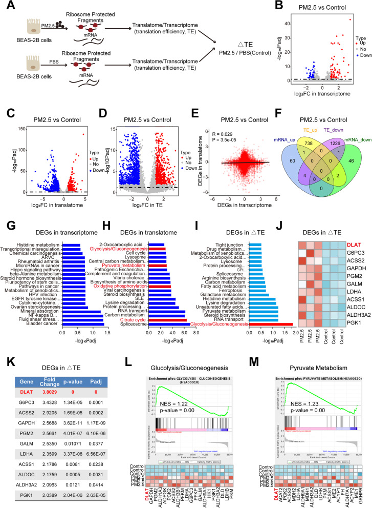
Methods: Paralleled ribosome sequencing (Ribo-seq) and RNA sequencing (RNA-seq) were performed to identify PM2.5-associated genes for further study. Quantitative real time-PCR (qRT-PCR), Western blot, and immunohistochemistry (IHC) were used to determine mRNA and protein expression levels in tissues and cells. The biological roles of PM2.5 and PM2.5-dysregulated gene were assessed by gain- and loss-of-function experiments, biochemical analyses, and Seahorse XF glycolysis stress assays. Human tissue microarray analysis and 18F-FDG PET/CT scans in patients with NSCLC were used to verify the experimental findings. Polysome fractionation experiments, chromatin immunoprecipitation (ChIP), and dual-luciferase reporter assay were implemented to explore the molecular mechanisms.

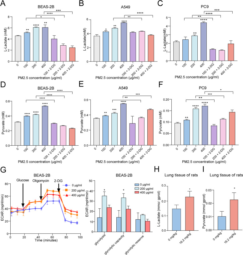
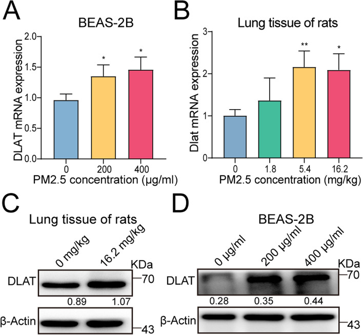
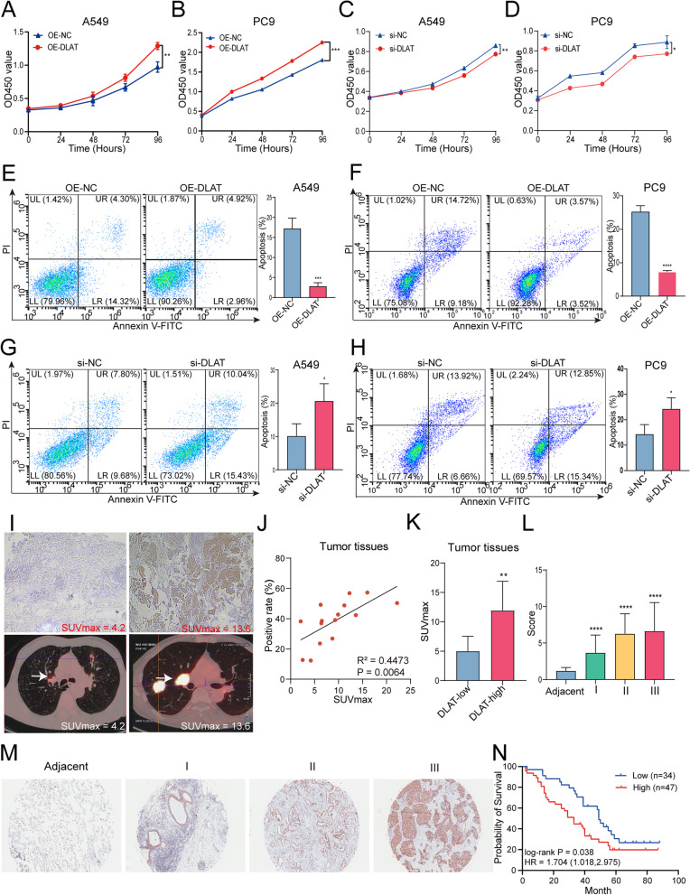
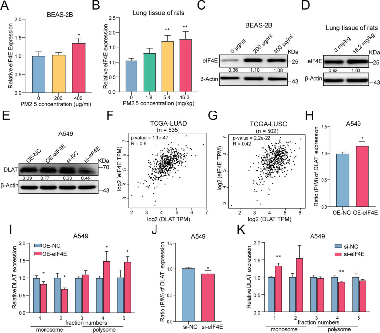
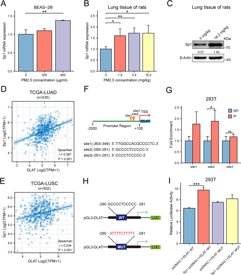
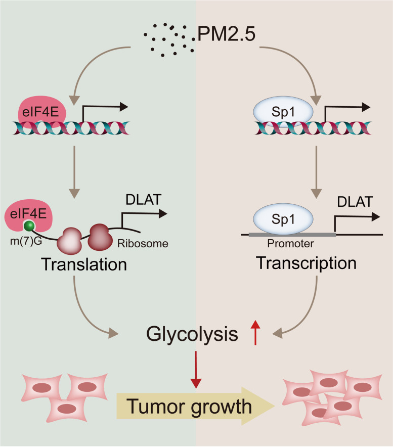
Results: We found that PM2.5 induced a translation shift towards glycolysis pathway genes and increased glycolysis metabolism, as evidenced by increased L-lactate and pyruvate concentrations or higher extracellular acidification rate (ECAR) in vitro and in vivo. Particularly, PM2.5 enhanced the expression of glycolytic gene DLAT, which promoted glycolysis but suppressed acetyl-CoA production and enhanced the malignancy of NSCLC cells. Clinically, high expression of DLAT was positively associated with tumor size, poorer prognosis, and SUVmax values of 18F-FDG-PET/CT scans in patients with NSCLC. Mechanistically, PM2.5 activated eIF4E, consequently up-regulating the expression level of DLAT in polysomes. PM2.5 also stimulated transcription factor Sp1, which further augmented transcription activity of DLAT promoter.

Conclusions: This study demonstrated that PM2.5-activated overexpression of DLAT and enhancement in glycolysis metabolism contributed to the tumorigenesis of NSCLC, suggesting that DLAT-associated pathway may be a therapeutic target for NSCLC.

Keywords: DLAT; Glycolysis reprograming; Non-small cell lung cancer (NSCLC); PM2.5; Sp1; Transcription; Translation; eIF4E.


Conflict of interest statement

The authors declare that they have no competing interests.

Figures

Fig. 1
Fig. 1
PM2.5 induces a translation efficiency (TE) shift towards up-regulation of glycolysis pathway genes. A Overview of experiments for RNA-seq and Ribo-seq in BEAS-2B cells following PM2.5 exposure. B Volcano map of differentially expressed genes (DEGs)in RNA-seq. Red and blue dots represent the upregulated and downregulated genes, respectively (FDR<0.05; |Log2(fold change)| >1). C Volcano plot of DEGs in Ribo-seq. D Volcano plot showing genes with significant changes in TE. E Genome-wide transcriptional and translational regulations showed very little correlation. F The four-way Venn diagram represented the different subsets of genes that are significantly upregulated or down-regulated at the TE and transcription levels. G KEGG pathway analysis of DEGs in transcriptome (RNA-seq). H Pathway analyses on DEGs in translatome revealed a shift towards glycolysis-related pathways. I Glycolysis/gluconeogenesis pathway was the only significantly enriched pathway in genes with TE changes. J Heatmap of DEGs in TE in the “Glycolysis/gluconeogenesis pathway”. K Fold change of TE for glycolytic genes between PM2.5-exposed cells and control cells. L Gene set enrichment analysis (GSEA) of Ribo-seq data showing the enrichment of glycolysis/gluconeogenesis pathway (upper) and the gene signature (lower) in PM2.5-exposed cells, with DLAT as the top up-regulated gene. M GSEA of Ribo-seq data revealing the enrichment of pyruvate metabolism pathway (upper) and the gene set in PM2.5-exposed cells, with DLAT ranking as the top gene. NES, normalized enrichment score
Fig. 2
Fig. 2
PM2.5 enhances glycolysis metabolism in vitro and in vivo. A PM2.5 enhanced L-lactate production in BEAS-2B cells, while 2-DG pretreatment reduced the generation of lactate in PM2.5-treated cells. B PM2.5 augmented L-lactate release from A549 cells in a dose response manner. C PM2.5 enhanced L-lactate production in PC9 cells. D PM2.5 increased pyruvate generation in BEAS-2B cells, and 2-DG pretreatment decreased pyruvate production in PM2.5-treated cells. E PM2.5 up-regulated pyruvate levels in A549 cells. F PM2.5 increased pyruvate levels in PC9 cells. G PM2.5 promoted ECAR levels in BEAS-2B cells. H PM2.5 exposure enhanced the production of L-lactate in lung issues of rats. I PM2.5 exposure promoted the generation of pyruvate in lung issues of rats. *P < 0.05, **P < 0.01, ***P < 0.001
Fig. 3
Fig. 3
PM2.5 increases the expression of glycolytic gene DALT in vitro and in vivo. A PM2.5 exposure enhanced the expression level of DLAT gene in BEAS-2B cells. B PM2.5 inhalation up-regulated DLAT gene expression in lung tissues of rats. C PM2.5 exposure promoted DLAT protein expression in BEAS-2B cells. Numerical numbers denoted the ratio of integrated optical density (IOD) to β-actin. D PM2.5 exposure increased DLAT protein expression in lungs of rats. *P < 0.05, **P < 0.01
Fig. 4
Fig. 4
DLAT enhances glycolysis metabolism in NSCLC cells. A DLAT overexpression increased L-lactate production in PC9 cells. B DLAT up-regulation enhanced L-lactate production in A549 cells. C DLAT overexpression increased pyruvate release from PC9 cells. D Up-regulation of DLAT promoted pyruvate release from A549 cells. E Knockdown of DLAT suppressed L-lactate generation from PC9 cells. F Down-regulation of DLAT inhibited L-lactate generation from A549 cells. G Inhibition of DLAT expression decreased pyruvate production in PC9 cells. H Decreased DLAT expression reduced pyruvate production in A549 cells. I Overexpression of DLAT enhanced ECAR levels in PC9 cells. J Up-regulation of DLAT increased ECAR levels in A549 cells. *P < 0.05, **P < 0.01
Fig. 5
Fig. 5
DLAT promotes the malignancy of NSCLC cells and correlates with glucose metabolism and poor prognosis in NSCLC patients. A, B Overexpression of DLAT promoted cell proliferation of NSCLC cells. C, D Knockdown of DLAT suppressed cell proliferation of A549 and PC9 cells. E, F Up-regulation of DLAT reduced apoptosis rate of PC9 and A549 cells. G, H Depletion of DLAT increased apoptosis rate of NSCLC cells. I Representative 18F-FDG PET/CT images in patients with NSCLC tumors exhibiting low or high expression of DLAT. J The expression levels (% of positive cells) of DLAT in tumor tissues were positively correlated with the SUVmax values in patients with NSCLC. K Analysis of SUVmax in the DLAT low and DLAT high groups. L DLAT expression levels in NSCLC tumor tissues increased as NSCLC progressed to more advanced stages. M Representative pictures showing that IHC signals of DLAT (brown staining) was increased along with the tumor stages of NSCLC. N Kaplan-Meier analysis showed that elevated expression of DLAT was associated with poorer overall survival (OS) in NSCLC patients. Data shown are mean ± SD. *P < 0.05, **P < 0.01, ***P < 0.001
Fig. 6
Fig. 6
PM2.5 activates the expression of eIF4E that subsequently increases the translation of DLAT in polysomes. A PM2.5 enhanced eIF4E gene expression in BEAS-2B cells in a dose-response manner. B PM2.5 increased eIF4E gene expression in lung tissues of rats. C PM2.5 up-regulated eIF4E protein expression in BEAS-2B cells. D PM2.5 increased eIF4E protein expression in lung tissues of rats. E Up-regulation of eIF4E increased the expression of DLAT protein and knockdown of eIF4E decreased DLAT protein expression. F The expression level of eIF4E was positively correlated with that of DLAT in LUAD tumor tissues (TCGA datasets). G Correlation between eIF4E and DLAT expression levels in LUSC (TCGA datasets). H Overexpression of eIF4E increased the polysome to monosome (P/M) ratio of DLAT mRNA expression, suggesting an increase of DLAT translation initiation. I Up-regulation of eIF4E increased the abundance of DLAT mRNA in polysome but reduced DLAT expression in monosome in A549 cells. J Knockdown of eIF4E decreased the polysome to monosome ratio of DLAT mRNA expression, indicating a reduction of DLAT translation initiation. K Downregulation of eIF4E decreased the abundance of DLAT mRNA in polysome but enhanced DLAT expression in monosome in A549 cells. *P<0.05, **P<0.01
Fig. 7
Fig. 7
PM2.5 activates the expression of transcription factor Sp1 which enhances the transcription of DLAT. A PM2.5 promoted Sp1 expression in a dose-response manner in BEAS-2B cells. B PM2.5 enhanced the expression of Sp1 in lung tissues of rats. C PM2.5 increased the expression of Sp1 protein in lung tissues of rats. D, E The expression level of Sp1 was positively correlated with that of DLAT in LUAD (lung adenocarcinoma) and LUSC (lung squamous carcinoma) in TCGA dataset. F In silico analysis identified Sp1 putative binding sites in the promoter region of DLAT. G ChIP assay showed the binding of Sp1 with DLAT promoter region. H Potential binding sequences for Sp1 were found in the DLAT promoter region. I Luciferase reporter assay verified that the binding of Sp1 with wild-type DLAT promoter significantly increased the luciferase activity. While binding of Sp1 with mutant DLAT promoter did not changes the intensity of luciferase signals. J Schematic view for the mechanisms of action of DLAT-mediated glycolysis reprograming in PM2.5-induced carcinogenesis. *P<0.05, **P<0.01, ***P<0.001.
Fig. 8
Fig. 8
Schematic view for the mechanisms of action of DLAT-mediated glycolysis reprograming in PM2.5-induced carcinogenesis

References

    1. Kang HR, Cho JY, Lee SH, Lee YJ, Park JS, Cho YJ, Yoon HI, Lee KW, Lee JH, Lee CT. Role of low-dose computerized tomography in lung cancer screening among never-smokers. J Thorac Oncol. 2019;14(3):436–444. doi: 10.1016/j.jtho.2018.11.002. - DOI - PubMed
    1. Casal-Mouriño A, Valdés L, Barros-Dios JM, Ruano-Ravina A. Lung cancer survival among never smokers. Cancer Lett. 2019;451:142–149. doi: 10.1016/j.canlet.2019.02.047. - DOI - PubMed
    1. Smolle E, Pichler M. Non-smoking-associated lung cancer: a distinct entity in terms of tumor biology, patient characteristics and impact of hereditary cancer predisposition. Cancers (Basel) 2019;11(2):204. doi: 10.3390/cancers11020204. - DOI - PMC - PubMed
    1. Lee YJ, Kim JH, Kim SK, Ha SJ, Mok TS, Mitsudomi T, Cho BC. Lung cancer in never smokers: change of a mindset in the molecular era. Lung Cancer. 2011;72(1):9–15. doi: 10.1016/j.lungcan.2010.12.013. - DOI - PubMed
    1. Fajersztajn L, Veras M, Barrozo LV, Saldiva P. Air pollution: a potentially modifiable risk factor for lung cancer. Nat Rev Cancer. 2013;13(9):674–678. doi: 10.1038/nrc3572. - DOI - PubMed

MeSH terms