

Chorioretinal abnormalities in idiopathic intracranial hypertension: case reports
- PMID: 35869502
- PMCID: PMC9308292
- DOI: 10.1186/s40942-022-00403-2
Chorioretinal abnormalities in idiopathic intracranial hypertension: case reports
Abstract
Background: Papilledema is the main ocular finding in patients with idiopathic intracranial hypertension (IIH) although several chorioretinal abnormalities may also occur and contribute to visual loss. The purpose of this paper is to describe two cases of chorioretinal abnormalities associated with idiopathic intracranial hypertension: one with choroidal folds and another with polypoidal choroidal vasculopathy, an extremely unusual ocular complication in the disease.
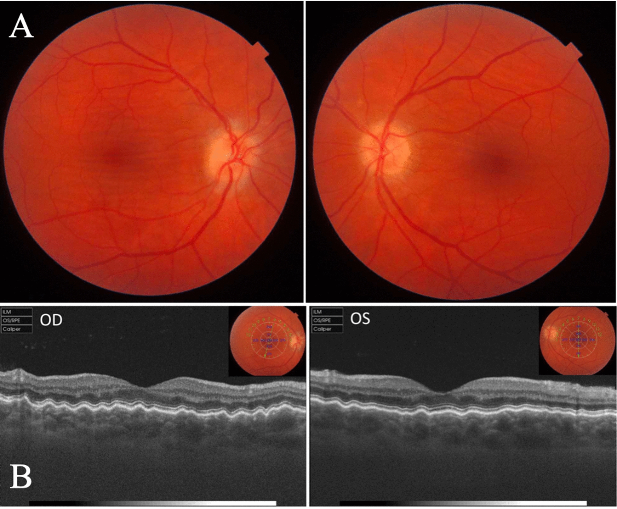
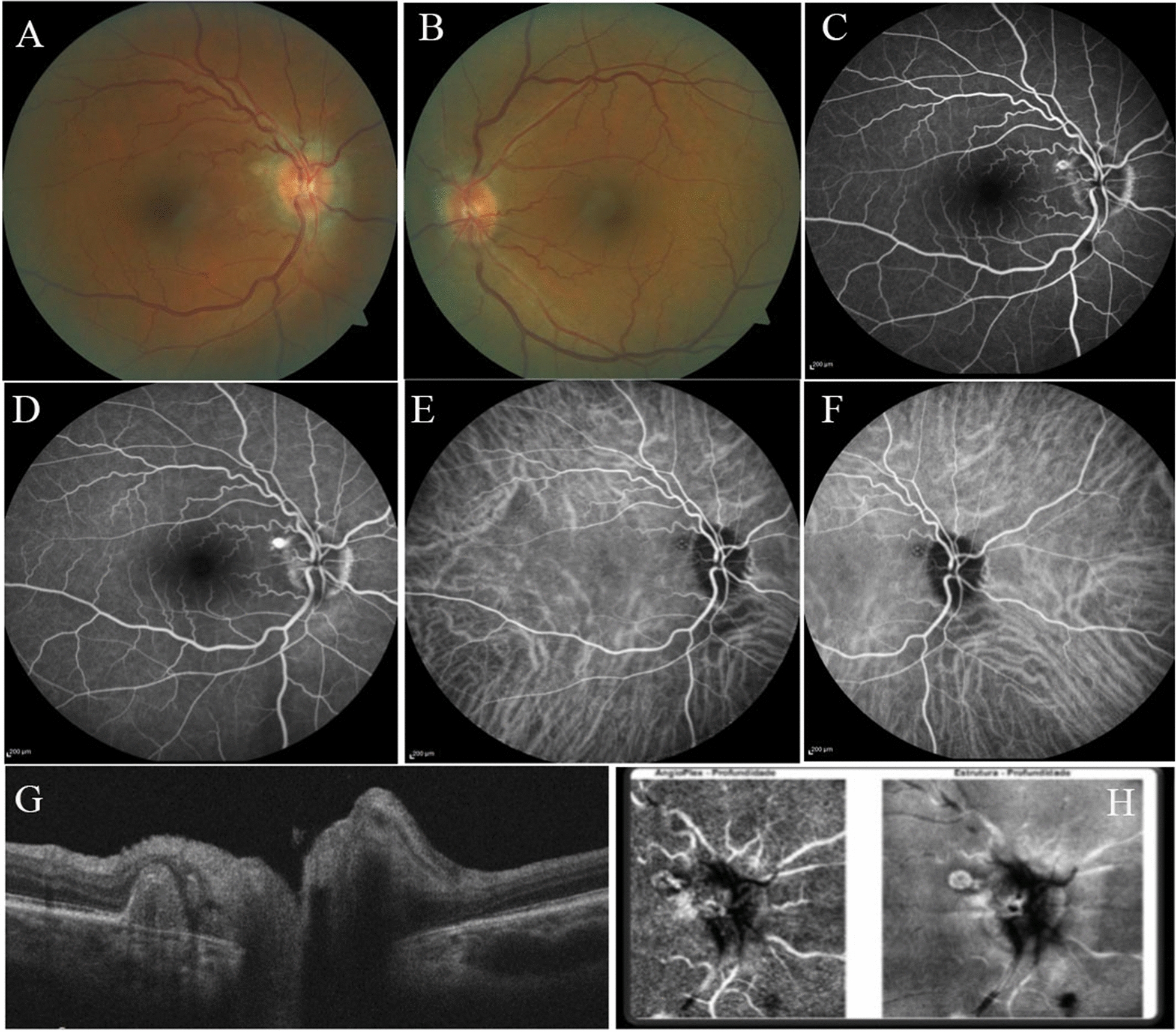
Case presentation: Case 1: A 47-year-old woman previous diagnosed with idiopathic intracranial hypertension treated with weight loss and acetazolamide that over the following 6 months had optic disc edema gradually resolved. The patient was follow-up for a period of 10 years and the papilledema disappeared, but choroidal folds remained unchanged. Case 2: A 61-year-old female patient was seen as a follow-up examination of a 5-year history of IIH that presented with papilledema. The patient was asymptomatic but fundoscopy evaluation revealed a yellowish white peripapillary subretinal nodular lesion temporally in OD. Multimodal imaging studies were made, and the patient was diagnosed with a rare and just recent described association of IIH and polypoidal choroidal vasculopathy.
Conclusion: Papilledema, RNFL and retinal ganglion cell loss are the most common structural complications of IIH, but chorioretinal complications are important findings and should be carefully evaluated in such patients. Awareness of such occurrence and the use of appropriated clinical and multimodal imaging studies are of great importance for its early detection, leading to proper treatment and prevention of further visual loss.
Keywords: Choroid; Choroidal folds; Choroidal neovascularization; Idiopathic intracranial hypertension; Papilledema; Retinal neovascularization.
© 2022. The Author(s).
Conflict of interest statement
None of the authors have any potential conflicts of interest to disclose.
Figures
References
-
- Monteiro MLR, Moura FC. Ophthalmic aspects of idiopathic intracranial hypertension syndrome (pseudotumor cerebri) Rev Bras Oftalmol. 2008;67(4):196–203. doi: 10.1590/S0034-72802008000400008. - DOI
LinkOut - more resources
Full Text Sources
