. 2022 Sep;162(5):417-429.
doi: 10.1111/jnc.15677. Epub 2022 Aug 4.

Subthalamic nucleus exclusively evokes dopamine release in the tail of the striatum

Affiliations

Subthalamic nucleus exclusively evokes dopamine release in the tail of the striatum

Kathryn L Todd et al. J Neurochem. 2022 Sep.

Abstract

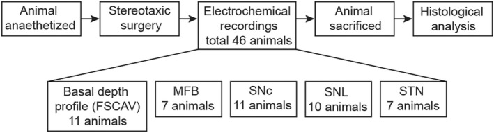
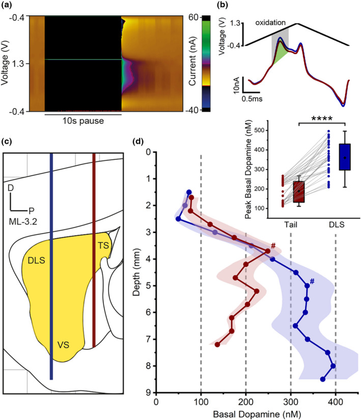
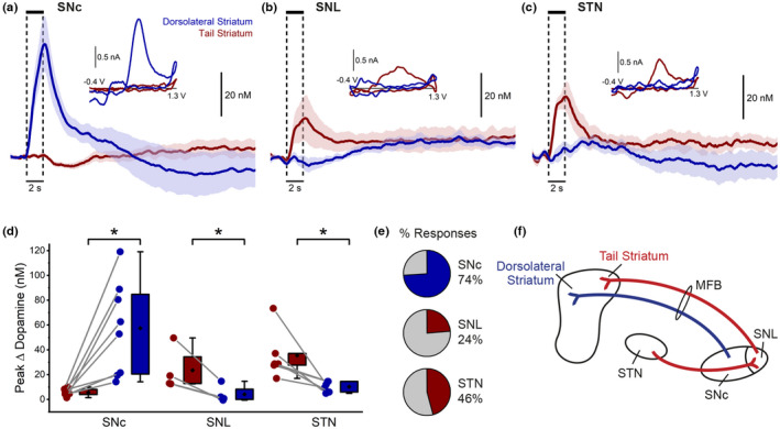
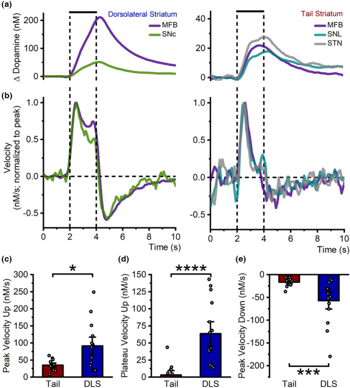
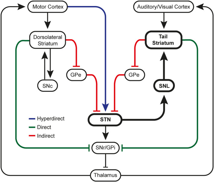
A distinct population of dopamine neurons in the substantia nigra pars lateralis (SNL) has a unique projection to the most caudolateral (tail) region of the striatum. Here, using two electrochemical techniques to measure basal dopamine and electrically evoked dopamine release in anesthetized rats, we characterized this pathway, and compared it with the 'classic' nigrostriatal pathway from neighboring substantia nigra pars compacta (SNc) dopamine neurons to the dorsolateral striatum. We found that the tail striatum constitutes a distinct dopamine domain compared with the dorsolateral striatum, with consistently lower basal and evoked dopamine, and diverse dopamine release kinetics. Importantly, electrical stimulation of the SNL and SNc evoked dopamine release in entirely separate striatal regions; the tail and dorsolateral striatum, respectively. Furthermore, we showed that stimulation of the subthalamic nucleus (STN) evoked dopamine release exclusively in the tail striatum, likely via the SNL, consistent with previous anatomical evidence of STN afferents to SNL dopamine neurons. Our work identifies the STN as an important modulator of dopamine release in a novel dopamine pathway to the tail striatum, largely independent of the classic nigrostriatal pathway, which necessitates a revision of the basal ganglia circuitry with the STN positioned as a central integrator of striatal information.

Keywords: basal ganglia; dopamine; electrochemistry; substantia nigra pars lateralis; subthalamic nucleus; tail striatum.


Conflict of interest statement

The authors declare no competing interests.

Figures

FIGURE 1
FIGURE 1
Experimental procedure. Stereotaxic surgery was performed under urethane anesthesia and electrodes were lowered into the relevant brain regions. Electrochemical recordings were conducted in one or both hemispheres. For FSCV recordings, stimulation site (MFB: medial forebrain bundle, SNc: substantia nigra pars compacta, SNL: substantia nigra pars lateralis, STN: subthalamic nucleus) was determined arbitrarily. Basal dopamine measurements (FSCAV) were taken across all experimental groups. Dorsolateral and tail striatum were recorded from sequentially (same hemisphere) in a random order. After experiments, animals were sacrificed by decapitation and brain tissue was stored in fixative for 48 hrs prior to histological validation of stimulation site (data not shown).
FIGURE 2
FIGURE 2
Basal dopamine profiles reveal distinct dorsolateral and tail striatum domains. (a) Color plot showing oxidation and reduction currents of dopamine adsorbed to the carbon‐fiber microelectrode after the 10 s pause in fast‐scan controlled adsorption voltammetry (FSCAV). (b) Voltammograms (10th scan after pause) showing dopamine oxidation current recorded in the tail (red) and dorsolateral (blue) striatum in response to the cyclic voltage ramp (top). Green area represents the integral of current (charge, pC) used to calculate dopamine concentration. (c) Representative sagittal section (ML −3.2) indicating rostral (AP 0; blue) and caudal (AP ‐1.8; red) recording tracts. DLS, dorsolateral striatum; TS, tail striatum; VS, ventral striatum. (d) Distinct depth profiles of basal dopamine in the rostral (blue; n = 6) and caudal (red; n = 7) recording tracts. Inset: Peak basal dopamine (#) was consistently higher in the dorsolateral striatum (DV ‐5.0) compared with tail striatum (DV ‐3.5) in all experiments (n = 35). Connecting lines indicate paired measurements (same hemisphere, sequentially measured). n = number of hemispheres. ****p < 0.0001.
FIGURE 3
FIGURE 3
MFB stimulation evokes dopamine release in both dorsolateral and tail striatum. (a) Schematic (sagittal section) showing carbon‐fiber microelectrode recording locations in the dorsolateral (DV ‐5.0) and tail (DV ‐3.5) striatum, and stimulation sites in the medial forebrain bundle (MFB), subthalamic nucleus (STN), substantia nigra pars compacta (SNc), and pars lateralis (SNL). Results for STN, SNc and SNL stimulation shown in Figure 4. (b) MFB stimulation (60 Hz, 120 pulses, 300 μA, 2 ms each phase; vertical lines) evoked pronounced dopamine release in the dorsolateral striatum (DLS; blue; n = 7), while recording in the tail striatum revealed much smaller release (red; n = 3), or no detectable release at all (gray; n = 4). n = number of hemispheres. Inset: Consistency (% of first response) of responses across repeated stimulations at 5 min intervals. (c) Color plot showing oxidation and reduction currents following MFB stimulation (vertical lines) recorded in the dorsolateral striatum. (d) Corresponding average cyclic voltammograms showing oxidation and reduction currents consistent with dopamine. (e) Peak dopamine release following MFB stimulation was significantly higher in the DLS compared to the tail striatum. Connecting lines indicate paired measurements (same hemisphere, sequentially measured). Gray connecting lines indicate pairs of recordings in which no dopamine release was detected in the tail striatum (see B). **p < 0.01. (f) MFB stimulation faithfully evoked dopamine release in the dorsolateral striatum (7 from 7 hemispheres; blue), but less reliably in the tail striatum (3 from 7 hemispheres; red).
FIGURE 4
FIGURE 4
SNL and STN stimulation exclusively evoke dopamine release in the tail striatum. (a–c) Stimulation of the substantia nigra pars compacta (a, SNc; n = 8) evoked dopamine release in the dorsolateral striatum (blue), with no release detected in the tail striatum (red). Conversely, stimulation of the substantia nigra pars lateralis (b, SNL; n = 4) or subthalamic nucleus (c, STN; n = 6) evoked dopamine release in the tail striatum, with no release detected in the dorsolateral striatum. n = number of hemispheres. Insets: Corresponding average cyclic voltammograms. (d) Comparison of peak dopamine release in the tail (red) and dorsolateral (blue) striatum. Connecting lines indicate paired measurements (same hemisphere, sequentially measured). *p < 0.05. (e) Stimulation of the SNc more reliably evoked dopamine release in the dorsolateral striatum compared with release in the tail striatum following SNL and STN stimulation. (f) Proposed schematic of distinct dopamine pathways from the SNc and SNL to the distinct dopamine domains in the dorsolateral and tail striatum, respectively, and selective activation of the SNL by the STN.
FIGURE 5
FIGURE 5
Distinct dopamine release kinetics in the tail and dorsolateral striatum. (a) Dopamine release in the dorsolateral (left) and tail (right) striatum had distinct profiles, independent of stimulation site (MFB: medial forebrain bundle, DLS n = 7, tail n = 3; SNc: substantia nigra pars compacta, n = 8; SNL: substantia nigra pars lateralis, n = 4; STN: subthalamic nucleus, n = 6; stimulation 60 Hz, 120 pulses, 300 μA, 2 ms each phase, vertical lines). (b) Velocity of dopamine release (first derivative, normalized to peak upward velocity) had distinct profiles between the dorsolateral (left) and tail (right) striatum with dopamine release in the dorsolateral striatum having obvious sustained release for the duration of the stimulation, while release in the tail striatum declined quickly after an initial phase despite ongoing stimulation. (c–e) Summary of grouped data showing that the tail striatum (red; n = 13) had a slower peak (c) and plateau (d) upward velocity as well as slower peak downward velocity (e), compared with the dorsolateral striatum (DLS, blue; n = 15). n = number of hemispheres. *p < 0.05, ***p < 0.001, ****p < 0.0001.
FIGURE 6
FIGURE 6
Proposed model of the basal ganglia. Cortical information from the motor cortex and auditory/visual cortex remains segregated in the dorsolateral and tail striatum dopamine domains, respectively. Information from the functionally distinct systems is integrated in the STN which receives direct motor input via the hyperdirect pathway and input from both striatal regions via the indirect pathway. The STN exclusively excites dopamine neurons in the SNL, which in turn modulates dopamine release in the tail striatum only (bold).

Similar articles

Cited by

References

    1. Abudukeyoumu, N. , Hernandez‐Flores, T. , Garcia‐Munoz, M. , & Arbuthnott, G. W. (2019). Cholinergic modulation of striatal microcircuits. The European Journal of Neuroscience, 49, 604–622. - PMC - PubMed
    1. Albin, R. L. , Young, A. B. , & Penney, J. B. (1989). The functional anatomy of basal ganglia disorders. Trends in Neurosciences, 12, 366–375. - PubMed
    1. Ammari, R. , Lopez, C. , Bioulac, B. , Garcia, L. , & Hammond, C. (2010). Subthalamic nucleus evokes similar long lasting glutamatergic excitations in pallidal, entopeduncular and nigral neurons in the basal ganglia slice. Neuroscience, 166, 808–818. - PubMed
    1. Atcherley, C. W. , Laude, N. D. , Parent, K. L. , & Heien, M. L. (2013). Fast‐scan controlled‐adsorption voltammetry for the quantification of absolute concentrations and adsorption dynamics. Journal of the American Chemical Society, 29, 14885–14892. - PubMed
    1. Baig, F. , Lawton, M. , Rolinski, M. , Ruffmann, C. , Nithi, K. , Evetts, S. G. , Fernandes, H. R. , Ben‐Shlomo, Y. , & Hu, M. T. (2015). Delineating nonmotor symptoms in early Parkinson's disease and first‐degree relatives. Movement Disorders, 30, 1759–1766. - PMC - PubMed

Publication types