

Visualising and quantifying microvascular structure and function in patients with heart failure using optical coherence tomography
- PMID: 35869823
- PMCID: PMC9541462
- DOI: 10.1113/JP282940
Visualising and quantifying microvascular structure and function in patients with heart failure using optical coherence tomography
Abstract
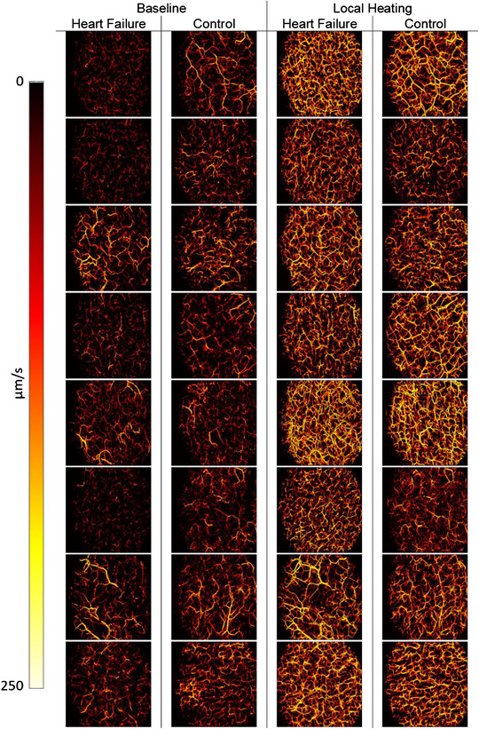
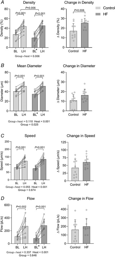
Heart failure (HF) is characterised by abnormal conduit and resistance artery function in humans. Microvascular function in HF is less well characterised, due in part to the lack of tools to image these vessels in vivo. The skin microvasculature is a surrogate for systemic microvascular function and health and plays a key role in thermoregulation, which is dysfunctional in HF. We deployed a novel optical coherence tomography (OCT) technique to visualise and quantify microvascular structure and function in 10 subjects with HF and 10 age- and sex-matched controls. OCT images were obtained from the ventral aspect of the forearm, at baseline (33°C) and after 30 min of localised skin heating. At rest, OCT-derived microvascular density (20.3 ± 8.7%, P = 0.004), diameter (35.1 ± 6.0 μm, P = 0.006) and blood flow (82.9 ± 41.1 pl/s, P = 0.021) were significantly lower in HF than CON (27.2 ± 8.0%, 40.4 ± 5.8 μm, 110.8 ± 41.9 pl/s), whilst blood speed was not significantly lower (74.3 ± 11.0 μm/s vs. 81.3 ± 9.9 μm/s, P = 0.069). After local heating, the OCT-based density, diameter, blood speed and blood flow of HF patients were similar (all P > 0.05) to CON. Although abnormalities exist at rest which may reflect microvascular disease status, patients with HF retain the capacity to dilate cutaneous microvessels in response to localised heat stress. This is a novel in vivo human observation of microvascular dysfunction in HF, illustrating the feasibility of OCT to directly visualise and quantify microvascular responses to physiological stimuli in vivo. KEY POINTS: Microvessels in the skin are critical to human thermoregulation, which is compromised in participants with heart failure (HF). We have developed a powerful new non-invasive optical coherence tomography (OCT)-based approach for the study of microvascular structure and function in vivo. Our approach enabled us to observe and quantify abnormal resting microvascular function in participants with HF. Patients with HF were able to dilate skin microvessels in response to local heat stress, arguing against an underlying structural abnormality. This suggests that microvascular functional regulation is the primary abnormality in HF. OCT can be used to directly visualise and quantify microvascular responses to physiological stimuli in vivo.
Keywords: heart failure; microvessels; optical imaging.
© 2022 The Authors. The Journal of Physiology published by John Wiley & Sons Ltd on behalf of The Physiological Society.
Figures
Similar articles
-
Visualizing and quantifying the impact of reactive hyperemia on cutaneous microvessels in humans.J Appl Physiol (1985). 2020 Jan 1;128(1):17-24. doi: 10.1152/japplphysiol.00583.2019. Epub 2019 Nov 14. J Appl Physiol (1985). 2020. PMID: 31725361
-
Novel Noninvasive Assessment of Microvascular Structure and Function in Humans.Med Sci Sports Exerc. 2019 Jul;51(7):1558-1565. doi: 10.1249/MSS.0000000000001898. Med Sci Sports Exerc. 2019. PMID: 30688767
-
Visualizing and quantifying cutaneous microvascular reactivity in humans by use of optical coherence tomography: impaired dilator function in diabetes.Am J Physiol Endocrinol Metab. 2020 Nov 1;319(5):E923-E931. doi: 10.1152/ajpendo.00233.2020. Epub 2020 Sep 21. Am J Physiol Endocrinol Metab. 2020. PMID: 32954827
-
Microvascular imaging of the skin.Phys Med Biol. 2019 Mar 21;64(7):07TR01. doi: 10.1088/1361-6560/ab03f1. Phys Med Biol. 2019. PMID: 30708364 Free PMC article. Review.
-
Quantitative depth-resolved microcirculation imaging with optical coherence tomography angiography (Part Ι): Blood flow velocity imaging.Microcirculation. 2018 Aug;25(6):e12375. doi: 10.1111/micc.12375. Microcirculation. 2018. PMID: 28419622 Review.
Cited by
-
Diagnostic and therapeutic optical imaging in cardiovascular diseases.iScience. 2024 Oct 22;27(11):111216. doi: 10.1016/j.isci.2024.111216. eCollection 2024 Nov 15. iScience. 2024. PMID: 39569375 Free PMC article. Review.
-
Improved microvascular imaging with optical coherence tomography using 3D neural networks and a channel attention mechanism.Sci Rep. 2024 Aug 1;14(1):17809. doi: 10.1038/s41598-024-68296-9. Sci Rep. 2024. PMID: 39090263 Free PMC article.
-
Cell diversity and signalling in the cardiovascular system.J Physiol. 2023 Jul;601(13):2537-2539. doi: 10.1113/JP284979. Epub 2023 May 27. J Physiol. 2023. PMID: 37211722 Free PMC article. No abstract available.
References
-
- Argarini, R. , McLaughlin, R. A. , Joseph, S. Z. , Naylor, L. H. , Carter, H. H. , Yeap, B. B. , Jansen, S. J. , & Green, D. J. (2020). Optical coherence tomography: a novel imaging approach to visualize and quantify cutaneous microvascular structure and function in patients with diabetes. BMJ Open Diabetes Research and Care, 8(1), e001479. - PMC - PubMed
-
- Argarini, R. , McLaughlin, R. A. , Naylor, L. H. , Carter, H. H. , & Green, D. J. (2020). Assessment of the human cutaneous microvasculature using optical coherence tomography: Proving Harvey's proof. Microcirculation, 27(2), e12594. - PubMed
-
- Atkinson, C. L. , Carter, H. H. , Thijssen, D. H. J. , Birk, G. K. , Cable, N. T. , Low, D. A. , Kerstens, F. , Meeuwis, I. , Dawson, E. A. , & Green, D. J. (2018). Localised cutaneous microvascular adaptation to exercise training in humans. European Journal of Applied Physiology, 118(4), 837–845. - PubMed
-
- Bauersachs, J. , Butler, J. , & Sandner, P. (2017). Heart Failure. Springer International Publishing, Champaign.
Publication types
MeSH terms
LinkOut - more resources
Full Text Sources
Medical
Research Materials
Miscellaneous