. 2022 Dec;45(6):2544-2558.
doi: 10.1007/s10753-022-01711-3. Epub 2022 Jul 23.

Interleukin-6 Downregulates the Expression of Vascular Endothelial-Cadherin and Increases Permeability in Renal Glomerular Endothelial Cells via the Trans-Signaling Pathway

Affiliations

Interleukin-6 Downregulates the Expression of Vascular Endothelial-Cadherin and Increases Permeability in Renal Glomerular Endothelial Cells via the Trans-Signaling Pathway

Yong-Chang Yang et al. Inflammation. 2022 Dec.

Abstract

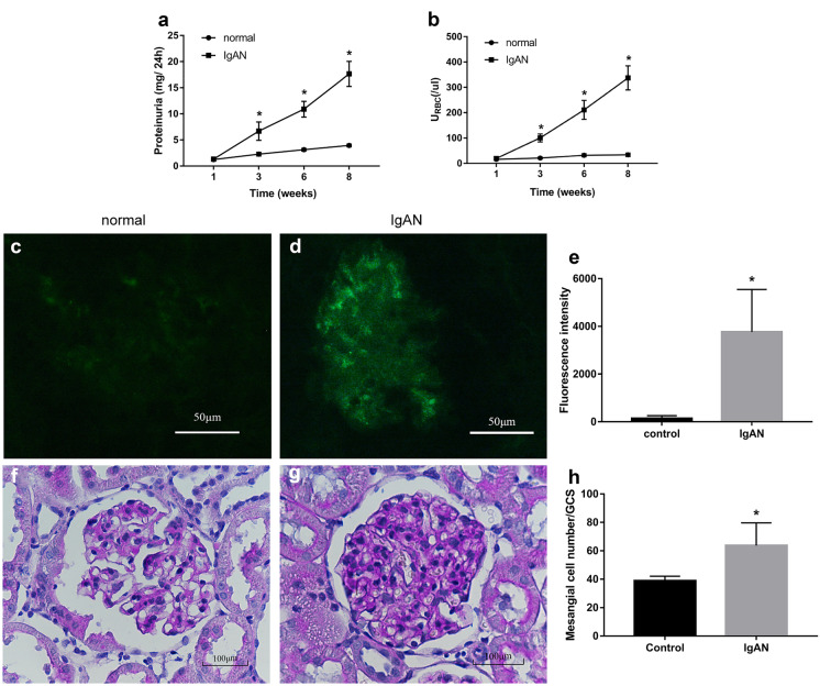
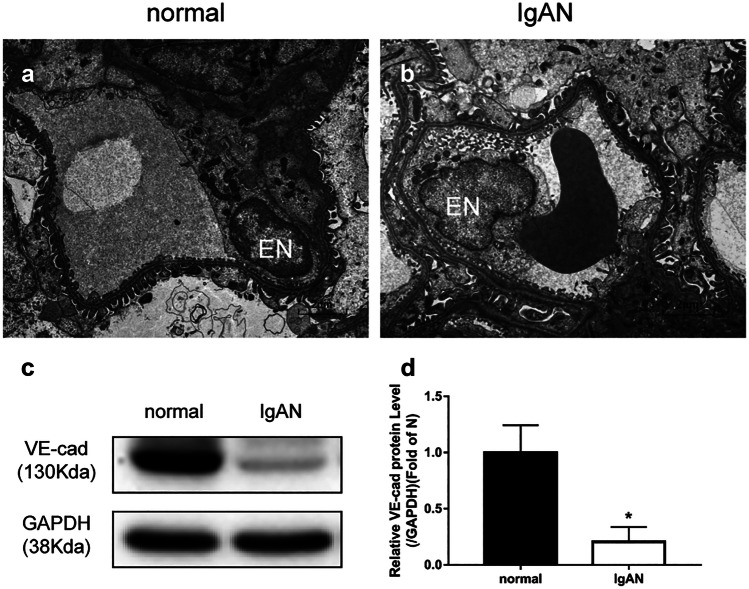
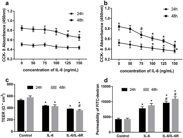
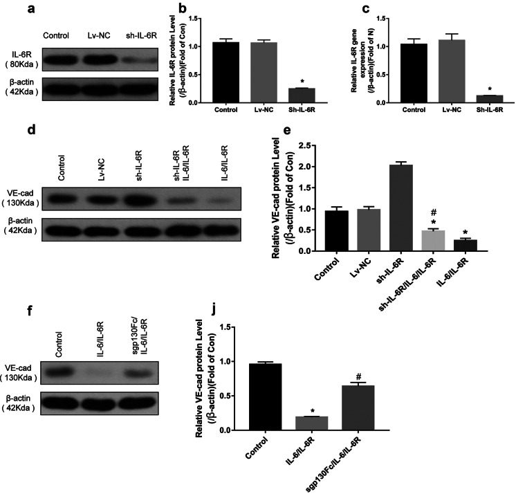
The pathogenesis of IgA nephropathy (IgAN) is still unknown, but reportedly, interleukin 6 (IL-6) is involved in this process. However, its role in damaging glomerular endothelial cells is still unclear. Therefore, in this study, to clarify the mechanism of the pathogenesis of IgAN, we investigated the effect of IL-6 on the permeability of glomerular endothelial cells. A rat model of IgAN was established, and the animals divided into two groups, namely, the normal and IgAN groups. Glomerular endothelial cell injury was evaluated via electron microscopy. Furthermore, IL-6-induced changes in the permeability of human renal glomerular endothelial cells (HRGECs) were measured via trans-endothelial resistance (TEER) measurements and fluorescein isothiocyanate-dextran fluorescence. Furthermore, vascular endothelial-cadherin (VE-cadherin) was overexpressed to clarify the effect of IL-6 on HRGEC permeability, and to determine the pathway by which it acts. The classical signaling pathway was blocked by silencing IL-6R and the trans-signaling pathway was blocked by sgp30Fc. In IgAN rats, electron microscopy showed glomerular endothelial cell damage and western blotting revealed a significant increase in IL-6 expression, while VE-cadherin expression decreased significantly in the renal tissues. IL-6/IL-6R stimulation also significantly increased the permeability of HRGECs (p < 0.05). This effect was significantly reduced by VE-cadherin overexpression (p < 0.01). After IL-6R was silenced, IL-6/IL-6R still significantly reduced VE-cadherin expression and sgp30Fc blocked the trans-signaling pathway as well as the upregulation of IL-6/IL-6R-induced VE-cadherin expression. This suggests that IL-6 mainly acts via the trans-signaling pathway. IL-6 increased the permeability of HRGECs by decreasing the expression of VE-cadherin via the trans-signaling pathway.

Keywords: IgA nephropathy; glomerular endothelial cells; interleukin 6; vascular endothelial-cadherin.


Conflict of interest statement

The authors declare no competing interests.

Figures

Fig. 1
Fig. 1
Hematuria, proteinuria, and IgA deposition in rats with IgAN nephropathy. From third week, rats in the IgAN group showed significantly higher (a) proteinuria and (b) hematuria than the normal group. In direct immunofluorescence, green fluorescence in the glomerulus represents IgA deposition. At high magnification (200×), (c) rats in the normal group showed faintly visible IgA deposition in their glomeruli, while (d) rats in the IgAN group showed a high amount of dazzling IgA deposition in their glomeruli. (e) Fluorescence intensity corresponding to the IgAN group significantly higher than that corresponding to the normal group (p < 0.01). PAS staining in paraffin sections showed (f) no proliferation of mesangial cells in the normal group, and (g) obvious proliferation of mesangial cells in the IgAN group. (h) Statistics showing significantly higher mesangial cell proliferation in the IgAN group than in the normal group (p < 0.01). These findings presented as the mean ± SD were analyzed by performing the t-test (n = 6). *p < 0.01, IgAN vs. normal group. IgAN, IgA nephropathy; URBC, urine red blood cells; SD, standard deviation
Fig. 2
Fig. 2
Glomerular endothelial cell injury in IgAN rats. Electron microscopy showed that a the glomerular endothelial cells were closely attached to the basement membrane for rats in the normal group (n = 6), but b were separated from the basement membrane for rats in the IgAN group (n = 4). c, d IgAN group showing significantly lower VE-cadherin expression than normal group based on western blot analysis (p < 0.01). *p < 0.01, IgAN vs. normal group. IgAN, IgA nephropathy; EN, endothelial cells; VE-cad, VE-cadherin; GAPDH, glyceraldehyde phosphate dehydrogenase.
Fig. 3
Fig. 3
Changes in IL-6 level. a IgAN group showing a significantly higher renal IL-6 level than the normal group based on western blot analysis (n = 7), b IgAN group showing a significantly higher serum IL-6 level than the normal group based on ELISA (n = 6). *p < 0.01, compared with the normal group, #p < 0.05, compared with the normal group. IgAN, IgA nephropathy; IL-6, interleukin 6.
Fig. 4
Fig. 4
Effects of IL-6 on HRGEC cell viability and permeability. a HRGEC cell viability was assessed via CCK-8 assay by exposing them to various concentrations of IL-6 (0, 50, 75, 100, 125, and 150 ng/mL) for 24 and 48 h. Cell proliferation was inhibited only at 48 h when the IL-6 concentration was greater than or equal to 125 ng/mL (p < 0.01, n = 5, *p < 0.01, compared with the control treatment). b HRGECs were exposed to various concentrations of IL-6 and IL-6R (IL-6: IL-6R = 1:15) for 24 and 48 h. At 24 h of stimulation, cell proliferation was inhibited when the IL-6 concentration was greater than or equal to 125 ng/mL (p < 0.01). At 48 h of stimulation, cell proliferation was inhibited when the IL-6 concentration was greater than or equal to 75 ng/mL (p < 0.05, n = 5, *p < 0.01, compared with the 0 ng/mL treatment; #p < 0.05, compared with control treatment). c Statistical analysis showing that IL-6 or IL-6/IL-6R significantly reduced TEER values at 24 and 48 h (p < 0.01). At 48 h, the TEER value corresponding to the IL-6/IL-6R group decreased to a greater extent than that corresponding to the IL-6 group (n = 3, *p < 0.01, compared with control; #p < 0.05, compared with IL-6 treatment). d FITC-dextran cell permeability analysis showed that IL-6 or IL-6/IL-6R increased HRGEC permeability at 24 and 48 h, and the IL-6 /IL-6R group induced a more significant increase in permeability both at 24 and 48 h (n = 3, *p < 0.01, compared with the control treatment; #p < 0.05, compared with the IL-6 treatment). Data are presented as mean ± SD. HRGEC, human renal glomerular endothelial cell; IL-6, interleukin-6; IL-6R, interleukin-6 receptor; CCK-8, Cell Counting Kit-8; TEER, transendothelial electrical resistance; SD, standard deviation.
Fig. 5
Fig. 5
IL-6 increases cell permeability by downregulating VE-cadherin expression. a, b Western blot analysis results showing that IL-6 or IL-6/IL-6R significantly reduced VE-cad expression, with IL-6/IL-6R showing a stronger downregulating effect in this regard than the IL-6-only treatment (p < 0.01). ce Green fluorescence represents VE-cadherin protein expression in different HRGEC treatment groups. c Control group showing continuously linear VE-cadherin expression in HRGECs. (d) Discontinuous linear decrease in VE-cadherin expression after IL-6 stimulation. e Almost absent intercellular VE-cadherin expression after IL-6/IL-6R stimulation. fh Significantly increased VE-cadherin protein and mRNA levels after lentivirus transfection, indicating successful VE-cadherin over-expression. i Compared with the control group, the IL-6/IL-6R-stimulated permeability of the cells in the Lv-VEcad group was still increased, but significantly lower than that corresponding to the IL-6/IL-6R group, suggesting that VE-cadherin overexpression can block the increased IL-6/IL-6R-induced permeability (n = 3); *p < 0.01, compared with the control; #p < 0.01, compared with the IL-6 group; $p < 0.01, compared with IL-6/IL-6R group. VE-cad, VE-cadherin; HRGECs, human renal glomerular endothelial cells; IL-6, interleukin-6; IL-6R, interleukin-6 receptor; Lv-NC, lentivirus-transfected negative control group; Lv-VEcad, lentivirus-transfected VE-cadherin over-expression group
Fig. 6
Fig. 6
IL-6 affects VE-cadherin expression in HRGEC cells via trans-signaling pathway. ac Significantly decreased IL-6R protein and mRNA expression levels after lentivirus transfection, indicating successful IL-6R silencing. d After IL-6R silencing, IL-6/IL-6R still showed the ability to downregulate VE-cadherin expression via the trans-signaling pathway. e The sh-IL-6R/IL-6/IL-6R group showing significantly lower VE-cadherin expression than the control group (p < 0.01), but higher than the IL-6 group (p < 0.01). f After the use of sgp130Fc to block the trans-signaling pathway, VE-cadherin expression significantly increased. j The sgp130Fc/IL-6/IL-6R group showing significantly higher VE-cadherin expression than the IL-6/IL-6R group (p < 0.01) (n = 3). *p < 0.01, compared with the control group; #p < 0.01, compared with the IL-6/IL-6R group. IL-6, interleukin-6; IL-6R, interleukin-6 receptor; sh-IL-6R, IL-6R silenced; Lv-NC, negative control.

References

    1. Julian. B.A., F.B. Waldo, A. Rifai, and J. Mestecky. 1988. IgA nephropathy, the most common glomerulonephritis worldwide. A neglected disease in the United States? American Journal of Medicine 84(1): 129–32. - PubMed
    1. Kusano T, Takano H, Kang D, Nagahama K, Aoki M, Morita M, et al. Endothelial cell injury in acute and chronic glomerular lesions in patients with IgA nephropathy. Human Pathology. 2016;49:135–144. doi: 10.1016/j.humpath.2015.10.013. - DOI - PubMed
    1. Yu WK, McNeil JB, Wickersham NE, Shaver CM, Bastarache JA, Ware LB. Vascular endothelial cadherin shedding is more severe in sepsis patients with severe acute kidney injury. Critical Care. 2019;23:18. doi: 10.1186/s13054-019-2315-y. - DOI - PMC - PubMed
    1. Dejana E, Vestweber D. The role of VE-cadherin in vascular morphogenesis and permeability control. Progress in Molecular Biology and Translational Science. 2013;116:119–144. doi: 10.1016/B978-0-12-394311-8.00006-6. - DOI - PubMed
    1. Giannotta M, Trani M, Dejana E. VE-cadherin and endothelial adherens junctions: Active guardians of vascular integrity. Developmental Cell. 2013;26:441–454. doi: 10.1016/j.devcel.2013.08.020. - DOI - PubMed