

Renal collision tumours: three additional case reports
- PMID: 35870918
- PMCID: PMC9308929
- DOI: 10.1186/s12894-022-01063-y
Renal collision tumours: three additional case reports
Abstract
Background: Multiple kidney tumours are frequently seen in hereditary syndromes and familial diseases. Renal collision tumours (RCT) are characterized by the simultaneous existence of different and unrelated tumour types within the same location in the kidney, forming a single, heterogenous lesion. RCT are uncommon histological entities with distinctive features. The most frequent subtypes include clear cell renal cell carcinoma (CCRCC), papillary renal cell carcinoma (PRCC), chromophobe renal cell carcinoma (CRCC), and collecting duct carcinoma (CDC).
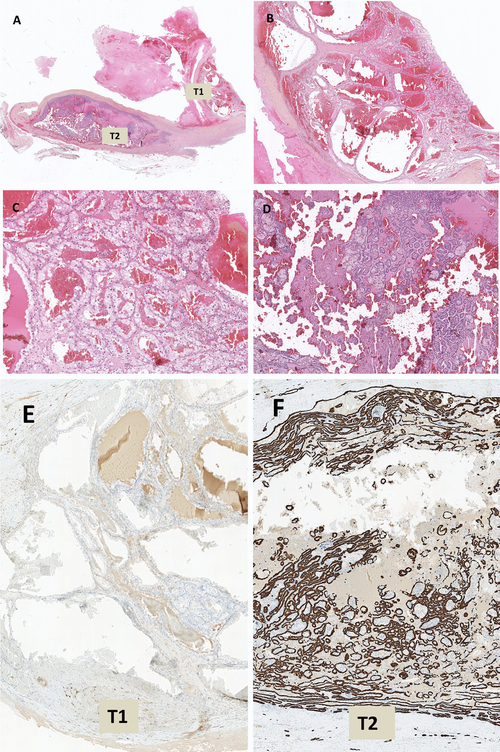
Case presentation: Here, we report three sporadic cases of RCT successfully treated by nephrectomy and confirmed by histological analysis. The first case was of a 64-year-old man diagnosed with RCT composed of a stage 2 nucleolar grade 3 CCRCC and a stage 1a nucleolar grade 2 type 1 PRCC. The second case was of a 68-year-old woman diagnosed with a combined nucleolar grade 2 type 1 PRCC and an angiomyolipoma (non-assessed stage), while the third case was of a 59-year-old woman diagnosed with a combined stage 1a nucleolar grade 3 CCRCC and a stage 1b CDC.
Conclusions: Due to the rarity of RCT, there are no standard guidelines for their management. Hence, the prognosis is considered to be associated with the most aggressive component, possibly the tumour with the highest nucleolar grade and stage. The histogenesis of RCT remains debated, and increase in knowledge regarding this can help enable the development of targeted therapies for advanced or metastatic tumours.
Keywords: Histogenesis; Histology; Kidney; Prognostic; Tumour.
© 2022. The Author(s).
Conflict of interest statement
The authors declare that they have no competing interests.
Figures


Similar articles
-
Type II papillary histology predicts poor outcome in patients with renal cell carcinoma and vena cava thrombus.BJU Int. 2012 Dec;110(11 Pt B):E673-8. doi: 10.1111/j.1464-410X.2012.11498.x. Epub 2012 Sep 14. BJU Int. 2012. PMID: 22973869
-
Evaluation of renal cell carcinoma histological subtype and fuhrman grade using 18F-fluorodeoxyglucose-positron emission tomography/computed tomography.Eur Radiol. 2017 Nov;27(11):4866-4873. doi: 10.1007/s00330-017-4875-z. Epub 2017 May 18. Eur Radiol. 2017. PMID: 28523353
-
Comparison of type I and II papillary renal cell carcinoma (RCC) and clear cell RCC.BJU Int. 2008 Nov;102(10):1381-4. doi: 10.1111/j.1464-410X.2008.07999.x. Epub 2008 Sep 8. BJU Int. 2008. PMID: 18782311
-
Collision kidney tumor with clear cell renal cell carcinoma and papillary type 1 renal cell carcinoma. A case report and review of the literature.Urologia. 2022 May;89(2):304-306. doi: 10.1177/03915603211001673. Epub 2021 Mar 12. Urologia. 2022. PMID: 33709826 Review.
-
Diagnostic and prognostic tissuemarkers in clear cell and papillary renal cell carcinoma.Cancer Biomark. 2010;7(6):261-8. doi: 10.3233/CBM-2010-0195. Cancer Biomark. 2010. PMID: 21694464 Review.
Cited by
-
Case report: A collision tumor of clear cell renal cell carcinoma and clear cell papillary renal cell tumor.Front Oncol. 2024 Feb 28;14:1284194. doi: 10.3389/fonc.2024.1284194. eCollection 2024. Front Oncol. 2024. PMID: 38482203 Free PMC article.
-
Uterine collision tumor. Case report and review of the literature.Rev Colomb Obstet Ginecol. 2023 Sep 30;74(3):225-236. doi: 10.18597/rcog.4011. Rev Colomb Obstet Ginecol. 2023. PMID: 37937912 Free PMC article. English, Spanish.
-
Supratentorial Collision Tumor of Hemangioblastoma and Metastatic Clear Cell Renal Cell Carcinoma in a Patient with von Hippel-Lindau Disease.Case Rep Oncol. 2023 Sep 18;16(1):919-929. doi: 10.1159/000531876. eCollection 2023 Jan-Dec. Case Rep Oncol. 2023. PMID: 37900808 Free PMC article.
References
-
- Cifuentes-C L, Martínez CH, García-Perdomo HA, Cifuentes-C L, Martínez CH, García-Perdomo HA. Synchronous and multiple renal cell carcinoma, clear cell and papillary: an approach to clinically significant genetic abnormalities. Int Braz J Urol. 2020;46:287–293. doi: 10.1590/s1677-5538.ibju.2019.0015. - DOI - PMC - PubMed
Publication types
MeSH terms
LinkOut - more resources
Full Text Sources
Medical
