. 2022 Jul 23;17(1):291.
doi: 10.1186/s13023-022-02441-3.

Autophagy-lysosome pathway alteration in ocular surface manifestations in Fabry disease patients

Affiliations

Autophagy-lysosome pathway alteration in ocular surface manifestations in Fabry disease patients

Marco Marenco et al. Orphanet J Rare Dis. .

Abstract

Background: Fabry disease (FD) is a rare X-linked, lysosomal storage disorder caused by mutations in the alpha-galactosidase gene and characterized by neurological, cutaneous, renal, cardiovascular, cochleo-vestibular and ocular manifestations. The aim of this study is to characterize morphological, functional and autophagy-lysosome pathway alterations of the ocular surface in FD patients.

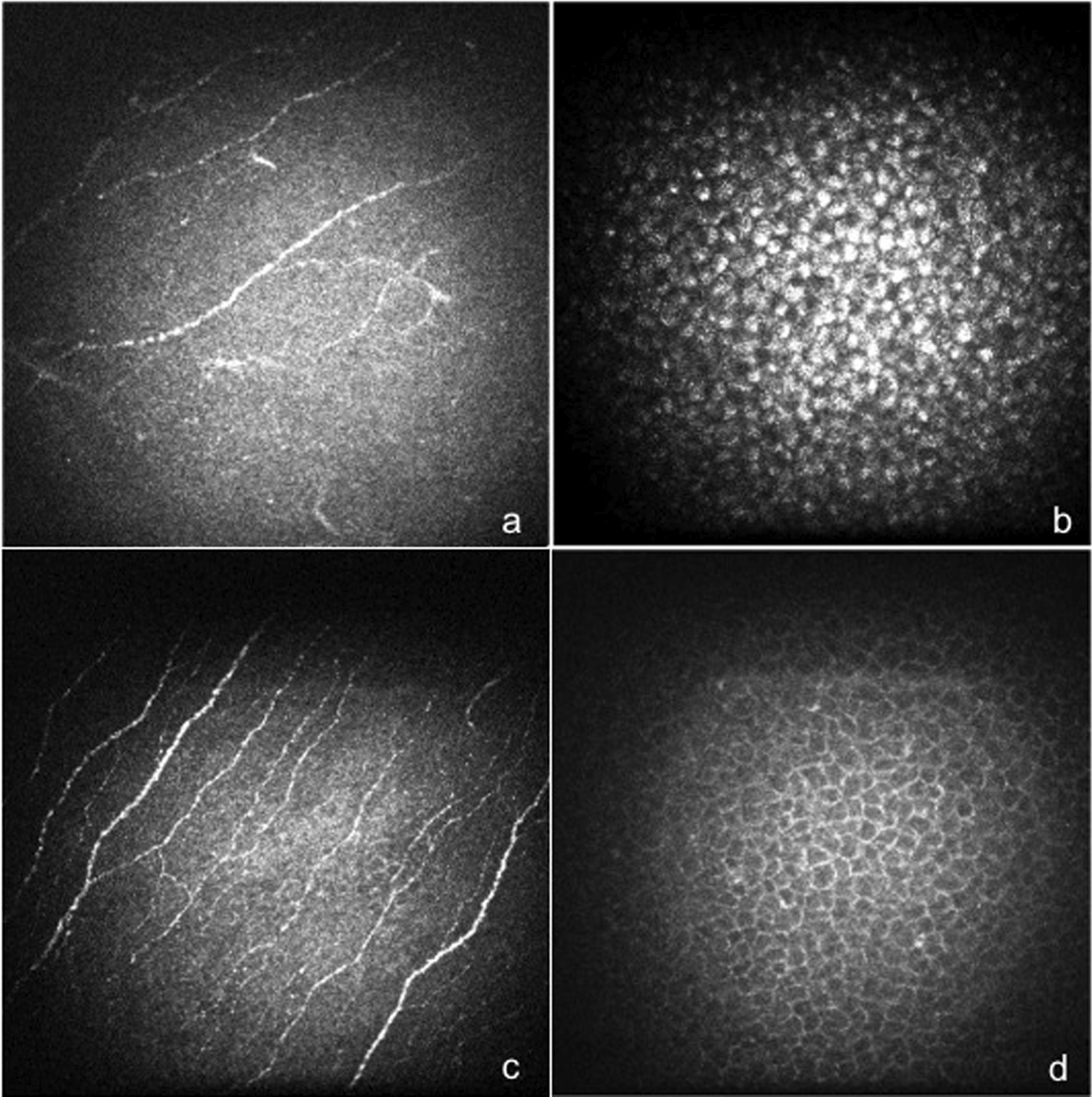
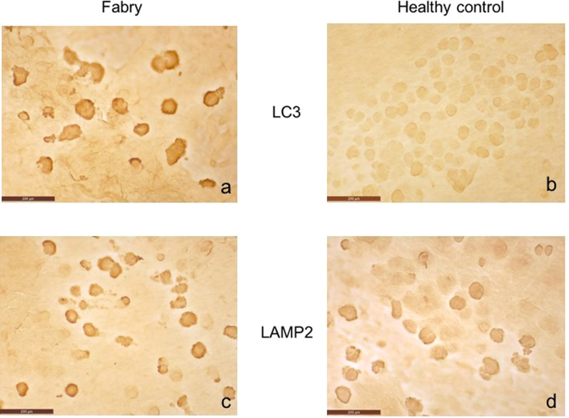
Methods: Eleven subjects with a diagnosis of FD and fifteen healthy control subjects were examined. All patients underwent ocular surface slit lamp examination, corneal aesthesiometry and in vivo confocal laser-scanning microscopy (CCM). Conjunctival impression cytology was performed in six FD patients and six controls, to assess for expression of two markers of the autophagy-lysosome pathway: the microtubule-associated protein light chain 3 (LC3) and lysosome-associated membrane protein 2 (LAMP2).

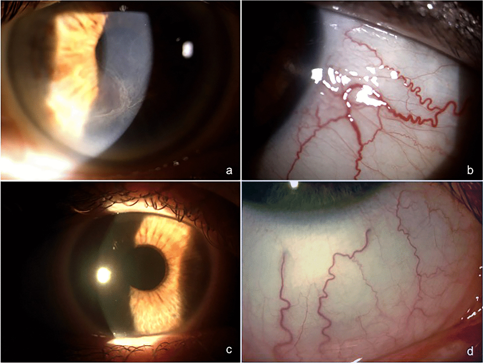
Results: Cornea verticillata and increased conjunctival vessel tortuosity were detected respectively in 67% and 33% of patients with FD. Compared with healthy subjects, patients affected by FD showed a significant reduction in corneal nerve fiber length, density and nerve branching on CCM and a significantly increased expression of LC3 on conjunctival impression cytology (p < 0.001). No changes were observed in the conjunctival expression of LAMP2 between the two groups.

Conclusions: This study shows that FD is associated with ocular surface alterations including corneal and conjunctival morphology, innervation and vascularization changes. Our data demonstrate an increased expression of LC3 protein in patients with FD, suggesting that alteration of the autophagy-lysosome pathway may play a role in the occurrence of ocular manifestations.

Keywords: Autophagy-lysosome pathway; Cornea; Fabry disease; LC3 protein; Lysosomal storage disorder.


Conflict of interest statement

The authors declare that they have no competing interests.

Figures

Fig. 1
Fig. 1
“Cornea verticillata and conjunctival vessels tortuosity” Slit lamp bio-microscopy image of a patient with FD showing the presence of cornea verticillata (a), a yellow–brown, whorl-like opacity caused by the storage of glycolipids within the basal corneal epithelial cells, and the increased tortuosity and aneurysmal dilatations of conjunctival vessels caused by accumulation of glycosphingolipids in the endothelial cells (b). Healthy cornea (c) and conjunctival vessels (d) of a control subject
Fig. 2
Fig. 2
“In vivo corneal confocal microscopy in Fabry Disease” In vivo corneal confocal microscopy examination showed significant decrease of NFL, NFD and NBD of the sub-basal nervous plexus (a) and an increase of reflectivity at the basal cells layer of the corneal epithelium due to intracellular highly reflective irregular material (b) in a patient with FD. Normal sub-basal nervous plexus (c) and normal reflectivity of the basal cells layer of corneal epithelium (d) of a healthy subject
Fig. 3
Fig. 3
“LC3 and LAMP2 expression on conjunctival epithelium” Immunohistochemistry of conjunctival impression cytology samples identified a statistically significant increase in mean expression of LC3 in FD group (a) when compared with healthy control group (b), while no statistically significant difference were observed in mean expression of LAMP2 between FD (c) healthy subjects (d)

Similar articles

Cited by

References

    1. Tuttolomondo A, Pecoraro R, Simonetta I, Miceli S, Pinto A, Licata G. Anderson-Fabry disease: a multiorgan disease. Curr Pharm Des. 2013;19(33):5974–5996. doi: 10.2174/13816128113199990352. - DOI - PubMed
    1. Ortiz A, Germain DP, Desnick RJ, et al. Fabry disease revisited: Management and treatment recommendations for adult patients. Mol Genet Metab. 2018;123(4):416–427. doi: 10.1016/j.ymgme.2018.02.014. - DOI - PubMed
    1. Nguyen TT, Gin T, Nicholls K, Low M, Galanos J, Crawford A. Ophthalmological manifestations of Fabry disease: a survey of patients at the Royal Melbourne Fabry Disease Treatment Centre. Clin Exp Ophthalmol. 2005;33(2):164–168. doi: 10.1111/j.1442-9071.2005.00990.x. - DOI - PubMed
    1. Mastropasqua L, Nubile M, Lanzini M, Carpineto P, Toto L, Ciancaglini M. Corneal and conjunctival manifestations in Fabry disease: in vivo confocal microscopy study. Am J Ophthalmol. 2006;141(4):709–718. doi: 10.1016/j.ajo.2005.11.053. - DOI - PubMed
    1. Sodi A, Ioannidis AS, Mehta A, Davey C, Beck M, Pitz S. Ocular manifestations of Fabry's disease: data from the Fabry outcome survey. Br J Ophthalmol. 2007;91(2):210–214. doi: 10.1136/bjo.2006.100602. - DOI - PMC - PubMed

Substances