. 2022 Jul 23;12(1):12620.
doi: 10.1038/s41598-022-16656-8.

Comparing dormancy in two distantly related tunicates reveals morphological, molecular, and ecological convergences and repeated co-option

Affiliations

Comparing dormancy in two distantly related tunicates reveals morphological, molecular, and ecological convergences and repeated co-option

Laurel S Hiebert et al. Sci Rep. .

Abstract

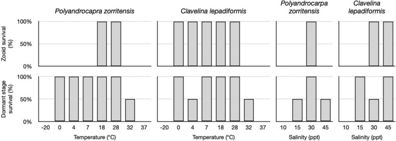
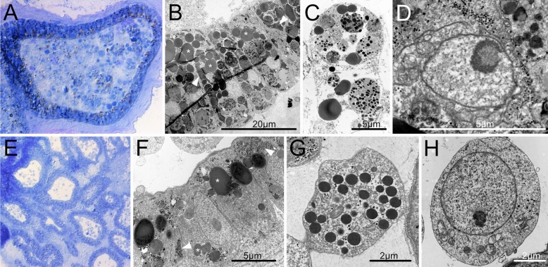
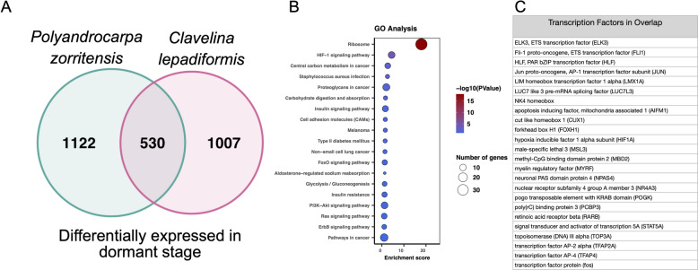
Many asexually-propagating marine invertebrates can survive extreme environmental conditions by developing dormant structures, i.e., morphologically simplified bodies that retain the capacity to completely regenerate a functional adult when conditions return to normal. Here, we examine the environmental, morphological, and molecular characteristics of dormancy in two distantly related clonal tunicate species: Polyandrocarpa zorritensis and Clavelina lepadiformis. In both species, we report that the dormant structures are able to withstand harsher temperature and salinity conditions compared to the adults. The dormant structures are the dominant forms these species employ to survive adverse conditions when the zooids themselves cannot survive. While previous work shows C. lepadiformis dormant stage is present in winters in the Atlantic Ocean and summers in the Mediterranean, this study is the first to show a year-round presence of P. zorritensis dormant forms in NW Italy, even in the late winter when all zooids have disappeared. By finely controlling the entry and exit of dormancy in laboratory-reared individuals, we were able to select and characterize the morphology of dormant structures associated with their transcriptome dynamics. In both species, we identified putative stem and nutritive cells in structures that resemble the earliest stages of asexual propagation. By characterizing gene expression during dormancy and regeneration into the adult body plan (i.e., germination), we observed that genes which control dormancy and environmental sensing in other metazoans, notably HIF-α and insulin signaling genes, are also expressed in tunicate dormancy. Germination-related genes in these two species, such as the retinoic acid pathway, are also found in other unrelated clonal tunicates during asexual development. These results are suggestive of repeated co-option of conserved eco-physiological and regeneration programs for the origin of novel dormancy-germination processes across distantly related animal taxa.


Conflict of interest statement

The authors declare no competing interests.

Figures

Figure 1
Figure 1
Consensus phylogeny of selected tunicate species indicating correlation between asexual cloning and dormancy. Orders are shown on the right. Species with clonal propagation are shown with “C” next to name. Species with capacity for dormancy are shown with a “D” next to name. The species in this study are boxed in a dashed line. Branch lengths and relationships are approximated from Alié et al. (for Stolidobranchia), Delsuc et al. (for Appendicularia, Thaliacea, Aplousobranchia, and Phlebobranchia except for Perophora), Tsagkogeorga et al. (2009) (for position of Perophora).
Figure 2
Figure 2
Asexual life cycles of Polyandrocarpa zorritensis and Clavelina lepadiformis. At the base of mature zooids of both P. zorritensis and C. lepadiformis, extensions of the blood vessels encased in tunic, called stolons, protrude along the substrate. Along the stolons, ramifications of the internal blood vessels develop that we call “pre-buds” here (and are also known as “budding nests” in P. zorritensis and “budding chambers” in C. lepadiformis). The pre-buds swell and the surrounding tunic thickens to form structures called “spherules” for P. zorritensis or may accumulate opaque mass inside the vessel to become what has been called “winter buds” in C. lepadiformis. These dormant forms go on to germinate into a new zooid, but they have the capacity to withstand harsh conditions before germinating. Note that the pre-bud can also initiate the budding process without passing through a dormant state, as indicated by the dotted line in each life cycle.
Figure 3
Figure 3
Photomicrographs of Polyandrocarpa zorritensis asexual stages. (A) Zooid. Siphons (white arrowheads) open for feeding. Stolons (black arrowhead marks one as an example) help attach the zooid to the substrate, and have begun to grow laterally. (B) Stolons attached firmly on a glass slide. Lower stolon starting to ramify (black arrowhead) where new pre-bud will likely develop. (C) Pre-bud still attached to “parental” zooid by stolon (black arrowhead), thus no bud tissues have begun to develop. Ramifications of vasculature seen within the structure through the tunic. (D) Early spherule, as indicated by thickened tunic, which obscures the vascular ramifications inside. This spherule remains attached to the zooid by the stolon, and thus budding has not yet initiated. (E) Detached spherule undergoes germination, as stolonal outgrowths form (black arrowhead). (F) Young zooid that arose from a hatched spherule. Siphons are open (white arrowheads), suggesting that filter feeding has been initiated. Stolon (black arrowhead) begins to grow outward. Scale bars are all 1 mm.
Figure 4
Figure 4
Photomicrographs of Clavelina lepadiformis asexual stages. (A) View of a feeding zooid with siphons (white arrowheads), branchial basket (“bb”), and intestine (“i”). (B) Stolons (marked with black arrowheads) protruding from zooid allow for attachment to substrate and for production of pre-buds. Mesenchymal septum (“ms”) indicated within stolon. (C) Stolons (marked with black arrowheads) undergo branching morphogenesis near the tips and begin to form the pre-buds (also called budding chambers in this species). (D) Early winter bud in a stolon still attached to the zooid. It has accumulated mass of trophocytes (“t”) inside as indicated by opaque white material. Stolon marked with white arrowhead. (E) Detached winter bud with a large mass of trophocytes (“t”). (F) Winter bud undergoing early steps of germination. The germinating bud (b) seen as a small transparent bump above the trophocyte mass. (G) Late germination stage showing young zooid (“z”) that arose from winter bud. Siphons are not yet visible. Remnants of winter bud remain and continue to supply nutrition to zooid until it begins to feed on its own or until it runs out of the supplied nutritive material. Scale bars are all 1 mm.
Figure 5
Figure 5
Field colony dynamics of Polyandrocarpa zorritensis in La Spezia, Italy. (A) Map shows collecting location in La Spezia in the NW of Italy. (B) Zooid collected from the harbor of La Spezia, with spherules attached along stolonal projections. Scale bar is 2 mm. (C) Close-up of dormant spherules. (D) Average surface seawater temperature in La Spezia (NOAA Sea Surface Temperature satellite data from AVHRR Pathfinder SST). Shaded area indicates the period that animals exist in the cryptic dormant form. The rest of the year, both zooids and spherules are found on the ropes in the harbor. (E) Graph showing the change in average spherule size (n = 45) over the year, with the largest spherules found in October, and the smallest spherules in May.
Figure 6
Figure 6
Dormant stages have broader capacity for survival, compared to zooids, in response to environmental stressors. Graphs of percent survival after 48 h exposure to the indicated temperatures and salinity (n = 2 per treatment) for P. zorritensis and C. lepadiformis.Top row shows survival rates of zooids. Bottom row shows survival rates for dormant structures (spherules for Polyandrocarpa zooirtensis and winter buds for Clavelina lepadiformis).
Figure 7
Figure 7
Cell types in dormant stages of Clavelina lepadiformis and Polyandrocarpa zorritensis include nutrient storage cells and putative stem cells. (AD) C. lepadiformis: (A) Methylene blue staining of winter bud histological section shows a thick layer of epithelial cells surrounded by tunic and enclosing abundant mesenchymal cells. (B) TEM image of epithelial cells, many with lipid and glycogen inclusions, labeled with asterisk and white arrowheads, respectively. (C) TEM image of a phagocyte with contents inside mesenchymal space. (D) Putative hemoblast cell inside mesenchymal space, as indicated by large nucleolus and small cell size (about 5 microns). (EH) P. zorritensis. (E) Methylene blue staining of spherule histological section shows numerous ampullary spaces lined with epithelium and filled with sparse hemocytes. (F) TEM close-up image of epithelial cells of ampulla, showing inclusions of lipids (asterisks) and glycogen (white arrowheads). (G) Example of a mesenchymal cell with inclusions. (H) TEM image of putative hemoblast cell within the mesenchymal space, as indicated by large nucleolus, small cell size, and round cell shape.
Figure 8
Figure 8
Germination transition in P. zorritensis. Transversal histological sections stained with hematoxylin and eosin of germinating spherule after 48 h at 24 °C. (A) The central vessel (black arrowhead) is distinguishable from the ampullary ramifications (one example marked with white arrowhead). (B) In a section through the center of the spherule, the vascular epidermis is invaginating (black arrowheads indicate location of the invaginations). White arrowhead indicates ampullary ramification.
Figure 9
Figure 9
Differentially expressed transcripts during life stages in Polyandrocarpa zorritensis and Clavelina lepadiformis. Heatmaps of differentially expressed transcripts across samples: (A) P. zorritensis and (D) C. lepadiformis; each row represents expression value of a single transcript as shown in the color key below plots, violet indicates low expression and yellow indicates high expression; columns show clustered life stage replicates, bars in red = zooid, blue = stolon, yellow = pre-bud, green = dormant, and magenta = germinating stage. Principal Component Analysis showing relationships among and across samples: (B) P. zorritensis and (E) C. lepadiformis. Key below indicates colored symbols for each life stage sample type. Number of differentially expressed transcripts in each pairwise comparison between sample types for each species: (C) P. zorritensis and (F) C. lepadiformis.
Figure 10
Figure 10
Shared up-regulated transcripts and pathways during dormancy in P. zorritensis and C. lepadiformis. (A) Venn diagram showing the number of orthologous upregulated transcripts during dormancy in P. zorritensis and C. lepadiformis. (B) KEGG pathway terms enriched in the overlapping set of 530 transcripts upregulated in both species. (C) Transcription factors upregulated in dormancy in both species.
Figure 11
Figure 11
Shared up-regulated transcripts and pathways during germination in P. zorritensis and C. lepadiformis. (A) Venn diagram showing the number of orthologous upregulated transcripts during germination in P. zorritensis and C. lepadiformis. (B) KEGG pathway terms enriched in the overlapping set of 674 transcripts upregulated in both species. (C) Transcription factors upregulated in germination in both species.

Similar articles

Cited by

References

    1. Hand, S.C. Metabolic dormancy in aquatic invertebrates. In Advances in Comparative and Environmental Physiology, Vol. 8 (ed. Gilles, R.) 1–50. 10.1007/978-3-642-75900-0_1 (1991).
    1. Cáceres CE. Dormancy in Invertebrates. Invertebr. Biol. 1997;116(4):371–383. doi: 10.2307/3226870. - DOI
    1. Wilsterman K, Ballinger MA, Williams CM. A unifying, eco-physiological framework for animal dormancy. Funct. Ecol. 2021;35:11–31. doi: 10.1111/1365-2435.13718. - DOI
    1. Bertolani R, Guidetti R, Altiero T, Nelson DR, Rebecchi L. Dormancy in Freshwater Tardigrades. In: Alekseev V, Pinel-Alloul B, editors. Dormancy in Aquatic Organisms. Theory, Human Use and Modeling. Monographiae Biologicae. Cham: Springer; 2019.
    1. Guidetti R, Altiero T, Rebecchi L. On dormancy strategies in tardigrades. J. Insect Physiol. 2011;57(5):567–576. doi: 10.1016/j.jinsphys.2011.03.003. - DOI - PubMed

Publication types