. 2022 Aug 12;50(14):8168-8192.
doi: 10.1093/nar/gkac596.

Double-stranded RNA drives SARS-CoV-2 nucleocapsid protein to undergo phase separation at specific temperatures

Affiliations

Double-stranded RNA drives SARS-CoV-2 nucleocapsid protein to undergo phase separation at specific temperatures

Christine A Roden et al. Nucleic Acids Res. .

Abstract

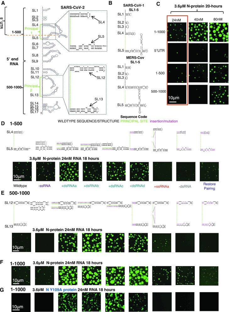
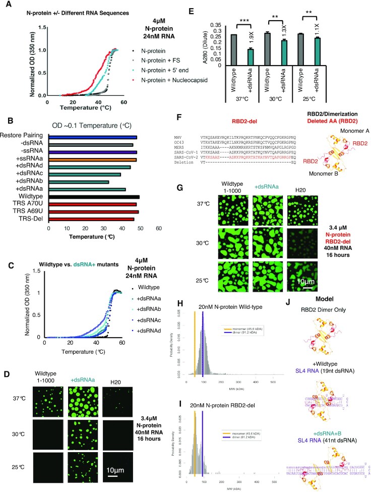
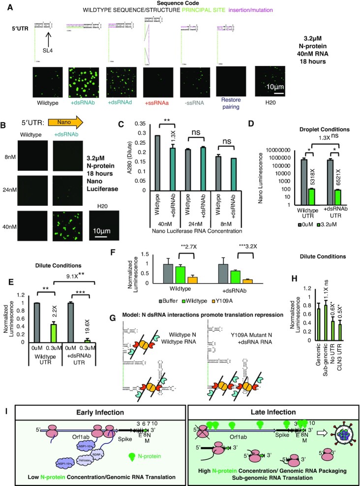
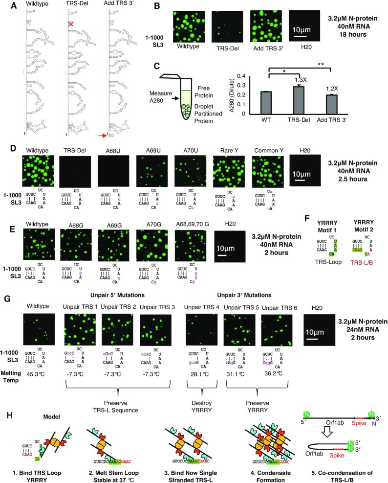
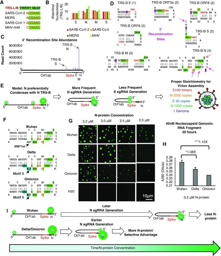
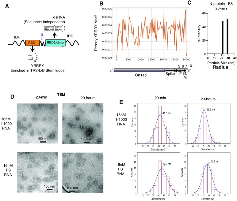
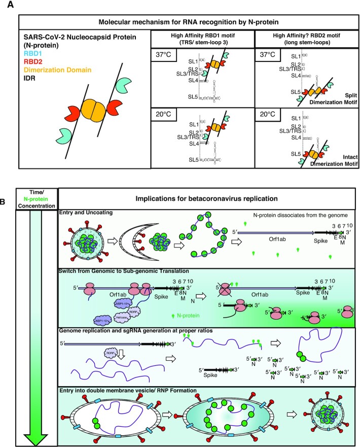
Nucleocapsid protein (N-protein) is required for multiple steps in betacoronaviruses replication. SARS-CoV-2-N-protein condenses with specific viral RNAs at particular temperatures making it a powerful model for deciphering RNA sequence specificity in condensates. We identify two separate and distinct double-stranded, RNA motifs (dsRNA stickers) that promote N-protein condensation. These dsRNA stickers are separately recognized by N-protein's two RNA binding domains (RBDs). RBD1 prefers structured RNA with sequences like the transcription-regulatory sequence (TRS). RBD2 prefers long stretches of dsRNA, independent of sequence. Thus, the two N-protein RBDs interact with distinct dsRNA stickers, and these interactions impart specific droplet physical properties that could support varied viral functions. Specifically, we find that addition of dsRNA lowers the condensation temperature dependent on RBD2 interactions and tunes translational repression. In contrast RBD1 sites are sequences critical for sub-genomic (sg) RNA generation and promote gRNA compression. The density of RBD1 binding motifs in proximity to TRS-L/B sequences is associated with levels of sub-genomic RNA generation. The switch to packaging is likely mediated by RBD1 interactions which generate particles that recapitulate the packaging unit of the virion. Thus, SARS-CoV-2 can achieve biochemical complexity, performing multiple functions in the same cytoplasm, with minimal protein components based on utilizing multiple distinct RNA motifs that control N-protein interactions.


Figures

Figure 1.
Figure 1.
dsRNA-driven condensation is independent of RBD1. (A) SHAPE based structure model of the first 1000 nucleotides of the SARS-CoV-2 genome. Light green letters indicate locations of preferential N-protein crosslinking (principal sites). Brackets indicate the fragments; 5′UTR, 1–500 nt and 500–1000 nt. Stem-loops are numbered (SL). Inset indicates locations of structure manipulations for the rest of the figure. Specifically, mutations altered the region containing SL4 and 5 of principal 1 and/or SL12 and 13 principal 2. (B) Comparison of SL1-5 of SARS-CoV-1 and MERS-CoV (56). (C) Representative images from phase separation experiments with 3.6 μM recombinant N-protein (green) and the corresponding RNA sequence 1–1000, 5′UTR, 1–500 and 500–1000 for 24, 40 and 80 nM RNA. Orange box indicates selected condition for (D–F). (D) Mutation series in the 1–500 context depicting the predicted structure of mutants directed against SL4 and 5 and the intervening single stranded sequence of principal site 1 (light green letters). N-protein is depicted in green. Mutation classes are as follows -ssRNA (purple), +dsRNA (teal), +ssRNA (orange). -dsRNA (grey), Restore pairing (blue). (E) The equivalent mutation series (as in D) for 500–1000 context (principal site 2 in light green letters) depicting the predicted structure of mutants directed against SL12 and 13 or the intervening single stranded sequence of principal site 2. (F) Combination of mutations from (D) and (E) in the context of 1–1000. N-protein is depicted in green. (D–F) Deletion of the single stranded regions of the principal sites do not significantly impact condensation (-ssRNA). Addition of dsRNA (teal) (+dsRNAa-d) enhances N-protein condensation. Addition of single stranded RNA (+ssRNAa orange) coding for HA tag in the center of the principal sites leads to a mild enhancement of condensation. Unpairing principal site adjacent stem-loops (grey -dsRNA) on the 5′ side reduces condensation. Restoration of wildtype RNA structure (blue Restore pairing) but with a different sequence restores condensation to wildtype levels. (G) Only those mutations that lead to an addition of dsRNA (+dsRNAa-d), retain the ability to induce phase separation following Y109A mutation and destruction of N-protein RBD1. For all images, scale bar indicates 10 μm all experiments show representative images from at least three replicates and two independent batches of RNA.
Figure 2.
Figure 2.
RNA sequence and structure encodes N-protein LCST behavior via RBD2. (A) Temperature dependent turbidity tests of N-protein alone (Black), N-protein with Frameshifting region RNA (FS) (Gray), and N-protein with 5′end RNA (1–1000nt) (Blue) and N-protein with Nucleocapsid RNA (Red). Addition of droplet forming RNAs, 5′end 1–1000nt and Nucleocapsid RNA to N-protein, lowers the transition temperature but solubilizing RNA (FS) does not. (B) Transition temperature comparison (repeat of the experiment shown in (A) of wildtype 5′end or 11 mutants in the context of 1–1000nt. Bar length indicates the temperature in°Celsius at which the turbidity of the solution reaches ∼0.1. Only those mutants which alter the dsRNA content (teal + dsRNA), lower the temperature at which OD reaches ∼0.1 indicative of increased solution turbidity. (C) Temperature dependent turbidity tests for N-protein plus wildtype 5′end RNA as well as the four more structured mutants (+dsRNA) which lower the transition temperature. (D) Validation of the turbidity assay using droplet imaging (Figure 2B and C). 3.4μM Wildtype N-protein was mixed with either 40nM of wildtype 5′end 1–1000 RNA, +dsRNAa (RBD1 independent Figure 1G) or water only added control (H20) and incubated at the indicated temperature 37, 30, or 25°C for a period of 20 hours prior to imaging. Consistent with previous results, +dsRNAa increases droplet size relative to wildtype at 37°C (Figure 1F) & induces condensation at lower temperatures. (E) A280 measurement of remaining N-protein in the dilute phase for (D). At all temperatures, +dsRNAa lowers A280 measurements relative to wildtype. Error bars mark standard deviation for the three replicates and * indicate significance students t test (*** P < 0.001, ** P < 0.01, *P < 0.05, ns not significant) with brackets showing comparison for the indicated statistical test. (F) Protein sequence conservation of N-protein RBD2 and structure model of the RBD2 dimerization domain for SARS-CoV-2 (red sequences/red ribbon) indicate the location of the deletion in the primary sequence tested in (G). (G) RBD2/Dimerization domain is required for proper N-protein LCST behavior at indicated temperature range. 3.4 μM of N-protein RBD-del (green) was mixed with 25nM of either wildtype 1–1000, +dsRNAa, or water only control and incubated at the indicated temperatures for 16 h. Droplet formation was observed in all conditions although RNA dependence was more evident at lower protein concentrations (Supplementary Figure S4G-H). (H, I) Mass photometry histograms showing the molecular weight (MW) distribution of detected particles for wild-type N-protein (H) or RBD-del N-protein (I). (H) Wildtype N-protein is a stable dimer in solution (250 mM NaCl pH 7.5 20 mM phosphate buffer 20 nM N-protein) but RBD2-del is mostly a monomer (I). (J) Model of N-protein RBD2/Dimerization domain interactions with dsRNA. Binding of the two RBD2s of the two monomers of N-protein to dsRNA facilitates dimerization dissociation with temperature facilitating dissociation for shorter stem-loops. For all images scale bar indicates 10 μm all experiments show representative images from at least 3 replicates.
Figure 3.
Figure 3.
Features which promote N-protein RBD2/dsRNA interactions repress translation. (A) Only +dsRNA (teal) mutants enhance condensation in the context of the 5′UTR fragment. All other mutations do not significantly alter condensation. 3.2 uM N-protein (green) 40nM RNA 18 hours of incubation. H2O is water only control. (B) Design of luciferase fusion to the 5′UTR of SARS-CoV-2 constructs. Only +dsRNAb UTR: Nano Luciferase undergoes condensation at the highest tested RNA concentration (40 nM/3.2 μM N-protein (green)) (C) A280 absorbance of the remaining protein in the dilute phase from (B). Error bars mark standard deviation for the three replicates and * indicate significance Student's t test (**P <0.01, ns not significant) with brackets showing comparison for the indicated statistical test. (D) In vitro translation assay results for nano luciferase wildtype or more structured fusion constructs. 20-min incubation with 3.2 μM N-protein prior to in vitro translation is sufficient to completely repress translation of nano luciferase. Error bars mark standard deviation for the three replicates and * indicate significance Student's t test (** P< 0.01, ns not significant) with brackets showing comparison for the indicated statistical test. (E) Presence of N-protein condensation promoting RNA structures is associated with reduced translation in dilute phase conditions. Normalized luminescence for nano luciferase constructs (no protein control fluorescent signal is set to 1). Nano luciferase +dsRNAb has a much greater reduction in normalized signal as compared to wildtype. (F) Y109A mutant protein which is deficient in RBD1 activity is better able to repress translation than wildtype protein in dilute conditions for both wildtype 5′UTR:Nano and +dsRNAb:Nano. (G) Model for N-protein mediated repression of translation via RNA affinity in the dilute phase (limiting protein conditions). Condensation at the structured SL5 inhibits translation and this preferentially occurs in the absence of RBD1 activity or following mutation which enhances RBD2 interactions with SL5 (addition of dsRNA). (H) TRS contain sequences (wildtype genomic UTR and subgenomic UTRs of the nucleocapsid gene are less repressed than sequences which do not contain a TRS such as Nano luciferase without a UTR and the 5′ UTR CLN3 from Ashbya gossypii. (I) Model for N-protein mediated repression of translation via RNA affinity in infection. High affinity sites in the 5′UTR are preferentially occupied by N-protein in early infection to shut down orf1ab translation and switch to packaging. Late-stage infection translation occurs preferentially in sub-genomic RNA.
Figure 4.
Figure 4.
TRS sequence/structure motif promotes N-protein condensation. (A) Cartoon of mutations depicted in (B) and (C). TRS-Del deletes SL3 TRS whereas add TRS 3′ adds an additional TRS element to the 3′ end of the RNA. (B) 3.2 μM N-protein (green) and 40 nM RNA following 18 hours of incubation for wildtype 1–1000, a mutation which deletes the entire TRS-stem-loop (TRS-del), or a mutation which appends an additional TRS to the 3′ end (Add TRS 3′). (C) Add TRS 3′ has lower A280 measurements then wildtype indicative of less protein in solution and more condensation whereas TRS-del is the opposite. Error bars mark standard deviation for the three replicates and * indicate significance Student's t test (***P < 0.001, **P < 0.01, ns not significant) with brackets showing comparison for the indicated statistical test. (D) 3.2 μM N-protein (green) and 40 nM RNA following 2.5 h of incubation for wildtype 1–1000, a mutation which deletes the entire TRS-stem-loop (TRS-del), A68U mutation, A69U mutation, A70U mutation and mutations which alter the sequence of the A flanking pyrimidines (Y’s) (C’s and U’s) to the most rare and common YYAAAY in the SARS-CoV-2 genome. Deletion of TRS-loop or alteration of the AAA of the loop but not the Y’s leads to a reduction in condensation. (E) 3.2 μM N-protein (green) and 40 nM RNA following 2 h of incubation for wildtype 1–1000, A68G mutation, A69G mutation, A70G mutation and mutations which alter the sequence of all three A’s (A68,69,70 G). Do not significantly alter condensation. Suggesting the motif recognized by N-protein is 3 purines flanked by pyrimidine. (F) 2 possible YRRRY motifs in SL3, the first is the Loop UAAAC and the second is contained in the TRS-L/B sequence ACGAAC. (G) 3.2 μM N-protein (green) and 24 nM RNA following 2 h of incubation for wildtype 1–1000, or mutations which unpair SL3 from the 5′ or 3′ sides. Unpairing SL3 generally enhances condensation (unpair TRS 1, 2, 3, 5 and 6) unless the YRRRY motif is destroyed (Unpair TRS 4). Melting temperature of mutant and wildtype stem loops was calculated using DINAMelt. (H) Model for N-protein-SL3 interactions which led to condensation. N-protein RBD1 recognizes the stem loop sequence of SL3, unwinding the stem loop which is stable at 37°C. The now single stranded SL3 is permissive for interaction with the second motif contained in the TRS-L sequence. Location of the two motifs in proximity facilitates condensate formation. Condensate formation at TRS-L/B sequences may promote the genome circularization interaction which is required for sub-genomic RNA generation.
Figure 5.
Figure 5.
Local YRRRY motif density may control sgmRNA generation ratio. (A) Sequence of TRS-L/B for model betacoronaviruses (red text) encompasses the YRRRY motif. (B) Similar number of identical nucleotides between TRS-L/B in model coronavirus for structural protein TRS-Bs. (C) Variation in abundance of sgmRNA reads in SARS-CoV-2 infected cells. Adapted from (48). (D) Sequence and structure of example TRS-B in SARS-CoV-2 genome. Adapted from (49). Red text is the TRS-B sequence ACGAAC. Green highlight is the adjacent YRRRY motifs contained in the stem loop. Of note Orf7ab TRS-B is not included as it is not structured. Magenta highlighted nucleotide is the location of the primary recombination site between TRS-L/and B. Pink arrows refer to less abundant but detectable recombination site. Indicating that nucleocapsid recombination site selection is more degenerate than that of other sgmRNA. Bracketed numbers refer to the number of YRRRY motifs. (E) Model of preferential sgmRNA generation dictated by local YRRRY motif abundance. N TRS-B contains more YRRRY motifs (4) than S TRS-B resulting in a higher propensity to form an N condensate at an N gene rather than S and preferential generation of N sgmRNA rather than S. This sgmRNA ratio may allow for proper protein abundance in the assembled virion where there is more absolute number of Nucleocapsid protein molecules than Spike. (F) Two independent mutations in Delta (UAAAAU → UAAAU) and Omicron (UAAAAU → UuAAAU) which create a fifth binding site in the in the TRS-B of N. Start codon of Nucleocapsid and overall structure of the TRS-B containing hairpin is not predicted to be altered. (G) Fragments of Nucleocapsid RNA containing the TRS-B of N have altered phase behavior following Delta or Omicron mutations in position 20871nt. Morphology is not significantly altered but sequence that contain a fifth binding site exit the phase diagram earlier indicative of higher affinity for RNA. (H) A280 absorbance in the dilute phase of the 3.2uM condition shown in figure G shows that sequences which contain five binding sites recruit more protein commensurate with altered phase behavior. Omicron recruits more protein to droplets than Delta and exits the phase diagram sooner. (I) Model for how Delta and Omicron mutations in position 20871 may provide a selective advantage for the virus by preferentially generating N sgmRNA earlier and more often at the expense of other sgmRNAs.
Figure 6.
Figure 6.
RNA sequence/ structure encodes N-protein genome interactions. (A) Model of RNA sequence preferences of SARS-CoV-2 N-protein RNA binding domains 1 (orange) and 2 (blue). RBD1 (teal box) binds TRS-like (YRRRY) sequences in a structure dependent manner. RBD2/dimerization domain (blue box) binds dsRNA in a sequence independent manner. (B) Density of YRRRY motif (orange) across the SARS-CoV-2 genome. (C) Dynamic light scattering of 16 nM FS RNA and 4 μM protein. Following 20-minutes of incubation results in particles of ∼21.9 or 29 nm radius (∼43.7–58 nm in diameter). (D) Representative TEM images of small clusters which form from a mixture of 4μM N-protein and either 16nM FS or 16 nM 1–1000 5′end when incubated for 20 min or 20 h at room temperature. Scale bar is 100 nM. (E) Quantification of small clusters as depicted in panel D. for 1–1000 5′end, or FS. Clusters shrink by ∼15% following 20 h of incubation.
Figure 7.
Figure 7.
Model: Molecular mechanism and implications for betacoronavirus replication. (A) N-protein's two RNA binding domains prefer two dsRNA dependent RNA stickers. RBD1 (teal) binds TRS stem–loop (and similar sequences) with high affinity. RBD2 (dark orange) binds long stem–loops in a temperature dependent manner. (B) Time dependent accumulation of N-protein specifies N-protein's multiple roles in betacoronavirus by tuned patterned affinity for the two dsRNA dependent RNA stickers. High affinity sites (genome ends) are occupied preferentially early in infection when N-protein concentrations are low. Low affinity sites (genome center) are occupied late in infection when N-protein concentrations are high. Occupation of high affinity sites at genome ends promotes the switch from genome translation to circularization and ultimately packaging.

Update of

Similar articles

Cited by

References

    1. Boeynaems S., Alberti S., Fawzi N.L., Mittag T., Polymenidou M., Rousseau F., Schymkowitz J., Shorter J., Wolozin B., Van Den Bosch L.et al. .. Protein phase separation: a new phase in cell biology. Trends Cell Biol. 2018; 28:420–435. - PMC - PubMed
    1. Hyman A.A., Weber C.A., Jülicher F.. Liquid-liquid phase separation in biology. Annu. Rev. Cell Dev. Biol. 2014; 30:39–58. - PubMed
    1. Alberti S., Gladfelter A., Mittag T.. Considerations and challenges in studying liquid-liquid phase separation and biomolecular condensates. Cell. 2019; 176:419–434. - PMC - PubMed
    1. Kar M., Dar F., Welsh T.J., Vogel L., Kühnemuth R., Majumdar A., Krainer G., Franzmann T.M., Alberti S., Seidel C.A.M.et al. .. Phase separating RNA binding proteins form heterogeneous distributions of clusters in subsaturated solutions. Proc. Natl. Acad. Sci. U.S.A. 2022; 119:e2202222119. - PMC - PubMed
    1. Mittag T., Pappu R.V.. A conceptual framework for understanding phase separation and addressing open questions and challenges. Mol. Cell. 2022; 82:2201–2214. - PMC - PubMed

Publication types

MeSH terms