. 2022 Aug 12;50(14):8240-8261.
doi: 10.1093/nar/gkac631.

mRNA- and factor-driven dynamic variability controls eIF4F-cap recognition for translation initiation

Affiliations

mRNA- and factor-driven dynamic variability controls eIF4F-cap recognition for translation initiation

Burak Çetin et al. Nucleic Acids Res. .

Abstract

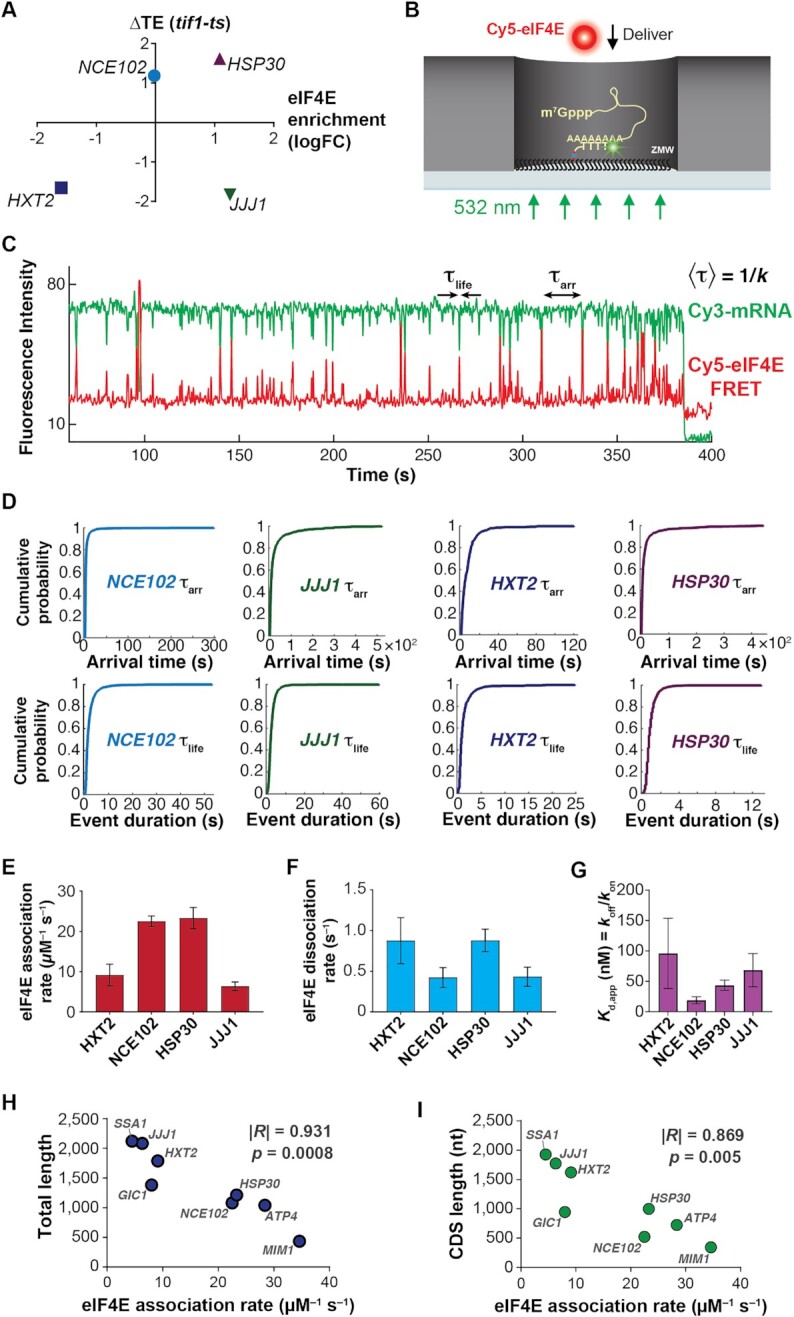
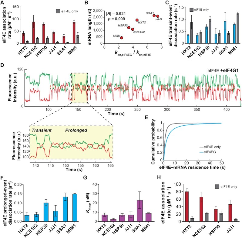
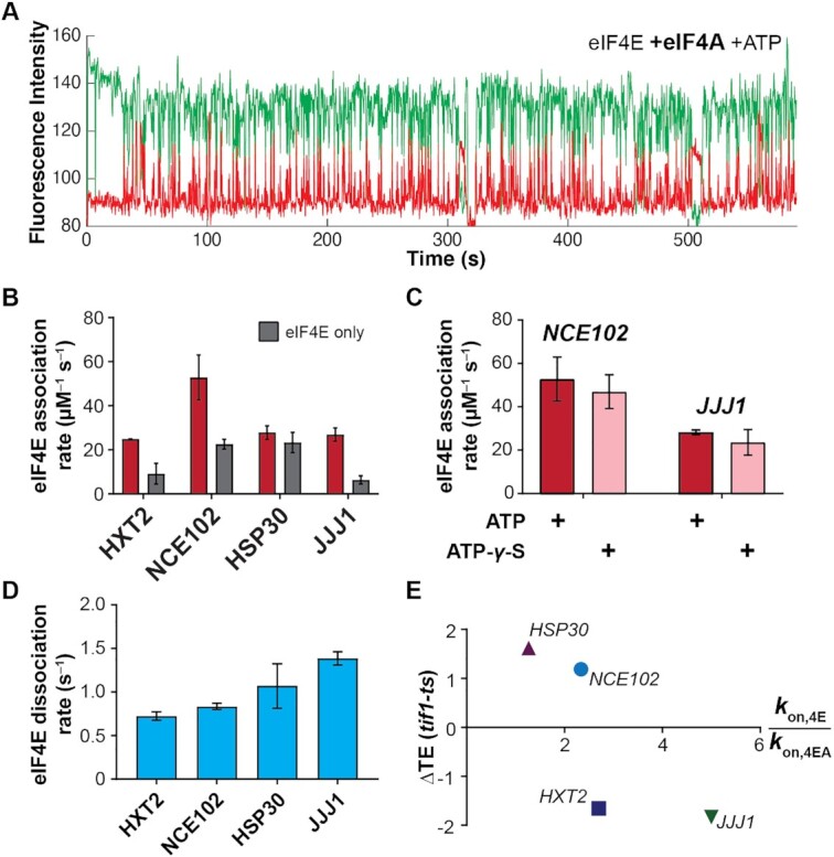
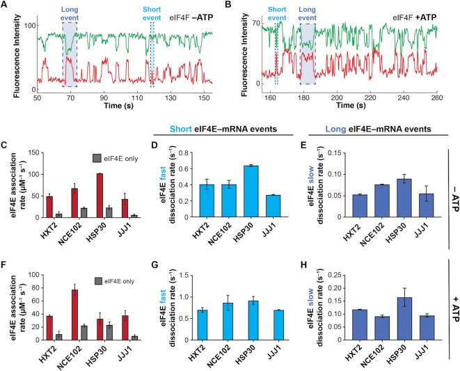
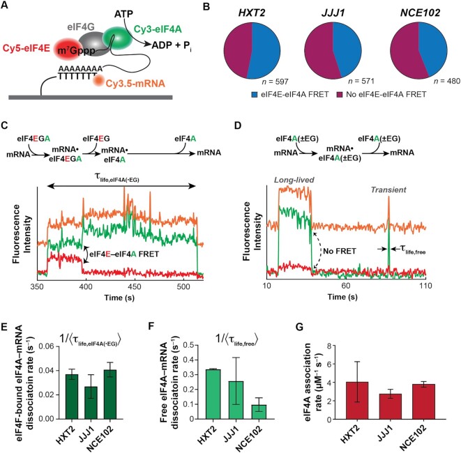
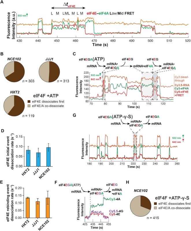
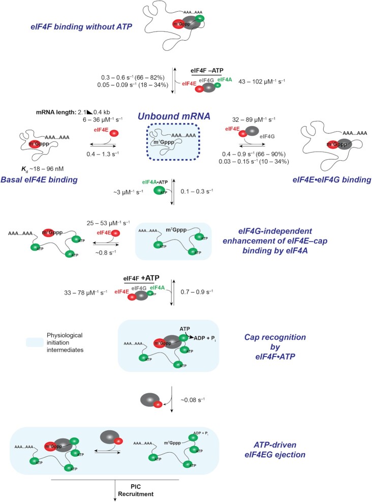
mRNA 5' cap recognition by eIF4F is a key element of eukaryotic translational control. Kinetic differences in eIF4F-mRNA interactions have long been proposed to mediate translation-efficiency differences between mRNAs, and recent transcriptome-wide studies have revealed significant heterogeneity in eIF4F engagement with differentially-translated mRNAs. However, detailed kinetic information exists only for eIF4F interactions with short model RNAs. We developed and applied single-molecule fluorescence approaches to directly observe real-time Saccharomyces cerevisiae eIF4F subunit interactions with full-length polyadenylated mRNAs. We found that eIF4E-mRNA association rates linearly anticorrelate with mRNA length. eIF4G-mRNA interaction accelerates eIF4E-mRNA association in proportion to mRNA length, as does an eIF4F-independent activity of eIF4A, though cap-proximal secondary structure still plays an important role in defining the final association rates. eIF4F-mRNA interactions remained dominated by effects of eIF4G, but were modulated to different extents for different mRNAs by the presence of eIF4A and ATP. We also found that eIF4A-catalyzed ATP hydrolysis ejects eIF4E, and likely eIF4E•eIF4G from the mRNA after initial eIF4F•mRNA complex formation, suggesting a mechanism to prepare the mRNA 5' end for ribosome recruitment. Our results support a role for mRNA-specific, factor-driven eIF4F association rates in kinetically controlling translation.


Figures

Figure 1.
Figure 1.
eIF4E interaction dynamics with full-length mRNAs depend on mRNA identity and length. (A) Selection of mRNAs with varying in-vivo enrichment in eIF4E•eIF4G and translation dependence on eIF4A, as measured by Costello et al. (2015), and Sen et al. (2015). (B) Schematic of single-molecule FRET experiment to detect binding of fluorescently-labeled eIF4E to surface-immobilized, fluorescently-labeled mRNA. (C) Sample smFRET trajectory showing eIF4E–mRNA interaction in the absence of other eIF4F components. (D) Representative cumulative distribution functions for eIF4E association with (top) and dissociation from (bottom) full-length mRNAs. (E) eIF4E–mRNA association rates quantified from exponential fitting of arrival-time and lifetime cumulative distribution functions. Error bars reflect the standard errors of the mean for three replicates of an experiment where the eIF4E–mRNA binding rate is measured across at least 100 mRNA molecules. (F) eIF4E–mRNA dissociation rates from the experiments in E. (G) eIF4E–mRNA equilibrium dissociation constants computed from the rates shown in E and F. (H) Dependence of eIF4E–mRNA association rate on mRNA length. The rates for JJJ1, HXT2, HSP30, NCE102, and MIM1 were measured in the present study. The rates for GIC1, SSA1 and ATP4, previously published in Çetin et al. (53), were added to include data points for correlation over a sufficient length range. (I) Dependence of eIF4E–mRNA association rate on coding-sequence length.
Figure 2.
Figure 2.
eIF4G1 accelerates eIF4E–mRNA binding in an mRNA-dependent manner and allows the interaction to persist on the translation initiation timescale. (A) eIF4E-mRNA association rates in the absence (grey) and presence (red) of full-length yeast eIF4G1. (B) Fold-stimulation of eIF4E–mRNA association rate by eIF4G1, as a function of mRNA length. (C) Kinetics of eIF4E–mRNA dissociation for transient binding events in the presence (blue) and absence (grey) of eIF4G1. (D) Representative single-molecule fluorescence trace for eIF4E–mRNA binding in the presence of eIF4G1. The inset shows representative transient and prolonged events on an expanded time axis. (E) Cumulative probability distributions of eIF4E–NCE102 mRNA event durations in the absence (grey) and presence (blue) of eIF4G1, showing appearance of slowly-dissociating events when eIF4G1 is present. (F) eIF4E–mRNA dissociation rates for long-lived binding events in the presence of eIF4G1. (G) Apparent equilibrium dissociation constants for the eIF4E–mRNA interaction in the presence of eIF4G1. (H) eIF4E–mRNA association rates in the presence (red) and absence (grey) of eIF4G11–452.
Figure 3.
Figure 3.
Free eIF4A with bound ATP stimulates eIF4E–mRNA association independently of eIF4G. (A) Representative single-molecule trace showing eIF4E–mRNA interaction in the presence of 2 μM eIF4A and 2.5 mM ATP. (B) eIF4E-mRNA association rates in the presence (red) and absence (grey) of eIF4A and ATP, compared with eIF4E-only rates. (C) eIF4E–mRNA association rates in the presence of eIF4A and ATP or ATP-γ-S, for the NCE102 and JJJ1 mRNAs. (D) eIF4E–mRNA dissociation rates in the presence of free eIF4A and ATP. (E) Relationship between translation-efficiency dependence on eIF4A and the fold-increase in eIF4E–mRNA association rate induced by free eIF4A and ATP.
Figure 4.
Figure 4.
The eIF4F complex discriminates eIF4E–mRNA interaction dynamics in an ATP-dependent manner. (A) Representative single-molecule fluorescence trace for eIF4E–mRNA interaction in the eIF4F complex without added ATP, on NCE102. (B) Representative trace for eIF4E–mRNA binding in the eIF4F complex with ATP, on NCE102. (C) eIF4E–mRNA association rates for the eIF4F complex without ATP (red), compared with the rates in the presence of eIF4E only (grey). (D) Dissociation rates of transient eIF4E–mRNA interactions in the eIF4F complex without ATP. (E) Dissociation rates of long-lived eIF4E–mRNA interactions in the eIF4F complex without ATP. (F) eIF4E–mRNA association rates in the eIF4F complex with ATP (red), compared with the rates in the presence of eIF4E only. (G) Dissociation rates of transient eIF4E–mRNA interactions in the eIF4F complex with ATP. (H) Dissociation rates of long-lived eIF4E–mRNA interactions in the eIF4F complex with ATP.
Figure 5.
Figure 5.
Three-color smFRET to probe eIF4F- and eIF4A-mRNA interaction dynamics. (A) Schematic of the three-color smFRET experiment with two donors (on eIF4A and mRNA) and one acceptor (eIF4E). A FRET signal between Cy5-labeled eIF4E and the Cy3.5-labeled mRNA is tracked at the same time as a FRET signal between Cy3-eIF4A and Cy5-eIF4E. (B) Relative incidence of eIF4A-mRNA binding occurring with and without FRET to eIF4E. n is the number of molecules analyzed to enumerate the event types on each mRNA. (C) Reaction pathway and representative smFRET trace showing concomitant mRNA binding of eIF4E and eIF4A with eIF4E–eIF4A FRET, consistent with eIF4F–mRNA binding, on JJJ1. The eIF4A–mRNA lifetime measured in panel E is indicated. (D) Reaction pathway and representative single-molecule fluorescence trace for eIF4A–mRNA binding without eIF4E–eIF4A FRET on JJJ1. These events result both from ‘free’ eIF4A–mRNA interaction (‘eIF4A(–EG)’), and eIF4F–mRNA interaction where eIF4E is unlabeled (‘eIF4A(+EG)’. The Cy3 and Cy5 signals were manually corrected by linear subtraction to equalize their background values, for clarity of presentation. (E) eIF4A–mRNA dissociation rates following eIF4A–mRNA binding with eIF4E, i.e. with observable eIF4E–eIF4A FRET as shown in panel C. The Cy3 and Cy5 signals were manually corrected by linear subtraction to equalize their background values, for clarity of presentation. (F) eIF4A–mRNA dissociation rates following eIF4A–mRNA binding without FRET to eIF4E. (G) eIF4A–mRNA association rates across all binding event types.
Figure 6.
Figure 6.
Dynamic coordination within eIF4F after cap recognition. (A) Representative smFRET trajectory showing event with ejection of eIF4E prior to eIF4A, and fluctuations in the eIF4F•mRNA conformation on JJJ1. (B) Relative incidence of initial eIF4E dissociation vs. eIF4E/eIF4A co-dissociation from eIF4F•mRNA complexes. n is the number of molecules analyzed to enumerate the event types on each mRNA. (C) Reaction pathway and annotated representative single-molecule fluorescence trajectory for eIF4F•mRNA complex formation and dynamics, observed by dual red/green illumination which directly reports on the presence of both Cy3-eIF4A and Cy5-eIF4E. (D) Rates for the initial eIF4E–mRNA dissociation event after eIF4F–mRNA complex formation. (E) eIF4E–mRNA dissociation rates for events where eIF4E rebinds mRNA following initial dissociation from eIF4F•mRNA. (F) Reaction pathway and representative single-molecule fluorescence trajectory for a four-color experiment where eIF4G is non-specifically labeled with Cy5.5, allowing its simultaneous detection with Cy3-eIF4A and Cy5-eIF4E. eIF4E and eIF4G fluorescence co-depart the mRNA. (G) Reaction pathway and annotated representative single-molecule fluorescence trajectory for eIF4F•mRNA complex dynamics with ATP-γ-S. The Cy3 and Cy5 signals were manually corrected by linear subtraction to equalize their background values, for clarity of presentation. (H) Relative incidence of eIF4E or eIF4A dissociation, or co-dissociation from the eIF4F•mRNA complex in the presence of ATP-γ-S.
Figure 7.
Figure 7.
Mechanistic model and summary of kinetic data for eIF4F–mRNA interaction dynamics.

Similar articles

Cited by

References

    1. Hershey J.W.B., Sonenberg N., Mathews M.B.. Principles of translational control: an overview. Cold Spring Harb. Perspect. Biol. 2012; 4:a011528. - PMC - PubMed
    1. Furuichi Y., Shatkin A.J.. Viral and cellular mRNA capping: past and prospects. Adv. Virus Res. 2000; 55:135–184. - PMC - PubMed
    1. Fraser C.S. Quantitative studies of mRNA recruitment to the eukaryotic ribosome. Biochimie. 2015; 114:58–71. - PMC - PubMed
    1. Marcotrigiano J., Gingras A.C., Sonenberg N., Burley S.K.. Cocrystal structure of the messenger RNA 5’ cap-binding protein (eIF4E) bound to 7-methyl-GDP. Cell. 1997; 89:951–961. - PubMed
    1. Siddiqui N., Sonenberg N.. Signalling to eIF4E in cancer. Biochem. Soc. Trans. 2015; 43:763–772. - PMC - PubMed

Publication types

MeSH terms