. 2022 Aug;44(8):1147-1156.
doi: 10.1007/s00276-022-02988-7. Epub 2022 Jul 24.

Phenotypical variability of the sigmoid sinus in translabyrinthine and retrosigmoid surgeries

Affiliations

Phenotypical variability of the sigmoid sinus in translabyrinthine and retrosigmoid surgeries

Maryna Al-Fauri et al. Surg Radiol Anat. 2022 Aug.

Abstract

Introduction: We hypothesized that the cranial phenotype influences the shape of the posterior cranial fossa and the relative position of the sigmoid sinus.

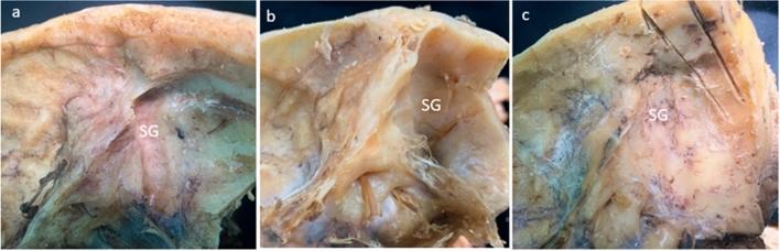
Materials and methods: The topography of the sigmoid sinus was studied on 26 magnetic resonance venograms and 35 embalmed cadavers by morphometric analysis, dissection, and photo modeling techniques.

Results: The data show that the transverse diameter of the posterior cranial fossa correlates positively with the laterolateral diameter of the skull. The majority of cases with the low-anterior position of the sigmoid sinus were recorded in the brachycephalic group (82%), while the high-posterior localization of the sigmoid sinus was typical for the dolichocephalic patients (63%). The results of the ANOVA test confirm the significance of differences.

Conclusions: The shape of the skull reflects the morphology of the posterior cranial fossa and influences the topographic characteristics of the sigmoid sinus that must be considered in the selection of surgical approach to the inner ear and pontocerebellar angle.

Keywords: Cranial phenotypes; Retrosigmoid approach; Sigmoid sinus; Translabyrinthine approach; Variability.


Conflict of interest statement

The authors declare no competing interests.

Not applicable.

Figures

Fig. 1
Fig. 1
Magnetic resonance venograms of the head demonstrate the measurements taken: a shortest distance between the posterior margin of the external acoustic meatus and the transverse-sigmoid sinus junction (DEj); b diameter of the transverse-sigmoid sinus junction and the diameter of the sigmoid sinus (dST and dSS, respectively); c distance between the internal occipital protuberance and the posterior margin of the foramen magnum (DP); the distance between the dorsum sellae and the anterior margin of the foramen magnum (DA); d latero-lateral (LL) and the anteroposterior (AP) diameters of the skull. The RadiAnt Pro (2021) software was employed to analyze the imaging data
Fig. 2
Fig. 2
Working window of the photo modeler platform with a skull base model is shown in this picture (Photo Modeler, 2021). All the reference points were set up in a plane parallel to the superior border of the pyramid of the temporal bone. After measuring the diameter of the sigmoid groove (dSG), the distances from its anterior (Dp1) and posterior (Dp2) margins to the porus acusticus (PA) were taken. Then, the Dp2 line was extended to the outer surface of the temporal bone (red arrow), and the distances between the posterior wall of the external acoustic meatus (EA), the tip of the mastoid process (MP), and the projected posterior margin of the sigmoid groove (Sp) were measured accordingly
Fig. 3
Fig. 3
Sagittal magnetic resonance venograms of the head demonstrate the topographic relationship of the transverse-sigmoid sinus junction and the external acoustic meatus by shapes of the skull. The inferior position of the sinuses (Type III) picked in brachycephalic patients (a), the superior position (Type I) dominated in the dolichocephalic patients (c), while patients with the normal shape of the head (b) showed the prevalence of intervening localization of the transverse-sigmoid sinus junction. For precise numerical data see Table 5
Fig. 4
Fig. 4
Cadaveric specimens of the skull base demonstrate the range of variability of the sigmoid groove (SG). Note the low localization and prominent indentation of the sigmoid groove into the pyramid of the temporal bone in specimen “a”, which belongs to a 68-year-old female with the cranial index (CI) equal to 83.6 percent. As an opposite, the shallow running up groove is barely visible on specimen “c” (a 56-year-old male, with CI = 72.1 percent). The specimen “b” belongs to a 63-year-old female with CI = 75.6 percent, and it shows the intermediate topography of the sigmoid groove
Fig. 5
Fig. 5
Summative scheme depicting the phenotypical characteristics of the topography of the sigmoid sinus (SS) and transverse-sigmoid sinus junction (TSj) projected on the mastoid process (MP) of the temporal bone. The transverse-sigmoid sinus junction is located lower and closer to the external acoustic meatus (EA) in the brachycephalic specimens (a) and is significantly higher and further from the EA in the dolichocephalic (c) ones. The mesocephalic heads (b) featured the intermediate position of the sinuses. The coordinates were collected and summarized using the photo modeling technique

References

    1. Al-Fauri M, Kelly P, Lee D, Hadidy A. Phenotypical variability of the internal acoustic canal in the middle cranial fossa surgery. J Neurol Surg B Skull Base. 2022 doi: 10.1055/a-1786-9026. - DOI - PMC - PubMed
    1. Boemo RL, Navarrete ML, Lareo S, Pumarola F, Chamizo J, Perelló E. Anatomical relationship between the position of the sigmoid sinus, tympanic membrane and digastric ridge with the mastoid segment of the facial nerve. Eur Arch Oto-Rhino-L. 2008;265:389–392. doi: 10.1007/s00405-008-0603-2. - DOI - PubMed
    1. Burulday V, Bayar MN, Kömürcü Erkmen SP, Akgül MH, Özdemir A. Important landmarks and distances for posterior fossa surgery measured by temporal MDCT. Neurosurg Rev. 2021;44(3):1533–1541. doi: 10.1007/s10143-020-01342-y. - DOI - PubMed
    1. Galal A, Caruso A, Laud L, Eldin OG, Baki F, Sanna M. Effect of sigmoid sinus position on the difficulty and approaches to cochlear implantation surgery. J Int Adv Otol. 2021;17(1):23–29. doi: 10.5152/iao.2020.8927. - DOI - PMC - PubMed
    1. Li D, Wang H, Fan Z, Fan Z. Complications in retrosigmoid cranial nerve surgery. Acta Otolaryngol. 2010;130(2):247–252. doi: 10.3109/00016480903092340. - DOI - PubMed

MeSH terms

LinkOut - more resources