Case Reports
. 2022;13(Suppl 3):307-310.
doi: 10.22088/cjim.13.0.307.

Cone Beam Computed Tomography (CBCT) findings of fungal sinusitis in post COVID-19 patient: A case report

Affiliations
Case Reports

Cone Beam Computed Tomography (CBCT) findings of fungal sinusitis in post COVID-19 patient: A case report

Farida Abesi et al. Caspian J Intern Med. 2022.

Abstract

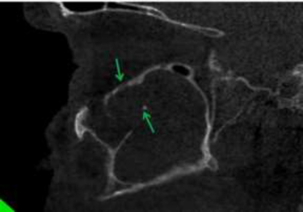
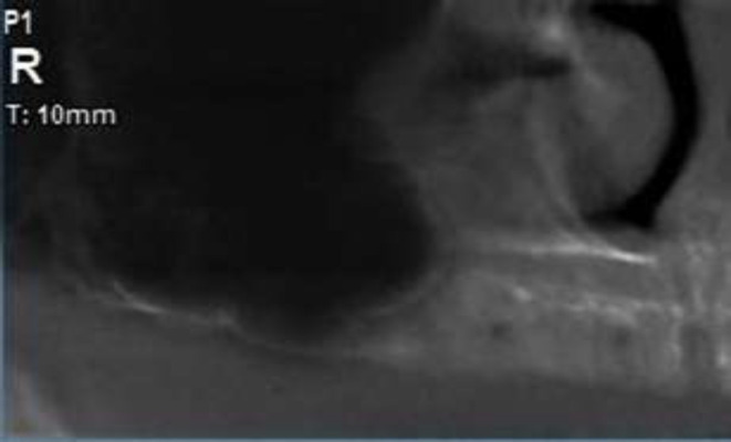
Background: Fungal infections of the paranasal sinus are increasingly recognized in both normal and immunocompromised individuals. It is necessary to distinguish invasive diseases from the non- invasive as the result and prognosis of sinus treatment different in each one. CBCT imaging could help us in this regard. In this case, we describe a fungal sinusitis according to Cone Beam Computed Tomography (CBCT) findings.

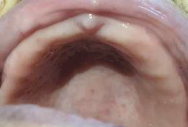
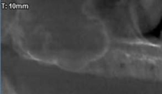
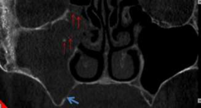
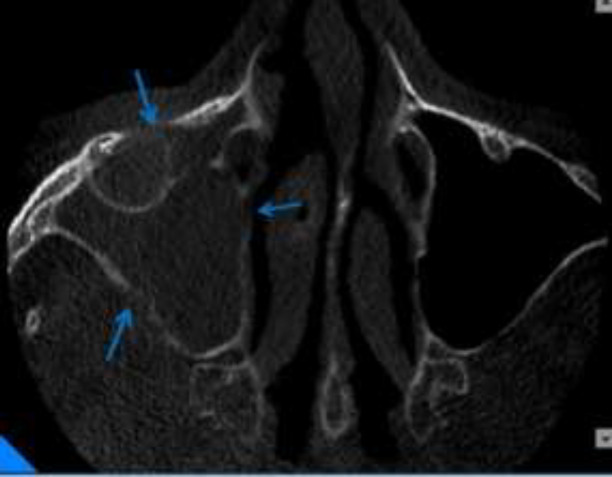
Case presentation: We present a case of a 48-year-old woman with diabetes mellitus referred to our Maxillofacial Radiology Center in Babol, Iran. The patient has been discharged from the hospital recently after recovering from COVID-19 Considering the background systemic disease (diabetes) and clinical and radiological findings (extension of bone destruction), fungal sinusitis (invasive form) was listed top in the differential diagnosis list , as it is the most common condition in post-COVID-19 patients.

Conclusion: CBCT images are very useful for diagnosing normal anatomy variations and sinus lesions especially bone lesions .In this case, its early diagnosis led to rapid recovery of the patient.

Keywords: Cone beam computed tomography (CBCT); Corona virus; Sinusitis.


Conflict of interest statement

None of the authors have any conflict of interest.

Figures

Figure 1
Figure 1
Photograph of right posterior maxillary showing normal palatal bone tissue and alveolar ridge
Figure 2
Figure 2
Cropped panoramic of the patient revealing no residual root and no infection but the right maxillary sinus showed increased mucosal thickening
Figure 3
Figure 3
Coronal view of CBCT showing complete opacification of right maxillary sinus with calcified particles (red arrows). Notice Invasion to lateral wall of middle turbinate and the right OMC was blocked. Also, there was noticeable of palatal bone erosion (blue arrow)
Figure 4
Figure 4
Axial view showing erosion of medial and posterolateral and anterior wall of right maxillary sinus in compared to intact and corticated left maxillary sinus walls (blue arrows)
Figure 5
Figure 5
Sagittal view showing erosion of inferior border of right orbit. Also notice to calcified particle. (Green arrows)
Figure 6
Figure 6
Cropped panoramic of the right maxillary sinus showing: the normal sinus without opacification

References

    1. Guo YR, Cao QD, Hong ZS, et al. The origin, transmission and clinical therapies on coronavirus disease 2019 (COVID-19) outbreak- an update on the status. Mil Med Res. 2020;7:11. - PMC - PubMed
    1. Young BE, Ong SWX, Kalimuddin S, et al. Epidemiologic features and clinical course of patients infected with SARS-CoV-2 in Singapore. JAMA. 2020;323:1488–94. - PMC - PubMed
    1. Zhang JJ, Dong X, Cao YY, et al. Clinical characteristics of 140 patients infected with SARS- CoV-2 in Wuhan, China. Allergy. 2020;75:1730–41. - PubMed
    1. Adhikari SP, Meng S, Wu YJ, et al. Epidemiology, causes, clinical manifestation and diagnosis, prevention and control of coronavirus disease (COVID-19) during the early outbreak period: a scoping review. Infect Dis Poverty. 2020;9:29. - PMC - PubMed
    1. Huang C, Wang Y, Li X, et al. Clinical features of patients infected with 2019 novel coronavirus inWuhan, China. Lancet. 2020;395:497–506. - PMC - PubMed

Publication types

LinkOut - more resources