

Case Report: Tetralogy of Fallot in a Chinese Family Caused by a Novel Missense Variant of MYOM2
- PMID: 35872890
- PMCID: PMC9300848
- DOI: 10.3389/fcvm.2022.863650
Case Report: Tetralogy of Fallot in a Chinese Family Caused by a Novel Missense Variant of MYOM2
Abstract
Background: Rare genetic variants have been identified to be important contributors to the risk of Tetralogy of Fallot (TOF), the most common cyanotic congenital heart disease (CHD). But relatively limited familial studies with small numbers of TOF cases have been reported to date. In this study, we aimed to identify novel pathogenic genes and variants that caused TOF in a Chinese family using whole exome sequencing (WES).
Methods: A Chinese family whose twins were affected by TOF were recruited for this study. A WES was performed for the affected twins, their healthy brother, and parents to identify the potential pathogenic mutated gene(s). Heterozygous variants carried by the twins, but not the unaffected brother, were retained. Public databases were used to assess the frequencies of the selected variants, and online prediction tools were accessed to predict the influences of these variants on protein function. The final candidate variant was further confirmed by Sanger sequencing in other members of the family.
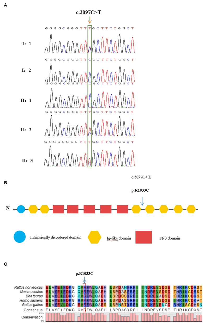
Results: After several filtering processes, a heterozygous missense variant in the MYOM2 gene (NM_003970.4:c.3097C>T:p.R1033C) was identified and confirmed by Sanger sequencing in the affected twins and their unaffected father, suggesting an inheritance pattern with incomplete penetrance. The variant was found to be extremely rare in the public databases. Furthermore, the mutated site was highly conserved among mammals, and as shown using multiple online prediction tools, this variant was predicted to be a detrimental variant.
Conclusion: We assessed a family with TOF caused by a rare heterozygous missense variant of MYOM2. Our findings not only further confirm the significant role of genetics in the incidence of TOF but also expand the spectrum of the gene variants that lead to TOF.
Keywords: MYOM2; Tetralogy of Fallot; heterozygous variant; incomplete penetrance; whole exome sequencing.
Copyright © 2022 Wang, Wang, Xie, Feng, Wei, Wang, Li, Pi and Gong.
Conflict of interest statement
The authors declare that the research was conducted in the absence of any commercial or financial relationships that could be construed as a potential conflict of interest.
Figures


References
-
- Diaz-Frias J, Guillaume M. Tetralogy of Fallot. Treasure Island (FL): Statpearls Publishing. (2021). - PubMed
Publication types
LinkOut - more resources
Full Text Sources