

Neoplasms of follicular helper T-cells: an insight into the pathobiology
- PMID: 35873103
- PMCID: PMC9301021
Neoplasms of follicular helper T-cells: an insight into the pathobiology
Abstract
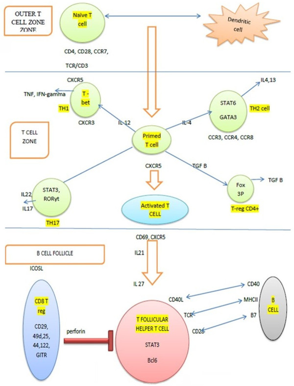
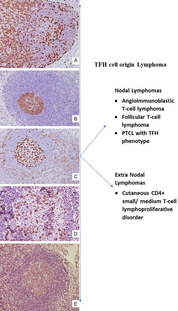
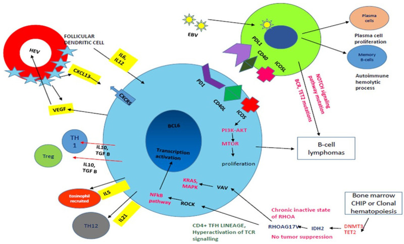
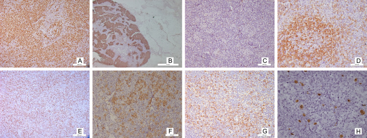
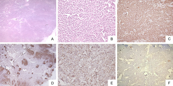
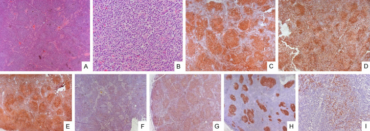
T-follicular helper cells (TFH) are a unique subset of T-cells with varied transcriptional profiles and functions. In the last 2016 WHO classification, lymphomas arising from TFH were included as a broad category and emphasis was given to separating them from other peripheral T cell lymphomas. The neoplasms derived from these mainly comprise angioimmunoblastic T-cell lymphoma, peripheral T-cell lymphoma with T-follicular helper cell phenotype, follicular T-cell lymphoma, and cutaneous CD4+ small-medium sized lymphoproliferative disorders. The TFH lymphomas comprise both indolent and aggressive forms. Additional immunohistochemistry to identify TFH cells like CD10, BCL6, ICOS, PD1, CXCL13 and mutations like RHOA, IDH2 is required for diagnosis and prognostication. The understanding of these has evolved over the years, and currently we review the updates and pathobiology of the above.
Keywords: AITL; Follicular helper T-cells; PTCL; follicular T helper cell lymphoma.
AJBR Copyright © 2022.
Conflict of interest statement
None.
Figures
Similar articles
-
Primary cutaneous peripheral T-cell lymphomas with a T-follicular helper phenotype: an integrative clinical, pathological and molecular case series study.Br J Dermatol. 2022 Dec;187(6):970-980. doi: 10.1111/bjd.21791. Epub 2022 Sep 2. Br J Dermatol. 2022. PMID: 35895386 Free PMC article.
-
Markers of Follicular Helper T Cells Are Occasionally Expressed in T-Cell or Histiocyte-Rich Large B-Cell Lymphoma, Classic Hodgkin Lymphoma, and Atypical Paracortical HyperplasiaA Diagnostic Pitfall For T-Cell Lymphomas of T Follicular Helper Origin.Am J Clin Pathol. 2021 Aug 4;156(3):409-426. doi: 10.1093/ajcp/aqaa249. Am J Clin Pathol. 2021. PMID: 33624021
-
Comprehensive analysis of clinical, pathological, and genomic characteristics of follicular helper T-cell derived lymphomas.Exp Hematol Oncol. 2021 May 14;10(1):33. doi: 10.1186/s40164-021-00224-3. Exp Hematol Oncol. 2021. PMID: 33990228 Free PMC article.
-
Recent Progress in the Understanding of Angioimmunoblastic T-cell Lymphoma.J Clin Exp Hematop. 2017;57(3):109-119. doi: 10.3960/jslrt.17019. J Clin Exp Hematop. 2017. PMID: 29279549 Free PMC article. Review.
-
Update on peripheral T-cell lymphomas with T-helper phenotype: Are there too many subtypes?Semin Diagn Pathol. 2020 Jan;37(1):24-31. doi: 10.1053/j.semdp.2019.12.005. Epub 2019 Dec 17. Semin Diagn Pathol. 2020. PMID: 31870687 Review.
Cited by
-
Interstitial Pneumonia Associated with Nodal T-follicular Helper Cell Lymphoma.Intern Med. 2024 Dec 1;63(23):3227-3231. doi: 10.2169/internalmedicine.3601-24. Epub 2024 Apr 9. Intern Med. 2024. PMID: 38599858 Free PMC article.
-
Clinical and prognostic significance of follicular helper and regulatory T cells in bladder cancer draining lymph nodes.Sci Rep. 2024 Sep 2;14(1):20358. doi: 10.1038/s41598-024-70675-1. Sci Rep. 2024. PMID: 39223192 Free PMC article.
-
Piceatannol Inhibits the Immunostimulatory Functions of Dendritic Cells and Alleviates Experimental Arthritis.Int J Mol Sci. 2025 Apr 11;26(8):3626. doi: 10.3390/ijms26083626. Int J Mol Sci. 2025. PMID: 40332204 Free PMC article.
-
Contribution of the Epstein-Barr virus to the oncogenesis of mature T-cell lymphoproliferative neoplasms.Front Oncol. 2023 Sep 14;13:1240359. doi: 10.3389/fonc.2023.1240359. eCollection 2023. Front Oncol. 2023. PMID: 37781191 Free PMC article. Review.
-
Spatially exploring RNA biology in archival formalin-fixed paraffin-embedded tissues.Cell. 2024 Nov 14;187(23):6760-6779.e24. doi: 10.1016/j.cell.2024.09.001. Epub 2024 Sep 30. Cell. 2024. PMID: 39353436
References
-
- Bellei M, Chiattone CS, Luminari S, Pesce EA, Cabrera ME, de Souza CA, Gabús R, Zoppegno L, Zoppegno L, Milone J, Pavlovsky A, Connors JM, Foss FM, Horwitz SM, Liang R, Montoto S, Pileri SA, Polliack A, Vose JM, Zinzani PL, Zucca E, Federico M. T-cell lymphomas in South America and Europe. Rev Bras Hematol Hemoter. 2012;34:42–47. - PMC - PubMed
-
- Pizzi M, Margolskee E, Inghirami G. Pathogenesis of peripheral T cell lymphoma. Annu Rev Pathol. 2018;13:293–320. - PubMed
-
- Vose J, Armitage J, Weisenburger D International T-Cell Lymphoma Project. International peripheral T-cell and natural killer/T-cell lymphoma study: pathology findings and clinical outcomes. J. Clin. Oncol. 2008;26:4124–4130. - PubMed
-
- Savage KJ, Ferreri AJ, Zinzani PL, Pileri SA. Peripheral T-cell lymphoma--not otherwise specified. Crit Rev Oncol Hematol. 2011;79:321–329. - PubMed
-
- Rizvi MA, Evens AM, Tallman MS, Nelson BP, Rosen ST. T-cell non-Hodgkin lymphoma. Blood. 2006;107:1255–1264. - PubMed
Publication types
LinkOut - more resources
Full Text Sources
Research Materials
Miscellaneous