. 2022 Jul 17:15:4047-4059.
doi: 10.2147/JIR.S370615. eCollection 2022.

Necroptosis-Mediated eCIRP Release in Sepsis

Affiliations

Necroptosis-Mediated eCIRP Release in Sepsis

Bridgette Reilly et al. J Inflamm Res. .

Erratum in

Abstract

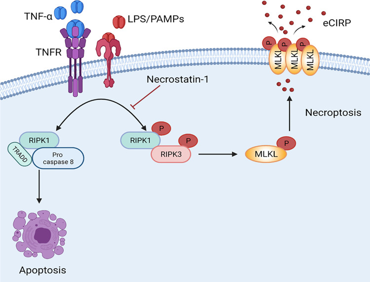
Introduction: Extracellular cold-inducible RNA-binding protein (eCIRP) is an endogenous pro-inflammatory mediator that exacerbates injury in inflammation and sepsis. The mechanisms in which eCIRP is released have yet to be fully explored. Necroptosis is a programmed cell death that is dependent on the activation of mixed lineage kinase domain-like pseudo kinase (MLKL) which causes the release of damage-associated molecular patterns. We hypothesize that eCIRP is released through necroptosis and intensifies inflammation in sepsis.

Methods: RAW264.7 cells were treated with pan-caspase inhibitor z-VAD (15 μM) 1 h before stimulation with LPS (1 μg/mL). Necroptosis inhibitor, Necrostatin-1 (Nec-1) (10 μM) was added to the cells with LPS simultaneously. After 24 h of LPS stimulation, cytotoxicity was determined by LDH assay. eCIRP levels in the culture supernatants and phospho-MLKL (p-MLKL) from cell lysates were assessed by Western blot. p-MLKL interaction with the cell membrane was visualized by immunofluorescence. Sepsis was induced in C57BL/6 mice by cecal ligation and puncture (CLP). Mice were treated with Nec-1 (1 mg/kg) or DMSO. 20 h post-surgery, serum and peritoneal fluid levels of eCIRP, TNF-α and IL-6 were determined by ELISA. H&E staining of lung tissue sections was performed.

Results: We found that in RAW264.7 cells, LPS+z-VAD induces necroptosis as evidenced by an increase in p-MLKL levels and causes eCIRP release. Nec-1 reduces both p-MLKL activation and eCIRP release in LPS+z-VAD-treated RAW264.7 cells. Nec-1 also inhibits the release of eCIRP, TNF-α and IL-6 in the serum and peritoneal fluid in CLP-induced septic mice. We predicted a transient interaction between eCIRP and MLKL using a computational model, suggesting that eCIRP may exit the cell via the pores formed by p-MLKL.

Conclusion: Necroptosis is a novel mechanism of eCIRP release in sepsis. Targeting necroptosis may ameliorate inflammation and injury in sepsis by inhibiting eCIRP release.

Keywords: Necrostatin-1; eCIRP; macrophage; necroptosis; sepsis.


Conflict of interest statement

The authors declared that they have no conflicts of interest in relation to this work.

Figures

Figure 1
Figure 1
Necroptosis induces eCIRP release from macrophages. RAW264.7 cells were pre-treated with or without z-VAD (15 μM) for 30 min and then stimulated with LPS (1 µg/mL) for 24 h. Cell culture supernatants and cell lysates were collected. (A and B) eCIRP present in the culture supernatants was quantified by Western blotting. (C and D) p-MLKL, MLKL, and β-actin were quantified by Western blotting. Representative Western blot images and quantitative bar diagrams are shown. (E) The release of LDH in the cell supernatants was measured by CyQUANTTM LDH cytotoxicity assay kit. Data are expressed as means ± SEM. Groups were compared by one-way ANOVA and Tukey’s multiple comparisons test. n=4–8 samples/group as shown in circles. *p<0.05 vs (-) LPS, (-) z-VAD. PS, Ponceau S red staining. Full blot images are shown in Supplementary Figure 1.
Figure 2
Figure 2
Necroptosis inhibitor necrostatin-1 inhibits eCIRP release from macrophages. RAW264.7 cells were pre-treated with z-VAD (15 μM) for 30 min and then stimulated with LPS (1 µg/mL) and Necrostatin-1 (Nec-1, 10 μM) for 24 hours, and cell culture supernatants and cell lysates were collected. (A and B) eCIRP present in the culture supernatants was quantified by Western blotting. (C and D) p-MLKL, and MLKL were quantified by Western blotting with β-actin as a loading control. Representative Western blot images and quantitative bar diagrams are shown. (E) The release of LDH in the cell supernatants was measured by CyQUANTTM LDH cytotoxicity assay kit. Data are expressed as means ± SEM. Groups were compared by one-way ANOVA and Tukey’s multiple comparisons test. n=3–10 samples/group. *p<0.05 vs (-) LPS, (-) z-VAD, (-) Nec-1; #p<0.05 vs (+) LPS, (+) z-VAD, (-) Nec-1. PS, Ponceau S red staining. Full blot images are shown in Supplementary Figure 2.
Figure 3
Figure 3
Necroptosis inhibitor necrostatin-1 inhibits the release of eCIRP and pro-inflammatory cytokines in sepsis. Sepsis was induced in C57BL/6 mice by CLP. Sham-operated animals served as control mice. C57BL/6 mice were injected i.p. with either necrostatin-1 (1 mg/kg) or vehicle in equivalent volumes immediately following CLP. At 20 h of CLP or sham operation, blood and peritoneal fluid were harvested from mice. (A) Serum levels of eCIRP were assessed by ELISA. (B and C) Serum levels of TNF-α, and Il-6 were assessed by ELISA. (D-F) Peritoneal fluid levels of eCIRP, TNF-α, and IL-6 were assessed by ELISA. n=4–9 mice/group (each circle represents one sample). *p<0.05 vs (-) CLP, (-) Nec-1; #p<0.05 vs (+) CLP, (-) Nec-1. Data are expressed as means ± SEM. Groups were compared by one-way ANOVA and Tukey’s multiple comparisons test.
Figure 4
Figure 4
Necroptosis attenuates lung injury in CLP-induced sepsis. Sepsis was induced via CLP in mice. Sham operated mice functioned as a control. C57BL/6 mice were injected i.p. with either necrostatin-1 (1 mg/kg) or vehicle in equivalent volumes immediately following CLP. 20 h post sham or CLP operation, lungs were collected. (A) The lungs were sliced into 5 μm sections and then stained with hematoxylin and eosin. Samples were examined using light microscopy. (B) Samples were given an injury score scored using a scoring system for acute lung injury in experimental animals as outlined by the American Thoracic Society. n=6 mice/group (each circle represents the tissue sample obtained from one mouse). *p<0.05 vs (-) CLP, (-) Nec-1; #p<0.05 vs (+) CLP, (-) Nec-1. Data are expressed as means ± SEM. Groups were compared by one-way ANOVA and Tukey’s multiple comparisons test.
Figure 5
Figure 5
p-MLKL co-localizes in the membrane, and MLKL and CIRP interact transiently. (A) RAW264.7 cells were pre-treated with z-VAD (15µM) for 30 min and then stimulated with LPS (1 µg/mL) for 24 h. After LPS stimulation, the cells were fixed and then immunostained with cell surface membrane marker (green), p-MLKL (red), and DNA (Hoechst 3334, blue), and the images were captured by confocal microscopy. Two confocal microscopy images in green and red fluorescence channels were tested for colocalization in ImageJ software with the colocalization finder plug-in. The plug-in calculates the Pearson correlation coefficient of the two original images and displays a correlation diagram, which is highlighted in white on a composite picture (bottom right of control and LPS-treated groups) of the two original images. The duplicated merge images indicated as colocalization panels highlight the areas in white, where green and red signals colocalize, as shown in the white arrow. (B) A computational model of MLKL’s (blue) interaction with CIRP (red) by using Iterative Threading ASSEmbly Refinement (I-TASSER) server.
Figure 6
Figure 6
Necroptosis induces eCIRP release from macrophage in Sepsis. Bacterial sepsis or endotoxemia induces phosphorylated RIPK1 and RIPK3, respectively, facilitates the activation of MLKL via phosphorylation. Then the p-MLKL oligomerizes and is inserted into the membrane to form a pore. During inflammation, CIRP is translocated from nuclear to cytoplasm, binds to p-MLKL in the cell membrane, and released through the MLKL pores or cell rupture. Necrostatin-1, an inhibitor of cell necroptosis, attenuates eCIRP release in sepsis.

Similar articles

Cited by

References

    1. Bianchi ME. DAMPs, PAMPs and alarmins: all we need to know about danger. J Leukoc Biol. 2007;81(1):1–5. doi:10.1189/jlb.0306164 - DOI - PubMed
    1. Denning NL, Aziz M, Gurien SD, Wang P. DAMPs and NETs in sepsis. Front Immunol. 2019;10:2536. doi:10.3389/fimmu.2019.02536 - DOI - PMC - PubMed
    1. Qiang X, Yang WL, Wu R, et al. Cold-inducible RNA-binding protein (CIRP) triggers inflammatory responses in hemorrhagic shock and sepsis. Nat Med. 2013;19(11):1489–1495. doi:10.1038/nm.3368 - DOI - PMC - PubMed
    1. Nishiyama H, Itoh K, Kaneko Y, et al. A glycine-rich RNA-binding protein mediating cold-inducible suppression of mammalian cell growth. J Cell Biol. 1997;137(4):899–908. doi:10.1083/jcb.137.4.899 - DOI - PMC - PubMed
    1. Yang WL, Sharma A, Wang Z, et al. Cold-inducible RNA-binding protein causes endothelial dysfunction via activation of Nlrp3 inflammasome. Sci Rep. 2016;6:26571. doi:10.1038/srep26571 - DOI - PMC - PubMed