

Bacterial diversity significantly reduces toward the late stages among filarial lymphedema patients in the Ahanta West District of Ghana: A cross-sectional study
- PMID: 35873398
- PMCID: PMC9297296
- DOI: 10.1002/hsr2.724
Bacterial diversity significantly reduces toward the late stages among filarial lymphedema patients in the Ahanta West District of Ghana: A cross-sectional study
Abstract
Background: Lymphatic Filariasis (LF), a neglected tropical disease, has been speculated to be complicated by secondary bacteria, yet a systematic documentation of these bacterial populations is lacking. Thus, the primary focus of this study was to profile bacteria diversity in the progression of filarial lymphedema among LF individuals with or without wounds.
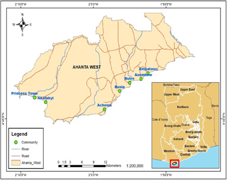
Methods: A cross-sectional study design recruited 132 LF individuals presenting with lymphedema with or without wounds from eight communities in the Ahanta West District in the Western Region, Ghana. Swabs from the lymphedematous limbs, ulcers, pus, and cutaneous surfaces were cultured using standard culture-based techniques. The culture isolates were subsequently profiled using Matrix-assisted Laser Desorption/Ionization Time of Flight Mass Spectrometry.
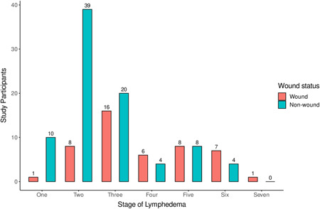
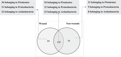
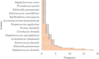
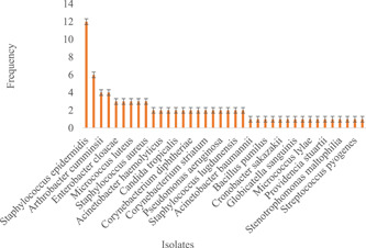
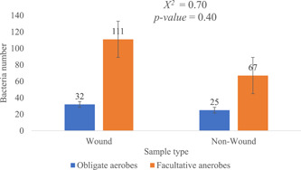
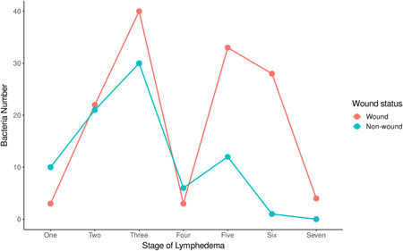
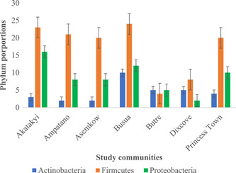
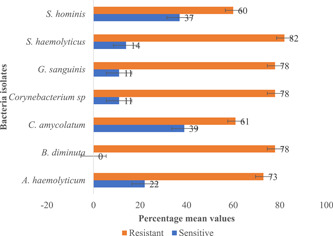
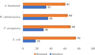
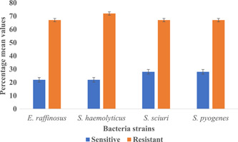
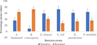
Results: Of the 132 LF participants recruited, 65% (85) had filarial lymphedema with no wounds. In total, 84% (235) of the bacterial isolates were identified. The remaining 16% (46) could not be identified with the method employed. Additionally, 129(55%) of the strains belonged to the phylum Firmicutes, while 61 (26%) and 45 (19%) represented Proteobacteria and Actinobacteria, respectively. Generally, irrespective of the samples type (i.e., wound sample and non-wound samples), there was a sharp increase of bacteria diversity from Stages 1 to 3 and a drastic decrease in these numbers by Stage 4, followed by another surge and a gradual decline in the advanced stages of the disease. The Shannon Diversity Index and Equitability for participants with and without wounds were (3.482, 0.94) and (3.023, 0.75), respectively. Further, Staphylococcus haemolyticus and Escherichia coli showed resistance to tetracycline, chloramphenicol, and penicillin.
Conclusion: The present study reveals a sharp decline in bacterial load at the late stages of filarial lymphedema patients. In addition, we report an emerging antimicrobial resistance trend of S. haemolyticus and E. coli against commonly used antibiotics such as tetracycline, chloramphenicol, and penicillin in communities endemic for LF in the Ahanta West District, Ghana. This could pose a huge challenge to the management of the disease; particularly as current treatments are not quite effective against the infection.
Keywords: MALDI‐TOF; antimicrobial resistance; lymphatic filariasis; lymphedema; microbiome.
© 2022 The Authors. Health Science Reports published by Wiley Periodicals LLC.
Conflict of interest statement
All authors have read and approved the final version of the manuscript. Alexander Kwarteng (EDCTP‐TMA2016 1561) had full access to all the data in this study and takes complete responsibility for the integrity of the data and the accuracy of the data analysis.
Figures
References
LinkOut - more resources
Full Text Sources
