. 2022 Jul 6:13:943197.
doi: 10.3389/fgene.2022.943197. eCollection 2022.

Strongly Truncated Dnaaf4 Plays a Conserved Role in Drosophila Ciliary Dynein Assembly as Part of an R2TP-Like Co-Chaperone Complex With Dnaaf6

Affiliations

Strongly Truncated Dnaaf4 Plays a Conserved Role in Drosophila Ciliary Dynein Assembly as Part of an R2TP-Like Co-Chaperone Complex With Dnaaf6

Jennifer Lennon et al. Front Genet. .

Abstract

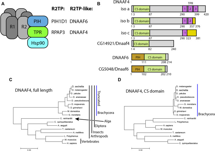
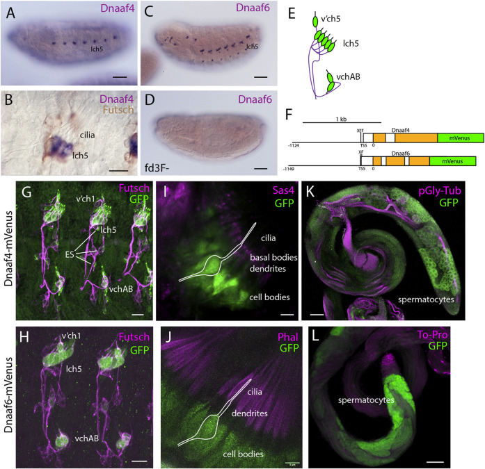
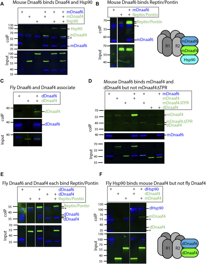
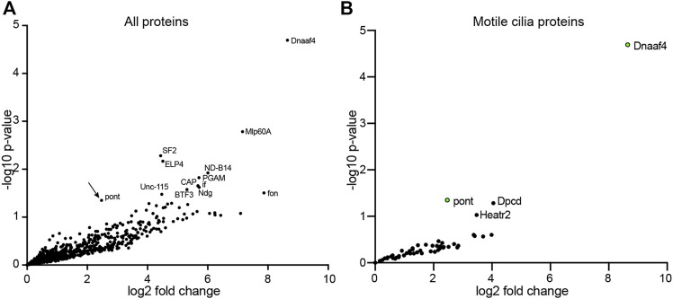
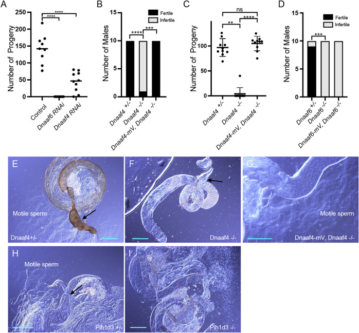
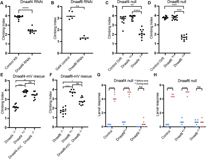
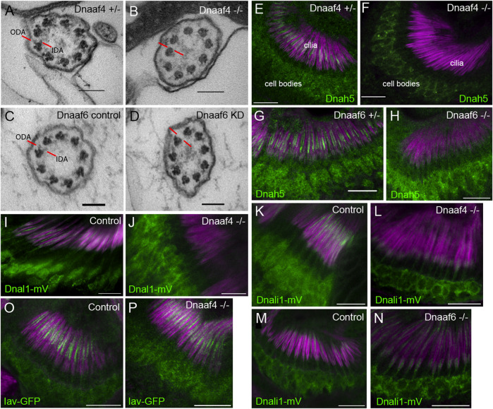
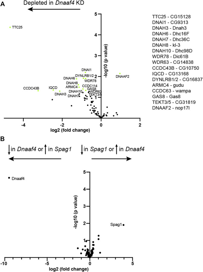
Axonemal dynein motors are large multi-subunit complexes that drive ciliary movement. Cytoplasmic assembly of these motor complexes involves several co-chaperones, some of which are related to the R2TP co-chaperone complex. Mutations of these genes in humans cause the motile ciliopathy, Primary Ciliary Dyskinesia (PCD), but their different roles are not completely known. Two such dynein (axonemal) assembly factors (DNAAFs) that are thought to function together in an R2TP-like complex are DNAAF4 (DYX1C1) and DNAAF6 (PIH1D3). Here we investigate the Drosophila homologues, CG14921/Dnaaf4 and CG5048/Dnaaf6. Surprisingly, Drosophila Dnaaf4 is truncated such that it completely lacks a TPR domain, which in human DNAAF4 is likely required to recruit HSP90. Despite this, we provide evidence that Drosophila Dnaaf4 and Dnaaf6 proteins can associate in an R2TP-like complex that has a conserved role in dynein assembly. Both are specifically expressed and required during the development of the two Drosophila cell types with motile cilia: mechanosensory chordotonal neurons and sperm. Flies that lack Dnaaf4 or Dnaaf6 genes are viable but with impaired chordotonal neuron function and lack motile sperm. We provide molecular evidence that Dnaaf4 and Dnaaf6 are required for assembly of outer dynein arms (ODAs) and a subset of inner dynein arms (IDAs).

Keywords: Drosophila; chaperone; ciliopathies; cilium; dynein; flagellum.


Conflict of interest statement

The authors declare that the research was conducted in the absence of any commercial or financial relationships that could be construed as a potential conflict of interest.

Figures

FIGURE 1
FIGURE 1
Drosophila and mammalian Dnaaf4/Dnaaf6 proteins. (A) Schematic showing the composition of R2TP and putative DNAAF4/6-containing R2TP-like complexes. Note that association of DNAAF4/6 with RUVBL1 and RUVBL2 is speculative. (B) Schematic showing the protein domains of human DNAAF4, DNAAF6 and their Drosophila orthologues. Human isoforms and protein structures are based on Maurizy et al. (2018). (C) Phylogenetic tree of DNAAF4 sequences from selected species including vertebrates, arthropods and the unicellular green alga, Chlamydomonas reinhardtii (established ciliary motility model organism). Higher dipterans (Brachycera) form a distinct group that correlates with gene truncation (blue bar). (D) When comparing CS domains alone, the tree structure remains similar, with Brachycera distinct from other taxa. Organisms included in this tree: Drosophila sechellia, D. melanogaster, D. yakuba, D. ananassae, D. pseudoobscura, D. mojavensis, D. grimshawi, Musca domestica, Glossina morsitans, Culex quinquefasciatus, Aedes aegypti, Tribolium castaneum, Apis mellifera, Chlamydomonas reinhardtii, Limulus polyphemus, Mus musculus and Homo sapiens.
FIGURE 2
FIGURE 2
Dnaaf4 and Dnaaf6 are both expressed in Drosophila motile cilia cells. (A–D) RNA in situ hybridisation (dark blue) conducted on late-stage whole-mount embryos. (A) Dnaaf4 probe, Dnaaf4 is expressed specifically in the chordotonal neurons. (B) Higher magnification indicates that this expression becomes restricted at a late stage to a subset of chordotonal neurons (lch5). Here the embryo has been counterstained with antibodies against Futsch (brown), which labels all sensory neurons. (C) Dnaaf6 shows expression in developing chordotonal neurons. (D) In an embryo homozygous mutant for fd3F, expression of Dnaaf6 is abolished. (E) Schematic of the arrangement of chordotonal neurons in embryonic abdominal segments. (F) Schematic illustrating mVenus fusion transgenes. Each includes 5’ flanking DNA containing potential binding sites for the transcription factors fd3f (F) and Rfx (X) (Dnaaf4: CTGTTCACTTG, GTTCACTTGCAGC; Dnaaf6: ACTAAATAAACAA, GTTGCCAGGAAA). (G–L) Expression of Dnaaf4-mVenus detected by anti-GFP antibodies. (G,H) Late embryos counterstained with anti-Futsch (magenta) show expression of both fusion genes in chordotonal neurons. In the case of Dnaaf4-mVenus, some expression is observed in some external sensory (ES) neurons. As this is not observed for the mRNA, it is likely an artefact of the expression construct. (I,J) In pupal antennae, both fusion genes are expressed in the cell bodies of chordotonal neurons that form Johnston’s Organ. A schematic of approximate neuronal location is shown. The counterstain (magenta) is the basal body marker Sas4. (I) or phalloidin (J), which marks the actin basket (scolopale) that surrounds the cilia. (K,L) In adult testes, both fusion genes are expressed in differentiating germline cells (spermatocytes and spermatids). Counterstains (magenta) are polyglycylated tubulin (K) or To-Pro (L). Scale bars are: (A,C,D,K,L) 50 µm (B,G,H) 10 µm (I,J) 5 µm. Number of samples imaged: (G) n = 7 (I) n = 9 (K) n = 8.
FIGURE 3
FIGURE 3
Drosophila and mouse Dnaaf4/Dnaaf6 complexes. Coimmunoprecipitations of tagged proteins expressed in S2 cells. In each case, the bait protein is FLAG-tagged (blue) and the prey protein is HA-tagged (green). Proteins are from Drosophila unless indicated. “Input” represents Western blot of whole cell extracts with bait/prey simultaneously detected (anti-FLAG + anti-HA). “coIP” represents FLAG-mediated coIP followed by simultaneous detection of FLAG- and HA-tagged proteins on Western blot. *indicates non-specific bands. (A) Mouse FLAG-Dnaaf6 protein associates with mouse HA-Dnaaf4 and Drosophila HA-Hsp90. (B) Mouse FLAG-Dnaaf6 protein binds Drosophila HA-Reptin/HA-Pontin. (C) Drosophila FLAG-Dnaaf6 and HA-Dnaaf4 associate. (D) Mouse FLAG-Dnaaf6 binds both mouse HA-Dnaaf4 and Drosophila HA-Dnaaf4, but is unable to bind the mouse Dnaaf4 protein with TPR domain deleted (HA-Dnaaf4ΔTPR). (E) Drosophila FLAG-Dnaaf6 and FLAG-Dnaaf4 are each capable of binding HA-Reptin/HA-Pontin. (F) Drosophila FLAG-Hsp90 is able to bind mouse HA-Dnaaf4 but not Drosophila HA-Dnaaf4.
FIGURE 4
FIGURE 4
Proteins preferentially associated with Dnaaf4 in Drosophila testes. Volcano plots of proteins detected by MS after affinity purification of Dnaaf4-mVenus, shown as relative abundance (fold change) compared with proteins associated with unrelated control protein (GAP43-mVenus). (A) All proteins, with those above threshold significance (-log10 (p-value)>1.3) labelled. Pontin of R2TP is significantly associated (arrow). (B) The same dataset filtered to extract proteins associated with motile cilia (zur Lage et al., 2021). Pontin is the only associated protein to reach statistical significance. However, two other proteins of interest are just below significance threshold: Dpcd and Heatr2 (Dnaaf5). Significance was determined using the Empirical Bayes method. n = 150 pairs of testes per replicate; 3 replicates per genotype.
FIGURE 5
FIGURE 5
Knockdown and Null mutants of Dnaaf4 and Dnaaf6 are male infertile. (A) Dnaaf4 and Dnaaf6 RNAi knockdown males (BamGal4) produce fewer progeny than control males. Progeny from individual males and median progeny value are shown. Knockdown of either gene significantly reduces progeny per male (p < 0.0001, One-way ANOVA followed by Sidak’s Test for multiple comparisons). (B,C) Fertility of Dnaaf4 null mutant males. (B) Proportion of males that are fully infertile. Most Dnaaf4 mutant males are infertile but this is rescued by the Dnaaf4-mVenus transgene (p = 0.001, Fisher’s exact test) (C) Number of progeny per male, showing that rescued homozygous males are fully fertile compared with heterozygotes (p > 0.9999, Kruskal–Wallis analysis followed by Dunn’s test for multiple comparisons). n = 10 males for each genotype. (C) Data for males in (B) plotted as number of progeny per male. A single Dnaaf4 homozygote gave progeny, perhaps due to being non-virgin at collection—40 progeny compared with a mean of 96.9 for heterozygotes. (D) Fertility assay results showing a decrease in the number of fertile males in the Dnaaf6 null mutant when compared to control groups (0.0001). Dnaaf6 rescue did not produce progeny (p < 0.0001) like that of the homozygous null mutants. n = 10 males per genotype. (E–I) Testes and associated male reproductive structures dissected from adult males and observed by light microscopy. Scale bars, 50 μm. (E) Dnaaf4 heterozygote testis showing S-shaped motile sperm emerging from large (sperm-filled) seminal vesicle (black arrow). (F) Dnaaf4 homozygote testis showing small (empty) seminal vesicle (black arrow) and absence of motile sperm. (G) Testis from Dnaaf4 homozygote with Dnaaf4-mVenus transgene showing rescue of motile sperm production. (H) Dnaaf6 heterozygote showing S-shaped motile sperm emerging from large (sperm-filled) seminal vesicle (black arrow). (I) Dnaaf6 homozygote testes homozygote testis showing absence of motile sperm.
FIGURE 6
FIGURE 6
Knockdown and Null mutants of Dnaaf4 and Dnaaf6 have defective chordotonal sensory function. (A–F) Adult climbing assays for proprioceptive ability. Plots (with median and individual values), each point is a batch of 8–12 females, n = 10 batches. (A,B) RNAi knockdown of Dnaaf4 and Dnaaf6 in sensory neurons (scaGal4) results in significant decrease in climbing ability. (C,D) Homozygote null adults for Dnaaf4 and Dnaaf6 have significantly decreased climbing ability compared with heterozygotes. (E,F) Rescue of null mutants. (E) Dnaaf4-mVenus transgene rescued the climbing ability of Dnaaf4 null mutant flies, showing a significant increase in climbing performance when compared to null (p = 0.0012), restoring climbing ability to the same level as the heterozygotes (p = 0.8130). (F) Dnaaf6-mVenus transgene partial restores climbing ability of Dnaaf6 null mutants (p = 0.0103), but not to levels seen in the heterozygote, although the latter difference does not reach significance (p = 0.1282). (G,H) Plots (with individual and median values) showing hearing assay performances for Dnaaf4 −/− and Dnaaf6 −/− larvae in comparison to heterozygote and wild-type (OrR) controls. Number of larvae contracting before and during a 1000-Hz tone was measured. Individual points are batches of 5 larvae, n = 5 batches. There is a significant difference between the number of larvae contracting before and during the tone (p < 0.0001) for control groups of both genotypes. There is no significant difference between the number of contractions occurring before and during the tone for Dnaaf4 or Dnaaf6 null mutants, indicating no behavioural response to stimulus. For climbing assays, significance was determined by Kruskal–Wallis followed by Dunn’s test for multiple comparisons. For hearing assay, significance was determined by two-way RM ANOVA and Sidak’s multiple comparisons test. Statistical significance on plots is indicated by asterisks: *, p ≤ 0.05; **, p ≤ 0.01; ***, p ≤ 0.001; ****, p ≤ 0.0001.
FIGURE 7
FIGURE 7
Defective dynein motor localisation in mutants. (A–D) TEM of chordotonal neurons in adult antennae, transverse sections of cilia showing 9 + 0 axonemal arrangement. (A) Control (Dnaaf4 +/− heterozygote) with ODAs and IDAs (red lines) on each microtubule doublet. (B) Dnaaf4 −/− homozygote showing severe loss of ODA and IDA structures from the microtubule doublets. (C) RNAi control (scaGal4, UAS-Dcr2, KK line) and (D) Dnaaf6 knockdown (scaGal4, UAS-Dcr2, UAS-Dnaaf6RNAi). The latter shows a reduction of ODA and IDA. (E–P) Immunofluorescence of ODA/IDA markers (green) in differentiating chordotonal neurons of pupal antennae. All are counterstained with phalloidin, detecting the scolopale structures surrounding the cilia (magenta). (E–H) ODA heavy chain Dnah5 localisation in cilia is lost from Dnaaf4 −/− and Dnaaf6 −/− homozygote mutants (F,H) compared to controls (E,G), despite presence of protein in the cell bodies. (I,J) ODA marker, Dnal1-mVenus shows a similar loss of ciliary localisation in Dnaaf4 −/− homozygote (J) relative to w - control (I). (K–N) IDA marker, Dnali1-mVenus shows a partial loss of ciliary localisation in Dnaaf4 −/− and Dnaaf6 −/− homozygotes (L,N) relative to heterozygote controls (K,M). (O,P) TRPV channel subunit Iav shows no difference in ciliary localisation between Dnaaf4 −/− homozygote (P) and w - control (O). Scale bars: (A–D) 100 nm, (E–P) 10 mm. Number of antennae imaged for IF: (E) n = 7; (F) 7; (G) 6; (H) 5; (I) 5; (J) 10; (K) 5; (L) 9; (M) 8; (N) 9; (O) 6; (P) 7.
FIGURE 8
FIGURE 8
Proteomic changes in Dnaaf4 mutant testes. (A) Volcano plot of motile cilia-associated proteins detected by MS in testes. To the left of the Y axis are proteins that are more less abundant in Dnaaf4RNAi KD (BamGal4, UAS-Dnaaf4RNAi) testes compared with BamGal4 control (depleted); to the right are proteins that are more abundant than in the control. Dnaaf4 protein itself is strongly depleted as expected (log2(FC) = −8.69, -log10 (p value) = 4.39) but for clarity it is not shown on plot. Proteins with -log10 (p value) > 1.3 (green points) are labelled with names of human homologues. The Drosophila gene names are shown to the right. n = 30 pairs of testes/replicate; 4 replicates per genotype. (B) Volcano plot comparing motile cilia-associated proteins detected in testes from Dnaaf4 knockdown testes compared with Spag1 knockdown testes (BamGal4, UAS-Spag1RNAi). The only proteins showing significant difference in abundance are Dnaaf4 and Spag1 themselves. Significance was determined using the Empirical Bayes method. n = 30 pairs of testes/replicate; 4 replicates per genotype.

Similar articles

Cited by

References

    1. Baker N. E., Yu S., Han D. (1996). Evolution of Proneural Atonal Expression during Distinct Regulatory Phases in the Developing Drosophila Eye. Curr. Biol. 6, 1290–1302. 10.1016/s0960-9822(02)70715-x - DOI - PubMed
    1. Bateman A., Martin M.-J., Orchard S., Magrane M., Agivetova R., Ahmad S., et al. (2021). UniProt: The Universal Protein Knowledgebase in 2021. Nucleic Acids Res. 49, D480–D489. 10.1093/nar/gkaa1100 - DOI - PMC - PubMed
    1. Bieder A., Einarsdottir E., Matsson H., Nilsson H. E., Eisfeldt J., Dragomir A., et al. (2020). Rare Variants in Dynein Heavy Chain Genes in Two Individuals with Situs Inversus and Developmental Dyslexia: A Case Report. BMC Med. Genet. 21, 87. 10.1186/s12881-020-01020-2 - DOI - PMC - PubMed
    1. Braschi B., Omran H., Witman G. B., Pazour G. J., Pfister K. K., Bruford E. A., et al. (2022). Consensus Nomenclature for Dyneins and Associated Assembly Factors. J. Cell Biol. 221, e202109014. 10.1083/jcb.202109014 - DOI - PMC - PubMed
    1. Chagot M.-E., Dos Santos Morais R., Dermouche S., Lefebvre D., Manival X., Chipot C., et al. (2019). Binding Properties of the Quaternary Assembly Protein SPAG1. Biochem. J. 476, 1679–1694. 10.1042/bcj20190198 - DOI - PubMed

LinkOut - more resources