. 2022 Jul 15:2022:6886752.
doi: 10.1155/2022/6886752. eCollection 2022.

Chlorpromazine and Promethazine (C+P) Reduce Brain Injury after Ischemic Stroke through the PKC- δ/NOX/MnSOD Pathway

Affiliations

Chlorpromazine and Promethazine (C+P) Reduce Brain Injury after Ischemic Stroke through the PKC- δ/NOX/MnSOD Pathway

Sichao Guo et al. Mediators Inflamm. .

Abstract

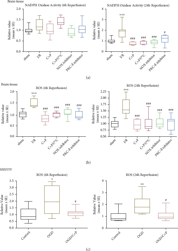
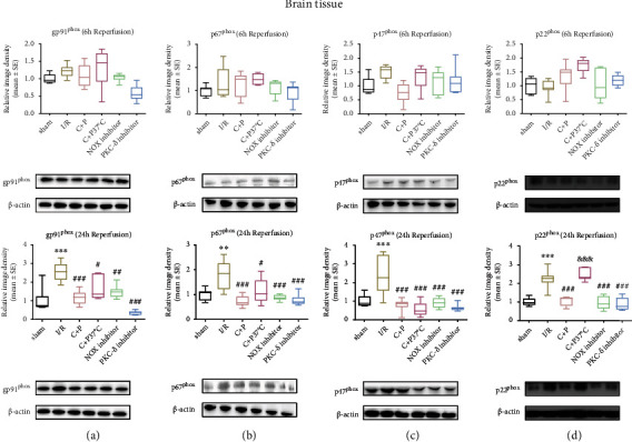
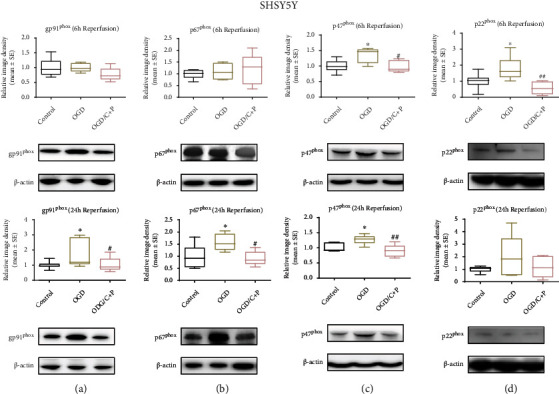
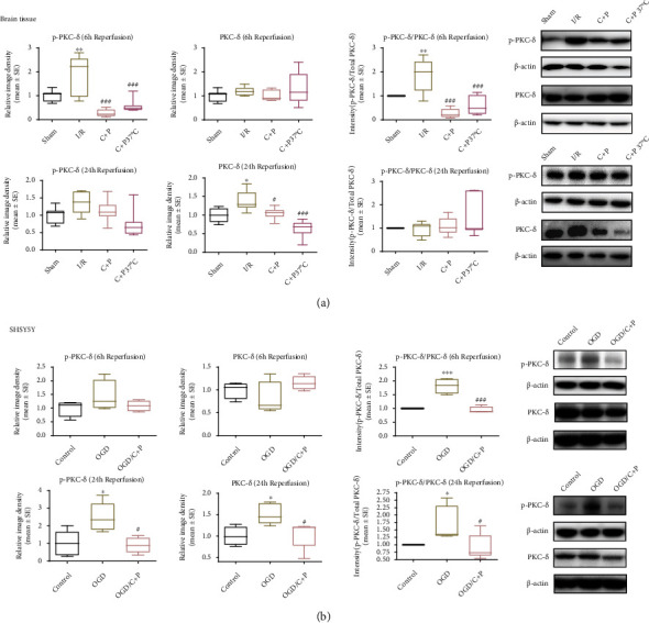
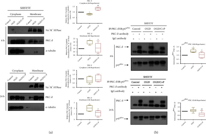
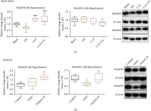
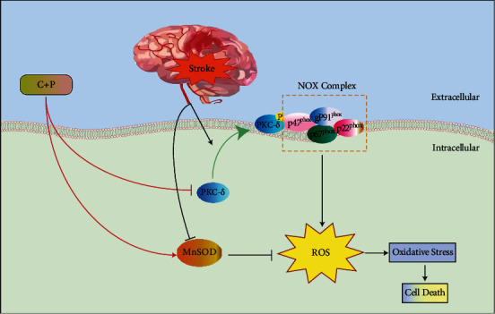
Cerebral ischemia-reperfusion (I/R) incites neurologic damage through a myriad of complex pathophysiological mechanisms, most notably, inflammation and oxidative stress. In I/R injury, nicotinamide adenine dinucleotide phosphate (NADPH) oxidase (NOX) produces reactive oxygen species (ROS), which promote inflammatory and apoptotic pathways, augmenting ROS production and promoting cell death. Inhibiting ischemia-induced oxidative stress would be beneficial for reducing neuroinflammation and promoting neuronal cell survival. Studies have demonstrated that chlorpromazine and promethazine (C+P) induce neuroprotection. This study investigated how C+P minimizes oxidative stress triggered by ischemic injury. Adult male Sprague-Dawley rats were subject to middle cerebral artery occlusion (MCAO) and subsequent reperfusion. 8 mg/kg of C+P was injected into the rats when reperfusion was initiated. Neurologic damage was evaluated using infarct volumes, neurological deficit scoring, and TUNEL assays. NOX enzymatic activity, ROS production, protein expression of NOX subunits, manganese superoxide dismutase (MnSOD), and phosphorylation of PKC-δ were assessed. Neural SHSY5Y cells underwent oxygen-glucose deprivation (OGD) and subsequent reoxygenation and C+P treatment. We also evaluated ROS levels and NOX protein subunit expression, MnSOD, and p-PKC-δ/PKC-δ. Additionally, we measured PKC-δ membrane translocation and the level of interaction between NOX subunit (p47phox) and PKC-δ via coimmunoprecipitation. As hypothesized, treatment with C+P therapy decreased levels of neurologic damage. ROS production, NOX subunit expression, NOX activity, and p-PKC-δ/PKC-δ were all significantly decreased in subjects treated with C+P. C+P decreased membrane translocation of PKC-δ and lowered the level of interaction between p47phox and PKC-δ. This study suggests that C+P induces neuroprotective effects in ischemic stroke through inhibiting oxidative stress. Our findings also indicate that PKC-δ, NOX, and MnSOD are vital regulators of oxidative processes, suggesting that C+P may serve as an antioxidant.


Conflict of interest statement

The authors declare no conflict of interest.

Figures

Figure 1
Figure 1
C+P lessened postischemic neurologic damage. (a) Alterations in body temperature post C+P administration at various points in time in 2-hour MCAO and after reperfusion (0–24 hours). Baseline temperatures were determined just before MCAO. Reperfusion was followed by C+P administration. ANOVA analyses specified that C+P significantly decreased body temperatures as early as 5 minutes and lasting up to 6 hours after the I/R onset (n = 7). (b) TTC stains showcase infarction volumes in MCAO rats regardless of C+P treatment at 48 hours of reperfusion. C+P administration or administration of the PKC-δ/NOX inhibitor significantly decreased infarct volumes (n = 7). (c) Neurologic deficits were determined using 5- or 12-point scoring systems at 48 hours of reperfusion. C+P administration or the administration of the PKC-δ/NOX inhibitor significantly reduced neurological deficits in all groups except those in the temperature-controlled C+P group, for which there was no effect on the 5-point scoring system (n = 7). (d) Cell death was significantly higher at 6 and 24 hours of reperfusion, demonstrated by ELISA. With C+P at 6 and 24 hours of reperfusion, cell death was significantly reduced. Administration of the PKC-δ/NOX inhibitor also suppressed cell death except in the NOX inhibitor group at 6 hours of reperfusion (n = 7). (e) Apoptotic cell death was significantly reduced with C+P administration regardless of temperature control at 24 hours of reperfusion, shown using the TUNEL assay (n = 5). Scale bar = 50 μm. ∗∗∗p < 0.001 versus the sham group; #p < 0.05, ##p < 0.01, and ###p < 0.001 versus the I/R group. Results are shown as mean ± SE.
Figure 2
Figure 2
C+P attenuated NOX activity and ROS level postreperfusion. Quantitative analysis of (a) NOX activity and (b) ROS level as determined in ischemic brain tissue. NOX activity was higher at 24 hours of reperfusion, and ROS levels were also higher at 6 and 24 hours of reperfusion. Decreased NOX activity levels at 24 hours of reperfusion and ROS at 6 and 24 hours of reperfusion were observed in C+P groups regardless of temperature control as well as in PKC-δ or NOX inhibitor groups (n = 7). ∗∗∗p < 0.001 versus the sham group; #p < 0.05, ###p < 0.001 versus the I/R group. Data is shown as the mean ± SE. Quantitative analysis of (c) ROS level in SHSY5Y cell was measured. C+P administration lowered the OGD-induced elevation of ROS at 6 and 24 hours of reoxygenation. p < 0.05, ∗∗p < 0.01 versus the OGD group; #p < 0.05 versus the OGD/C+P group. Results are shown as mean ± SE of three independent experiments.
Figure 3
Figure 3
C+P decreased NOX subunit protein levels after stroke in ischemic brain tissue. (a) gp91phox, (b) p67phox, (c) p47phox, and (d) p22phox assessed via the Western blot technique at 6 and 24 hours of reperfusion. Levels of gp91phox, p67phox, p47phox, and p22phox rose after I/R but decreased after C+P treatment and administration of the PKC-δ or NOX inhibitor at 24 hours of reperfusion, with the exception of p22phox in the C+P at the 37°C group at 24 hours of reperfusion. There was a larger decrease in p22phox expression in the non-temperature-controlled C+P versus the temperature-controlled C+P group at 24 hours of reperfusion (n = 7). ∗∗p < 0.01 and ∗∗∗p < 0.001 versus the sham group; #p < 0.05, ##p < 0.01, and ###p < 0.001 versus the I/R group; &&&p < 0.001 versus the C+P group. Results are shown as mean ± SE. The representative immunoblots are visualized.
Figure 4
Figure 4
C+P reduced protein levels of NOX subunits after OGD/R in SHSY5Y cells. (a) gp91phox, (b) p67phox, (c) p47phox, and (d) p22phox, identified by Western blot at 6 and 24 hours of reoxygenation. Levels of gp91phox and p67phox protein increased after OGD but decreased after C+P therapy at 24 hours of reoxygenation. Moreover, p47phox levels increased after OGD, but C+P lowered this expression at 6 and 24 hours of reoxygenation. OGD induced the elevation of p22phox, and there was a greater reduction in p22phox expression in the OGD/C+P treatment group versus the OGD group at 6 hours of reoxygenation. p < 0.05 versus the OGD group; #p < 0.05, ##p < 0.01 versus the OGD/C+P group. Results are shown as mean ± SE of three independent experiments. The representative immunoblots are presented.
Figure 5
Figure 5
C+P decreased activation of PKC-δ induced by cerebral ischemia or OGD/R. (a) Representative bands of phosphorylated and total protein expressions of PKC-δ in brain tissue identified via Western blot at 6 and 24 hours of reperfusion. β-Actin served as an internal control. Bar graphs demonstrate semiquantitative levels of PKC-δ, identified using band density analysis. MCAO enhanced PKC-δ phosphorylation. C+P treatment reduced PKC-δ phosphorylation as compared with the stroke group at 6 hours of reperfusion. Total PKC-δ protein expression increased, but C+P reduced its level at 24 hours of reperfusion. Moreover, the ratio of p-PKC-δ/PKC-δ significantly increased post-MCAO but decreased with C+P treatment at 6 hours of reperfusion (n = 7). p < 0.05, ∗∗p < 0.01 versus the sham group; #p < 0.05, ###p < 0.001 versus the I/R group. Results are presented as mean ± SE. (b) Representative bands of both phosphorylated and total PKC-δ levels in SHSY5Y cells identified via Western blot at 6 and 24 hours of reoxygenation. β-Actin served as an internal control. Bar graphs display semiquantitative levels of PKC-δ as determined by band density analysis. The p-PKC-δ/PKC-δ ratio was significantly higher after OGD/R; however, it decreased after C+P administration at 6 hours of reoxygenation. Furthermore, both PKC-δ phosphorylation and the total levels of PKC-δ were significantly higher after OGD/R; however, C+P annulled this trend at 24 hours of reoxygenation. Similarly, OGD/R facilitated the raise in the p-PKC-δ/PKC-δ ratio, but C+P decreased this trend at 24 hours of reoxygenation. p < 0.05, ∗∗∗p < 0.001 versus the OGD group; #p < 0.05, ###p < 0.001 versus the OGD/C+P group. Results are shown as mean ± SE of three independent experiments.
Figure 6
Figure 6
C+P suppressed the translocation of PKC-δ and interaction of PKC-δ with p47phox induced by OGD/R in SHSY5Y cells. (a) Representative bands of cytosolic and membrane PKC-δ identified using Western blot at 6 and 24 hours of reperfusion. Bar graphs show semiquantitative PKC-δ levels in the cytosol and membrane determined using band density analysis. Cytosolic PKC-δ expression decreased but increased in the membrane in the OGD/R group at 6 hours after reoxygenation as compared to the control group. Administration of C+P negated the changes of OGD/R-induced PKC-δ expression. No differences were detected in cytosolic PKC-δ expression in the OGD/R group at 24 hours of reoxygenation, but OGD/R increased membranous PKC-δ expression as compared to the control group. C+P attenuated the raised cellular membrane PKC-δ concentration. p < 0.05, as compared to the OGD group; #p < 0.05, ##p < 0.01, as compared to the OGD/C+P group. All the quantifications were normalized to α-tubulin (cytosol) or Na+/K+ ATPase (membrane), respectively. Results are shown as mean ± SE of three independent experiments. (b) Representative bands of coimmunoprecipitation of PKC-δ and p47phox in SHSY5Y cells, identified via the Western blot technique at 6 and 24 hours of reoxygenation. Bar graphs show the ratios of p47phox/PKC-δ identified using band density analysis. IgG antibody served as the control. In comparison to IgG control, a significant enhancement in p47phox was detected in the immunoprecipitation complex with the PKC-δ antibody from cell lysates, which suggests a heightened interaction of PKC-δ and p47phox. Moreover, the interaction of PKC-δ and p47phox increases after 6 hours of reoxygenation, while C+P treatment reduced the interaction. Although the interaction between the control and the OGD group was not significant at 24 hours of reoxygenation, the interaction did increase, and C+P treatment lowered the interaction between PKC-δ and p47phox. p < 0.05 versus the OGD group; #p < 0.05, ##p < 0.01 versus the OGD/C+P group. The data is represented as mean ± SE of three independent experiments.
Figure 7
Figure 7
C+P facilitated the expression of MnSOD after cerebral ischemia or OGD/R. (a) Representative bands of MnSOD in brain tissue as identified using Western blot at 6 and 24 hours of reperfusion. β-Actin served as an internal control. Semiquantitative levels of MnSOD are displayed in bar graphs, identified using band density analysis. MnSOD protein levels decreased after I/R. C+P administration significantly accelerated MnSOD expression at 6 hours of reperfusion (n = 7). p < 0.05 versus the sham group; ###p < 0.001 versus the I/R group. The data is presented as mean ± SE. (b) Representative bands of MnSOD expression in SHSY5Y cells as identified using Western blot at 6 and 24 hours of reoxygenation. β-Actin protein served as an internal control. Bar graphs show semiquantitative levels of MnSOD as identified using band density analysis. MnSOD protein levels decreased at 24 hours of reoxygenation. However, C+P therapy elevated MnSOD expression at 6 and 24 hours of reoxygenation. p < 0.05 versus the OGD group; #p < 0.05 versus the OGD/C+P group. Results are shown as mean ± SE of three independent experiments.
Figure 8
Figure 8
Schematic illustration of neuroprotection of C+P through the PKC-δ/NOX/MnSOD pathway. After I/R, PKC-δ translocated to the cell membrane is phosphorylated and activated. PKC-δ interacted with p47phox which facilitates the activation of NOX. Moreover, MCAO also induced the elevated expression of NOX subunits gp91phox, p47phox, p67phox, and p22phox, resulting in NOX activation. NOX activation further increased ROS production and oxidative stress. Additionally, MnSOD expression is repressed after stroke. C+P suppressed both PKC-δ and NOX activation and promoted MnSOD expression, resulting in the reduced ROS production and oxidative stress.

Similar articles

Cited by

References

    1. Shimizu Y., Harashima A., Munesue S., et al. Neuroprotective effects of endogenous secretory receptor for advanced glycation end-products in brain ischemia. Aging and Disease . 2020;11(3):547–558. doi: 10.14336/AD.2019.0715. - DOI - PMC - PubMed
    1. Rajah G. B., Ding Y. Experimental neuroprotection in ischemic stroke: a concise review. Neurosurgical Focus . 2017;42(4):p. E2. doi: 10.3171/2017.1.FOCUS16497. - DOI - PubMed
    1. Ding Y., Zhang Y., Zhang W., Shang J., Xie Z., Chen C. Effects of lipoic acid on ischemia-reperfusion injury. Oxidative Medicine and Cellular Longevity . 2021;2021:15. doi: 10.1155/2021/5093216.5093216 - DOI - PMC - PubMed
    1. Hollist M., Morgan L., Cabatbat R., Au K., Kirmani M. F., Kirmani B. F. Acute stroke management: overview and recent updates. Aging and Disease . 2021;12(4):1000–1009. doi: 10.14336/AD.2021.0311. - DOI - PMC - PubMed
    1. Geng X., Li F., Yip J., et al. Neuroprotection by chlorpromazine and promethazine in severe transient and permanent ischemic stroke. Molecular Neurobiology . 2017;54(10):8140–8150. doi: 10.1007/s12035-016-0280-x. - DOI - PubMed