Review
. 2022 Jul 7:16:895000.
doi: 10.3389/fncir.2022.895000. eCollection 2022.

Medial Septum Modulates Consciousness and Psychosis-Related Behaviors Through Hippocampal Gamma Activity

Affiliations
Review

Medial Septum Modulates Consciousness and Psychosis-Related Behaviors Through Hippocampal Gamma Activity

L Stan Leung et al. Front Neural Circuits. .

Abstract

Abnormally high-amplitude hippocampal gamma activity (30-100 Hz) in behaving animals is seen after a hippocampal seizure, following injection of phencyclidine (PCP) or ketamine, and transiently in a delirium stage during induction of general anesthesia. High-amplitude hippocampal gamma activity in behaving rats is associated with hyperactive behavior and impairment in sensorimotor gating and sensory gating. The medial septum is necessary for the high-amplitude gamma activity and abnormal behaviors observed following a hippocampal seizure or injection of PCP/ketamine. Glutamatergic projection of the hippocampus to the nucleus accumbens (NAC) and dopaminergic transmission in NAC is necessary for abnormal behaviors. Large hippocampal gamma waves are suggested to contribute to seizure-induced automatism following temporal lobe seizures, and the schizophrenia-like symptoms induced by PCP/ketamine. Low-amplitude gamma activity is found during general anesthesia, associated with loss of consciousness in humans and loss of righting reflex in animals. Local inactivation or lesion of the medial septum, NAC, and brain areas connected to the septohippocampal-NAC system attenuates the increase in hippocampal gamma and associated behavioral disruptions induced by hippocampal seizure or PCP/ketamine. Inactivation or lesion of the septohippocampal-NAC system decreases the dose of anesthetic necessary for gamma decrease and loss of consciousness in animals. Thus, it is proposed that the septohippocampal-NAC system serves to control consciousness and the behavioral hyperactivity and neural dysfunctions during psychosis.

Keywords: gamma waves; general anesthesia; hippocampal seizure; ketamine; schizophrenia.


Conflict of interest statement

The authors declare that the research was conducted in the absence of any commercial or financial relationships that could be construed as a potential conflict of interest.

Figures
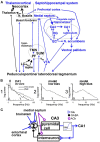
Figure 1
Figure 1
(A) Schematic neural circuit for arousal and psychotic behaviors in septohippocampal and thalamocortical systems. Peduncolopontine tegmentum and lateral dorsal tegmentum project to monoaminergic nuclei [locus ceruleus (LC), raphe, and ventral tegmental area (VTA)] and hypothalamic areas [perifornical area (PF), tuberomammillary nucleus (TMN), and supramammillary area (SUM)] which also project to the basal forebrain (medial septum and n. basalis) and thalamus. The limbic cortices of hippocampus, entorhinal cortex (EC) receive afferents from the medial septum; cingulate and prefrontal cortices receive afferents from both the medial septum and n. basalis. Limbic cortices project to the nucleus accumbens (NAC) and then ventral pallidum, and back to the brainstem. NAC and ventral pallidum mediate behaviors. (B) 1. Power spectrum of hippocampal local field potentials recorded in CA1 stratum radiatum during awake immobility (imm) and walking in the rat; power was measured in (mV)2/Hz, and theta peak power corresponds to a theta amplitude of ~ 1 mV peak-to-peak. 2. Hippocampal residue spectrum (without theta peaks) is generated by model with low bias; and 3. with high bias. (C) Recurrent feedback model of pyramidal cells exciting inhibitory interneurons through a non-linear element, with bias level determined by background activity, and feedback gain modulated by the medial septum, adapted from Leung (1982). Glu, glutamatergic; GABA, gamma aminobutyric acid, ACh, acetylcholine.
Figure 2
Figure 2
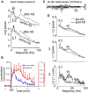
Hippocampal seizure and gamma. (A) Electrically induced after discharge (AD) in the hippocampus induced large gamma activity at electrodes across hippocampal CA1 area; L1 electrode placed at stratum radiatum, and L2 at near the alveus. (B) Minute by minute horizontal locomotion (infrared beans interrupts/min) and increase in hippocampal gamma (30–60 Hz) from baseline after a hippocampal AD at time 0; mean plus SE (n = 4 rats). (C) Characteristic pattern of hippocampal AD was observed at 17 min after injection of GABAB receptor antagonist CGP35348 (110 μg intraventricular, icv). (D) Power spectra at L1 and L2 electrodes across CA1, and L1-L2 coherence z-transform spectrum, recorded at 13 min after the CGP35348-induced AD (dark traces), overlaid with those during baseline walking (light traces). Power and coherence were high at theta (8.2 Hz) and gamma (peak at ~40 Hz). Power at the theta peak corresponded to ~ 1 mV of theta amplitude at L1 electrode. (A) After Leung (1987), (C,D) after Leung et al. (2005).
Figure 3
Figure 3
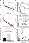
Hippocampal gamma and behaviors induced by 0.6 mg/kg s.c. ketamine and control. (A) Power spectra of local field potentials recorded in CA1 stratum radiatum after intraseptal 0.6 μl saline (Sal), compared with baseline walking; note increase of 30–100 Hz gamma after ketamine. (B) Same as A except preceded by intraseptal infusion of 0.25 μg/0.6 μl muscimol (Mus) instead of Sal; both theta and gamma were decreased after ketamine; intraseptal Mus only reduced theta peak and 30–100 Hz gamma as compared to baseline. (C) Prepulse inhibition (PPI) of the acoustic startle response (10–30 min post-ketamine/ saline s.c.) shows low PPI after intraseptal Sal plus ketamine (Ket) as compared to Sal plus saline s.c.; low PPI induced by Ket was normalized by pretreatment with intraseptal Mus. (D) Time course of average 30–70 Hz hippocampal gamma change with time after Ket injection, preceded by either intraseptal Sal or Mus infusion; gamma amplitude of ~0.1 mV = 2 log power units. (E) Average gamma (30–70 Hz) change after intraseptal infusion of Sal or Mus only. (F) Horizontal movements measured by interrupts of infrared beams show lower movements after septal infusion of muscimol as compared to saline. After Ma and Leung (2007).

Similar articles

Cited by

References

    1. Adler L. E., Waldo M. C., Freedman R. (1985). Neurophysiologic studies of sensory gating in schizophrenia: comparison of auditory and visual responses. Biol. Psych. 20, 1284–1296. - PubMed
    1. Alkire M. T., Hudetz A. G., Tononi G. (2008). Consciousness and anesthesia. Science 322, 876–880. 10.1126/science.1149213 - DOI - PMC - PubMed
    1. Allen T. A., Narayanan N. S., Kholodar-Smith D. B., Zhao Y., Laubach M., Brown T. H. (2008). Imaging the spread of reversible brain inactivations using fluorescent muscimol. J. Neurosci. Methods 171, 30–38. 10.1016/j.jneumeth.2008.01.033 - DOI - PMC - PubMed
    1. Anderson T., Hu B., Pittman Q., Kiss Z. H. (2004). Mechanisms of deep brain stimulation: an intracellular study in rat thalamus. J. Physiol. 559, 301–313. 10.1113/jphysiol.2004.064998 - DOI - PMC - PubMed
    1. Arcaro J., Ma J., Chu L., Kuo M. C., Mirsattari S., Leung L. S. (2016). The hippocampus participates in a pharmacological rat model of absence seizures. Epilepsy. Res. 120, 79–90. 10.1016/j.eplepsyres.2015.12.007 - DOI - PMC - PubMed

Publication types

Grants and funding