. 2022 Jul 7:13:913673.
doi: 10.3389/fphys.2022.913673. eCollection 2022.

Age-Associated Dysregulation of Integrin Function in Vascular Smooth Muscle

Affiliations

Age-Associated Dysregulation of Integrin Function in Vascular Smooth Muscle

Krishna Raj Ojha et al. Front Physiol. .

Abstract

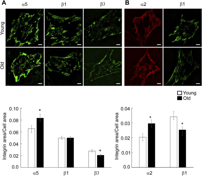
Arterial aging results in a progressive reduction in elasticity of the vessel wall and an impaired ability of aged blood vessels to control local blood flow and pressure. Recently, a new concept has emerged that the stiffness and decreased contractility of vascular smooth muscle (VSM) cells are important contributors to age-induced arterial dysfunction. This study investigated the hypothesis that aging alters integrin function in a matrix stiffness-dependent manner, which contributes to decreased VSM contractility in aged soleus muscle feed arteries (SFA). The effect of RGD-binding integrins on contractile function of cannulated SFA isolated from young (4 months) and old (24 months) Fischer 344 rats was assessed by measuring constrictor responses to norepinephrine, phenylephrine, and angiotensin II. Results indicated that constrictor responses in presence of RGD were impaired in old compared to young SFA. VSM cells isolated from young and old SFA were used for functional experiments using atomic force microscopy and high-resolution imaging. Aging was associated with a modulation of integrin β1 recruitment at cell-matrix adhesions that was matrix and substrate stiffness dependent. Our data showed that substrate stiffening drives altered integrin β1 expression in aging, while soft substrates abolish age-induced differences in overall integrin β1 expression. In addition, substrate stiffness and matrix composition contribute to the modulation of SMα-actin cytoskeleton architecture with soft substrates reducing age effects. Our results provide new insights into age-induced structural changes at VSM cell level that translates to decreased functionality of aged resistance soleus feed arteries.

Keywords: actin; aging; atomic force microscopy; integrins; vascular smooth muscle.


Conflict of interest statement

The authors declare that the research was conducted in the absence of any commercial or financial relationships that could be construed as a potential conflict of interest.

Figures

FIGURE 1
FIGURE 1
The role of integrin signaling in agonist-induced constrictor responses with age was assessed in the presence of a functional integrin blocking RGD peptide, a non-blocking RGE peptide, and control no treatment condition. Maximal constrictor responses to NE (10–4 M), PE (10–4 M), and Ang II (10–7 M) in SFA are presented. n = 5–12 rats per group. Data shown are mean ± SE. Significance was evaluated at p < 0.05. *Values are significantly different from young. # Values are significantly different from age-matched control and RGE group.
FIGURE 2
FIGURE 2
PCR assays were performed on VSM cells isolated from young and old SFA. Relative mRNA expression levels from young and old VSM cells were expressed relative to young VSM cells for each gene. Data are shown as mean ± SD (n = 3-4 animals). Significance was evaluated at p < 0.05. *Values are significantly different from young.
FIGURE 3
FIGURE 3
Representative TIRF images of VSM cells isolated from young and old Fischer 344 rats fluorescently labeled for integrin α5, β1 and β3 are shown. Scale bar represents 10 μm. Quantitative measurements (n = 29–39) are presented as mean ± SE. *Significance was evaluated at p < 0.05.
FIGURE 4
FIGURE 4
(A) Representative TIRF images of VSM cells plated on fibronectin and fluorescently labeled for integrin α5, β1 and β3 are shown. (B) Representative TIRF images of VSM cells plated on collagen-I and fluorescently labeled for integrin α2 (red) and β1 (green) are shown. Scale bar represents 10 μm. Quantitative measurements (n = 15–50) are presented as mean ± SE. Significance was evaluated at p < 0.05.
FIGURE 5
FIGURE 5
(A) Adhesion assay of VSM cells to fibronectin was performed in the presence of a functional integrin blocking RGD peptide and a non-blocking RGE peptide. Negative control experiments were performed using untreated cells plated on substrates blocked with BSA, and specific matrix without cells. Data are presented as mean ± SEM. (B) Functional AFM measurements of integrin α5β1 binding force to fibronectin for young and old VSM cells treated with a functional integrin blocking RGD peptide and a non-blocking RGE peptide are shown. For comparisons, force peak values whose confidence intervals did not overlap were considered significantly different (p < 0.05). (C) Percent of adhesion events for each treatment is shown.
FIGURE 6
FIGURE 6
Representative confocal images of VSM cells plated on (A) rigid and (B) soft substrates functionalized with fibronectin and collagen-I, respectively, are shown. VSM cells have been fluorescently labeled for integrin β1. Scale bar represents 10 μm. Quantitative measurements of overall fluorescence intensity (n = 28–68) are presented as mean ± SE. Significance was evaluated at p < 0.05. *Values are significantly different from young. # Values are significantly different from age matched VSM cells on different matrices. $ Values are significantly different between matrices.
FIGURE 7
FIGURE 7
Representative confocal images of VSM cells plated on soft substrates functionalized with fibronectin (A) and collagen-I (B) are shown. VSM cells have been fluorescently labeled for integrin β1. Scale bar represents 10 μm. Quantification of fluorescence intensity relative to inner vs. outer cell area shows a strong recruitment of integrin β1 at cell edges for old cells plated on fibronectin (n = 28–30), with a modest relative change for cells plated on collagen-I (n = 40–50). Data are presented as mean ± SE. Significance was evaluated at p < 0.05. *Values are significantly different from young. #Values are significantly different from age matched VSM cells.
FIGURE 8
FIGURE 8
(A) Representative confocal images of VSM cells plated on rigid substrates functionalized with fibronectin or collagen-I are shown. VSM cells have been fluorescently labeled for SMα-actin. Scale bar represents 10 μm. Quantitative measurements of overall fluorescence intensity and actin fiber segmentation (n = 34–68) are presented as mean ± SE. Significance was evaluated at p < 0.05. (B) Representative confocal images of VSM cells plated on soft substrates functionalized with fibronectin or collagen-I are shown. VSM cells have been fluorescently labeled for SMα-actin. Scale bar represents 10 μm. Quantitative measurements of overall fluorescence intensity and actin fiber segmentation (n = 22–42) are presented as mean ± SE. Significance was evaluated at p < 0.05. *Values are significantly different from young. $ Values are significantly different from age matched VSM cells on different matrices. # Values are significantly different between matrices.

References

    1. AlGhatrif M., Strait J. B., Morrell C. H., Canepa M., Wright J., Elango P., et al. (2013). Longitudinal Trajectories of Arterial Stiffness and the Role of Blood Pressure. Hypertension 62 (5), 934–941. 10.1161/hypertensionaha.113.01445 - DOI - PMC - PubMed
    1. Barton M., Cosentino F., Brandes R. P., Moreau P., Shaw S., Lüscher T. F. (1997). Anatomic Heterogeneity of Vascular Aging. Hypertension 30 (4), 817–824. 10.1161/01.hyp.30.4.817 - DOI - PubMed
    1. Brassard P., Amiri F., Thibault G., Schiffrin E. L. (2006). Role of Angiotensin Type-1 and Angiotensin Type-2 Receptors in the Expression of Vascular Integrins in Angiotensin II-Infused Rats. Hypertension 47 (1), 122–127. 10.1161/01.hyp.0000196272.79321.11 - DOI - PubMed
    1. Briones A. M., Salaices M., Vila E. (2007). Mechanisms Underlying Hypertrophic Remodeling and Increased Stiffness of Mesenteric Resistance Arteries from Aged Rats. Journals Gerontology Ser. A Biol. Sci. Med. Sci. 62 (7), 696–706. 10.1093/gerona/62.7.696 - DOI - PubMed
    1. Bunni M. A., Kramarenko, Walker L., Raymond J. R., Garnovskaya M. N. (2011). Role of Integrins in Angiotensin II-Induced Proliferation of Vascular Smooth Muscle Cells. Am. J. Physiology-Cell Physiology 300 (3), C647–C656. 10.1152/ajpcell.00179.2010 - DOI - PMC - PubMed

LinkOut - more resources