

Multisystem Inflammatory Syndrome in Children and Long COVID: The SARS-CoV-2 Viral Superantigen Hypothesis
- PMID: 35874696
- PMCID: PMC9300823
- DOI: 10.3389/fimmu.2022.941009
Multisystem Inflammatory Syndrome in Children and Long COVID: The SARS-CoV-2 Viral Superantigen Hypothesis
Abstract
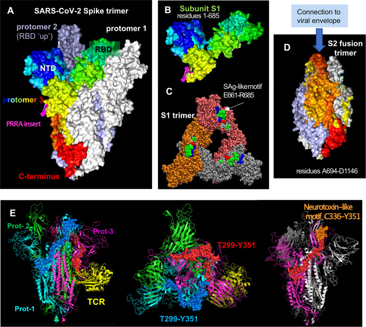
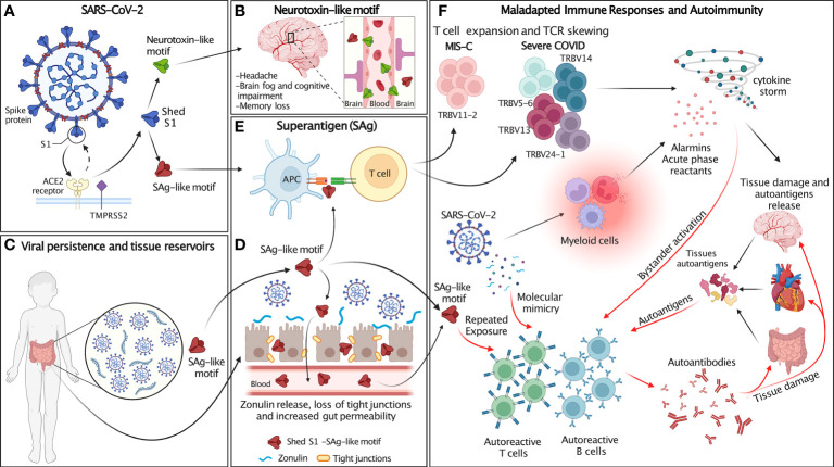
Multisystem inflammatory syndrome in children (MIS-C) is a febrile pediatric inflammatory disease that may develop weeks after initial SARS-CoV-2 infection or exposure. MIS-C involves systemic hyperinflammation and multiorgan involvement, including severe cardiovascular, gastrointestinal (GI) and neurological symptoms. Some clinical attributes of MIS-C-such as persistent fever, rashes, conjunctivitis and oral mucosa changes (red fissured lips and strawberry tongue)-overlap with features of Kawasaki disease (KD). In addition, MIS-C shares striking clinical similarities with toxic shock syndrome (TSS), which is triggered by bacterial superantigens (SAgs). The remarkable similarities between MIS-C and TSS prompted a search for SAg-like structures in the SARS-CoV-2 virus and the discovery of a unique SAg-like motif highly similar to a Staphylococcal enterotoxin B (SEB) fragment in the SARS-CoV-2 spike 1 (S1) glycoprotein. Computational studies suggest that the SAg-like motif has a high affinity for binding T-cell receptors (TCRs) and MHC Class II proteins. Immunosequencing of peripheral blood samples from MIS-C patients revealed a profound expansion of TCR β variable gene 11-2 (TRBV11-2), which correlates with MIS-C severity and serum cytokine levels, consistent with a SAg-triggered immune response. Computational sequence analysis of SARS-CoV-2 spike further identified conserved neurotoxin-like motifs which may alter neuronal cell function and contribute to neurological symptoms in COVID-19 and MIS-C patients. Additionally, autoantibodies are detected during MIS-C, which may indicate development of post-SARS-CoV-2 autoreactive and autoimmune responses. Finally, prolonged persistence of SARS-CoV-2 RNA in the gut, increased gut permeability and elevated levels of circulating S1 have been observed in children with MIS-C. Accordingly, we hypothesize that continuous and prolonged exposure to the viral SAg-like and neurotoxin-like motifs in SARS-CoV-2 spike may promote autoimmunity leading to the development of post-acute COVID-19 syndromes, including MIS-C and long COVID, as well as the neurological complications resulting from SARS-CoV-2 infection.
Keywords: MIS-C multisystem inflammatory syndrome in children; SARS-CoV-2; long COVID; neurotoxin-like segment; post-acute sequelae of COVID-19 (PASC); superantigen; superantigen-like motif.
Copyright © 2022 Noval Rivas, Porritt, Cheng, Bahar and Arditi.
Conflict of interest statement
The authors declare that the research was conducted in the absence of any commercial or financial relationships that could be construed as a potential conflict of interest.
Figures
Similar articles
-
Kawasaki Disease and Multisystem Inflammatory Syndrome in Children: Common Inflammatory Pathways of Two Distinct Diseases.Rheum Dis Clin North Am. 2023 Aug;49(3):647-659. doi: 10.1016/j.rdc.2023.03.002. Epub 2023 Mar 17. Rheum Dis Clin North Am. 2023. PMID: 37331738 Free PMC article. Review.
-
Superantigenic character of an insert unique to SARS-CoV-2 spike supported by skewed TCR repertoire in patients with hyperinflammation.Proc Natl Acad Sci U S A. 2020 Oct 13;117(41):25254-25262. doi: 10.1073/pnas.2010722117. Epub 2020 Sep 28. Proc Natl Acad Sci U S A. 2020. PMID: 32989130 Free PMC article.
-
SARS-CoV-2 Spike Does Not Possess Intrinsic Superantigen-like Inflammatory Activity.Cells. 2022 Aug 15;11(16):2526. doi: 10.3390/cells11162526. Cells. 2022. PMID: 36010602 Free PMC article.
-
HLA class I-associated expansion of TRBV11-2 T cells in multisystem inflammatory syndrome in children.J Clin Invest. 2021 May 17;131(10):e146614. doi: 10.1172/JCI146614. J Clin Invest. 2021. PMID: 33705359 Free PMC article.
-
Phenotype, Susceptibility, Autoimmunity, and Immunotherapy Between Kawasaki Disease and Coronavirus Disease-19 Associated Multisystem Inflammatory Syndrome in Children.Front Immunol. 2021 Feb 26;12:632890. doi: 10.3389/fimmu.2021.632890. eCollection 2021. Front Immunol. 2021. PMID: 33732254 Free PMC article.
Cited by
-
The results of a unique dietary supplement (nutraceutical formulation) used to treat the symptoms of long-haul COVID.Front Nutr. 2022 Oct 25;9:1034169. doi: 10.3389/fnut.2022.1034169. eCollection 2022. Front Nutr. 2022. PMID: 36386945 Free PMC article.
-
SARS-CoV-2 spike does not interact with the T cell receptor or directly activate T cells.Proc Natl Acad Sci U S A. 2024 Jul 30;121(31):e2406615121. doi: 10.1073/pnas.2406615121. Epub 2024 Jul 23. Proc Natl Acad Sci U S A. 2024. PMID: 39042676 Free PMC article.
-
Youth experiences with and perspectives on long covid.BMC Public Health. 2023 Oct 20;23(1):2059. doi: 10.1186/s12889-023-16899-8. BMC Public Health. 2023. PMID: 37864192 Free PMC article.
-
Kawasaki Disease and Multisystem Inflammatory Syndrome in Children: Common Inflammatory Pathways of Two Distinct Diseases.Rheum Dis Clin North Am. 2023 Aug;49(3):647-659. doi: 10.1016/j.rdc.2023.03.002. Epub 2023 Mar 17. Rheum Dis Clin North Am. 2023. PMID: 37331738 Free PMC article. Review.
-
Multi-Inflammatory Syndrome in Children (MIS-C) in 2023: Is It Time to Forget about It?Children (Basel). 2023 May 31;10(6):980. doi: 10.3390/children10060980. Children (Basel). 2023. PMID: 37371212 Free PMC article.
References
Publication types
MeSH terms
Substances
Supplementary concepts
Grants and funding
LinkOut - more resources
Full Text Sources
Medical
Research Materials
Miscellaneous